RCOM RADIOLOGICAL CASE OF THE MONTH
Applied Radiology — Vol. 31 , Issue 5 , pp. 47 -49
DOI: 10.37549/AR1088
Published: May 1, 2002
Categories
CASE SUMMARY
A 60-year-old Hispanic man with history of diabetes mellitus, status post left-leg below-the-knee amputation presented with several hours of worsening diffuse abdominal pain. Patient was discharged against medical advice. Figure 1 illustrates the scout film.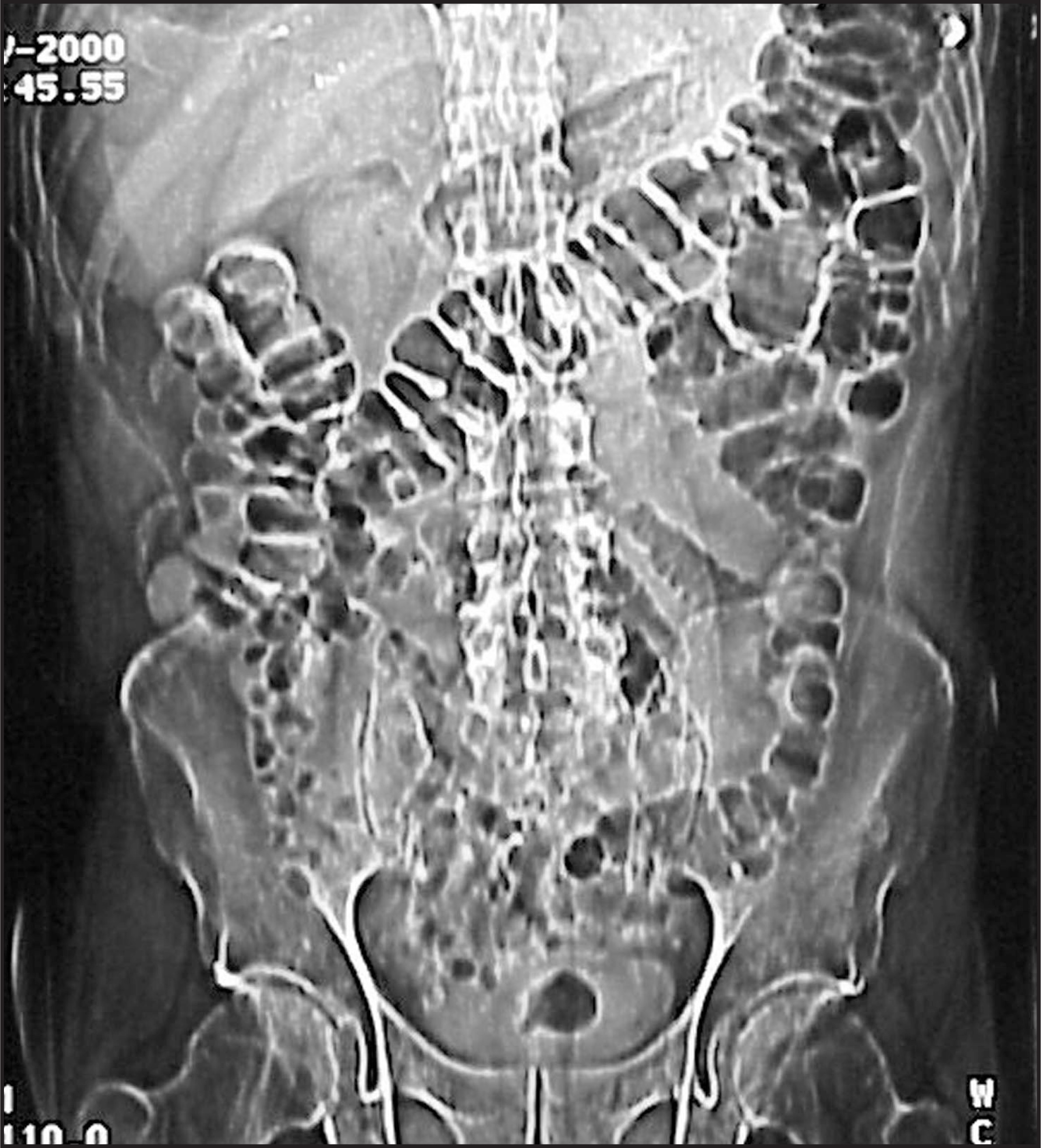
DIAGNOSIS
Lumbar hernia of the Petit’s subtype
IMAGING FINDINGS
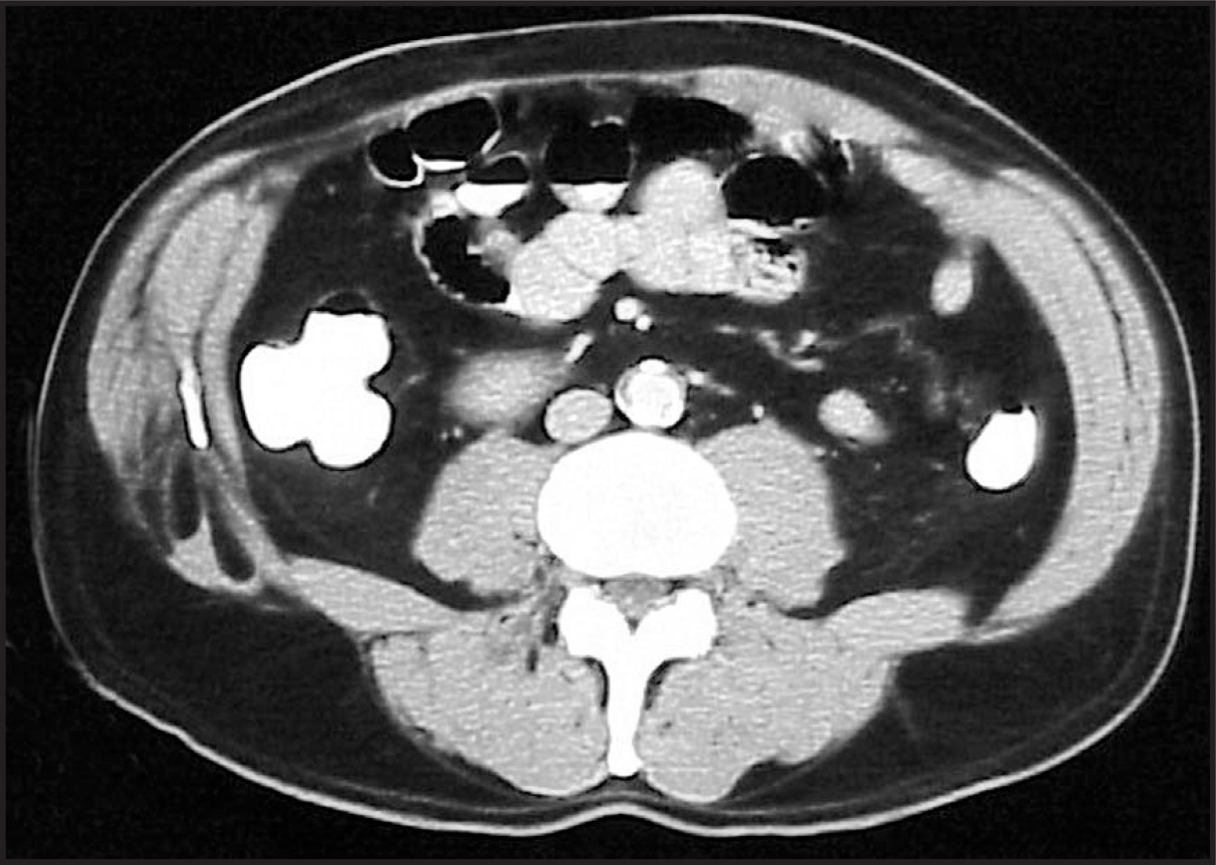
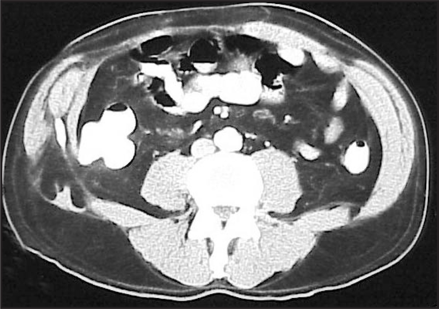
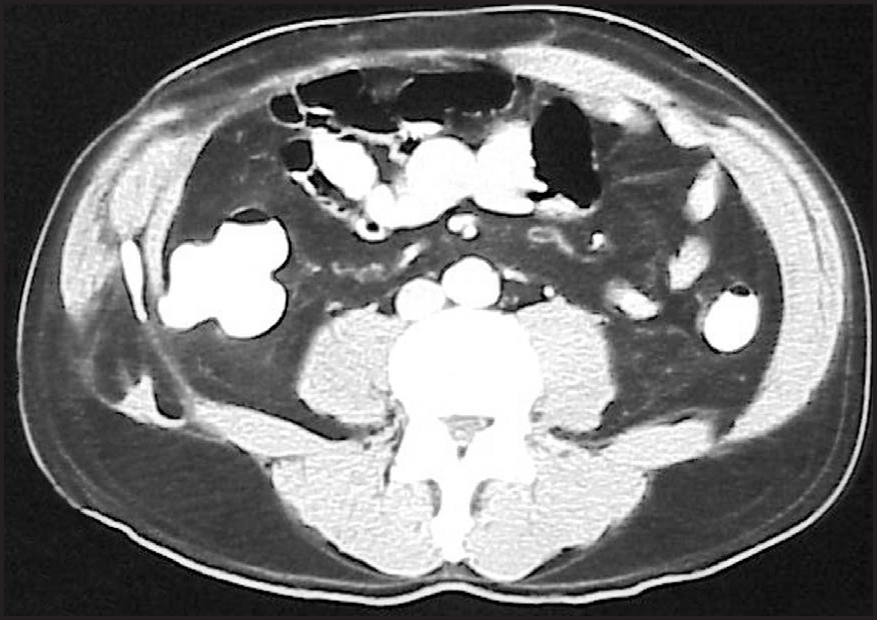
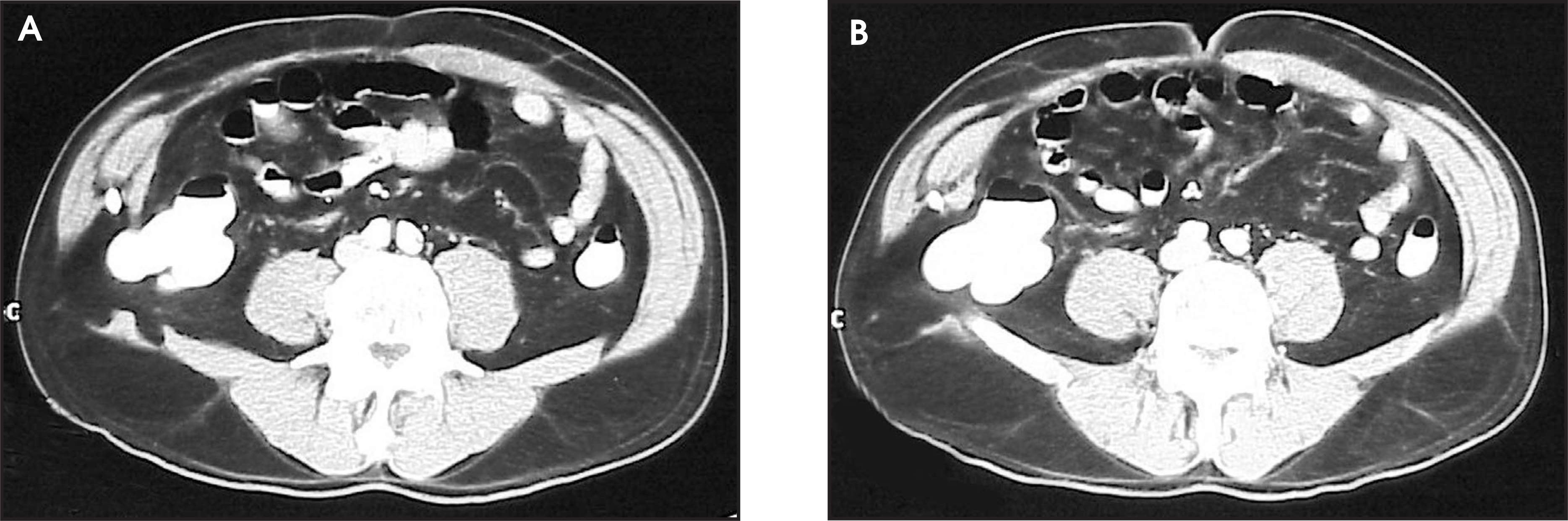
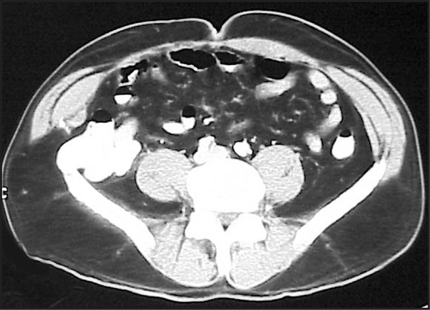
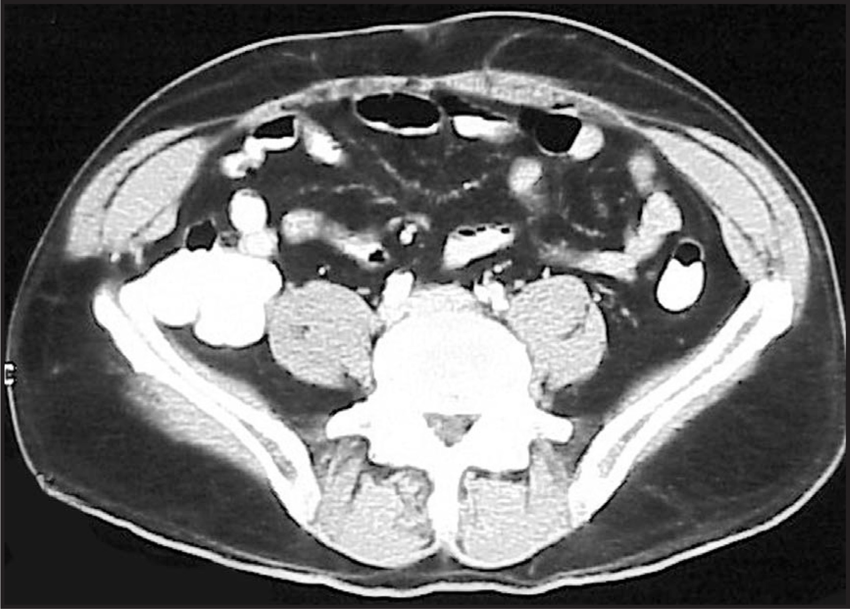
CT revealed a right lower lateral body wall defect containing intra-abdominal fat, contrast-opacified cecum, and vermiform appendix (figures 2 through 8). The study was obtained utilizing 7-mm contiguous sections through the abdomen with a pitch of 1.5 and 10-mm sections through the pelvis with a pitch of 1.0 following administration of oral and intravenous contrast. The scanner was a Siemens Somatom Plus 4 (Siemens Medical Systems, Erlangen, Germany).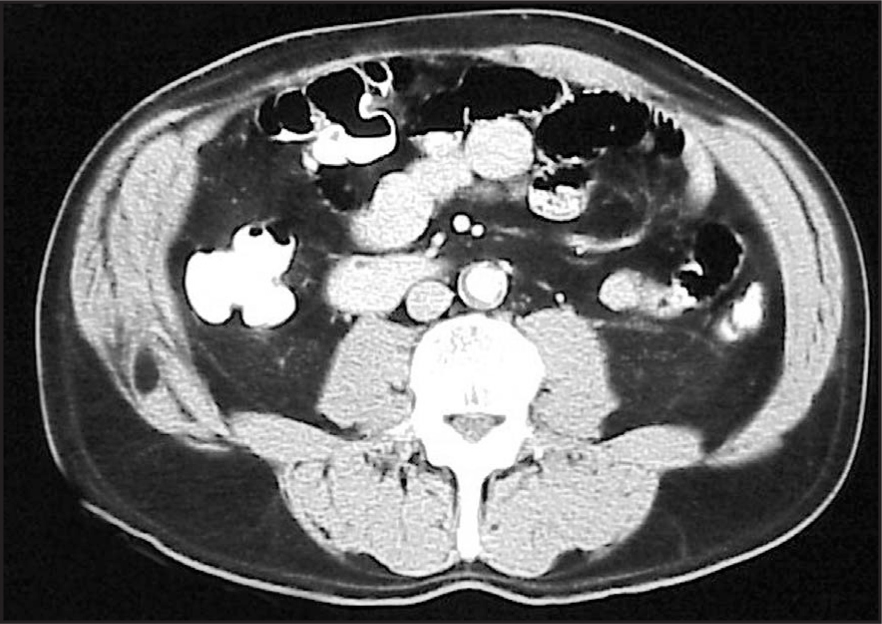
DISCUSSION
Of the various types of abdominal hernias, the most unique are arguably the lumbar hernias. There are approximately 300 cases reported in the medical literature.1 Most lumbar hernias are of the Grynfeltt-Lesgaft subtype, which is defined by an inverted triangle bound by the 12th rib superiorly, the internal oblique muscle anteriorly, and the erector spinae muscle posteriorly.2-4 Even more infrequent is the Petit’s subtype. Petit’s triangle is bound by the iliac crest inferiorly, by the external oblique muscle anteriorly, and by the latissimus dorsi muscle posteriorly.2-4 The existence of this space is found in the adult consistently, but not in the infant, therefore suggesting its development as a by-product of body growth.
When these defects occur in the pediatric population, they are thought to be congenital and are often associated with the lumbo-costo-vertebral syndrome. This entity is composed of caudal regression anomalies, diaphragmatic hernia, ureteropelvic junction obstruction, cloacal exstrophy, and lipomeningocele.
The great majority of these hernias, however, occur in the adult population, especially on the left side of the body and in middle-aged men. Spontaneous lumbar hernias may represent true attenuation of abdominal fascial layers secondary to the aging process. These hernias can also occur iatrogenically following the removal of the iliac crest for bone graft.5
The contents inside these defects may include retroperitoneal fat, kidney, or a bowel loop. Associated symptoms may range from low back pain to intestinal obstruction.2 Bowel incarceration occurs in up to 25% of cases and strangulation in up to 10%.
References
- Nyhus L, Condon R. Hernia.. 1995:412-424.
- Gore R, Levine M. Textbook of Gastrointestinal Radiology.. 2000:2002-2003.
- Schwartz S. Principles of Surgery.. 1999:1606.
- Townsend C. Sabiston Textbook of Surgery.. 2001:798.
- Morris P, Malt R. Oxford Textbook of Surgery.. 1994:1411-1412.
Citation
. RCOM RADIOLOGICAL CASE OF THE MONTH. Applied Radiology. 2002;31(5):47-49. doi:10.37549/AR1088.