RCOM RADIOLOGICAL CASE OF THE MONTH
Applied Radiology — Vol. 31 , Issue 11 , pp. 63 -65
DOI: 10.37549/AR1132
Published: November 1, 2002
Categories
CASE SUMMARY
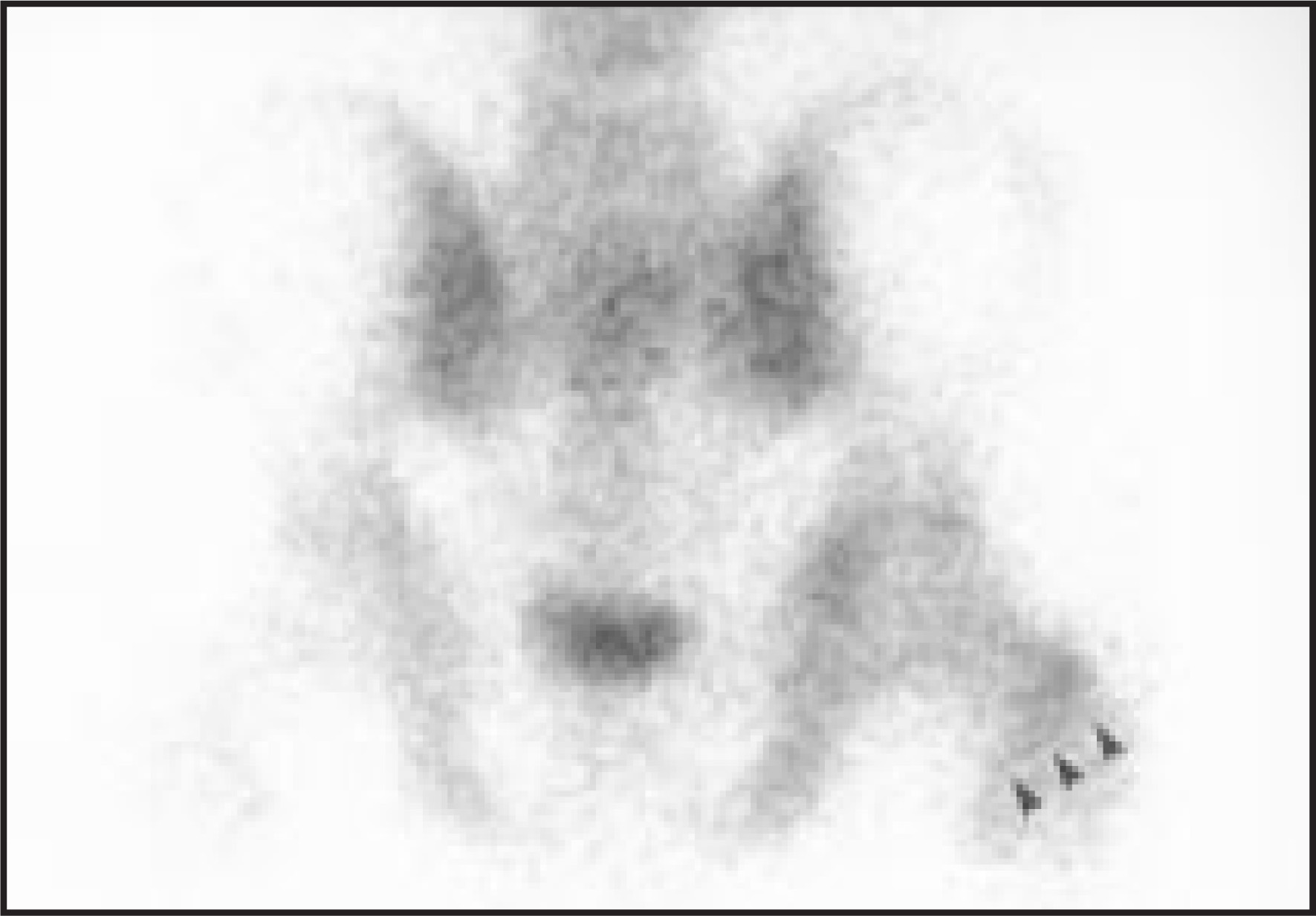
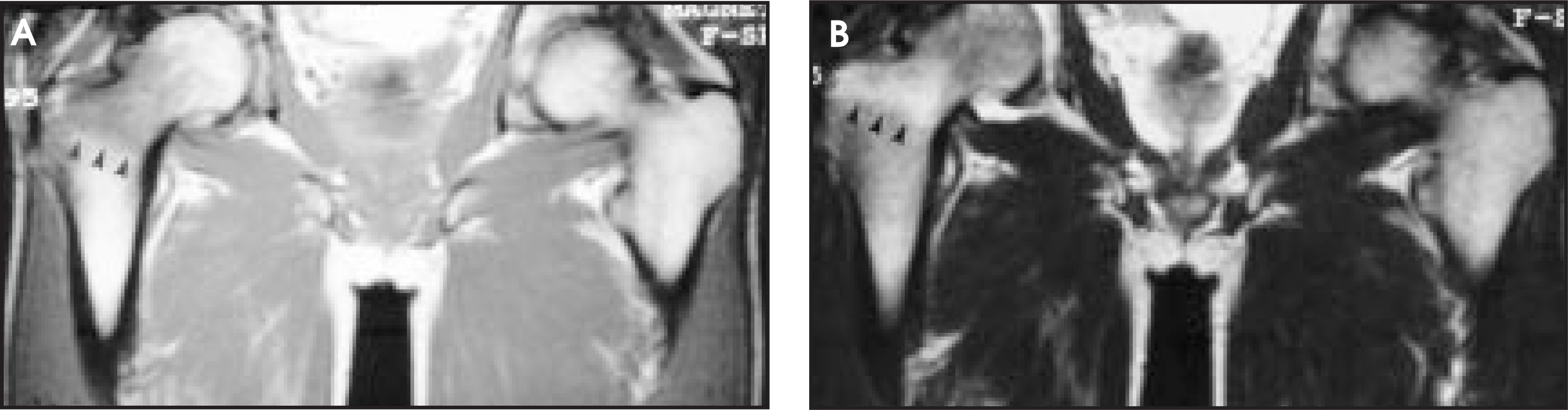
A 34-year-old white man presented with right hip pain of 1-year duration. He is a mechanic and has been an avid footbag player for several years. He denies any specific trauma. On physical examination, pain was present with any range of motion but was more severe with external and internal rotation and straight-leg raise. He was treated with non-weight-bearing, non-steroidal anti-inflammatory drugs, and analgesics for 1 year, but the pain was refractory. Plain radiograph (Figure 1), radionuclide bone scan (Figure 2), and MRI of the right hip (Figure 3) were obtained.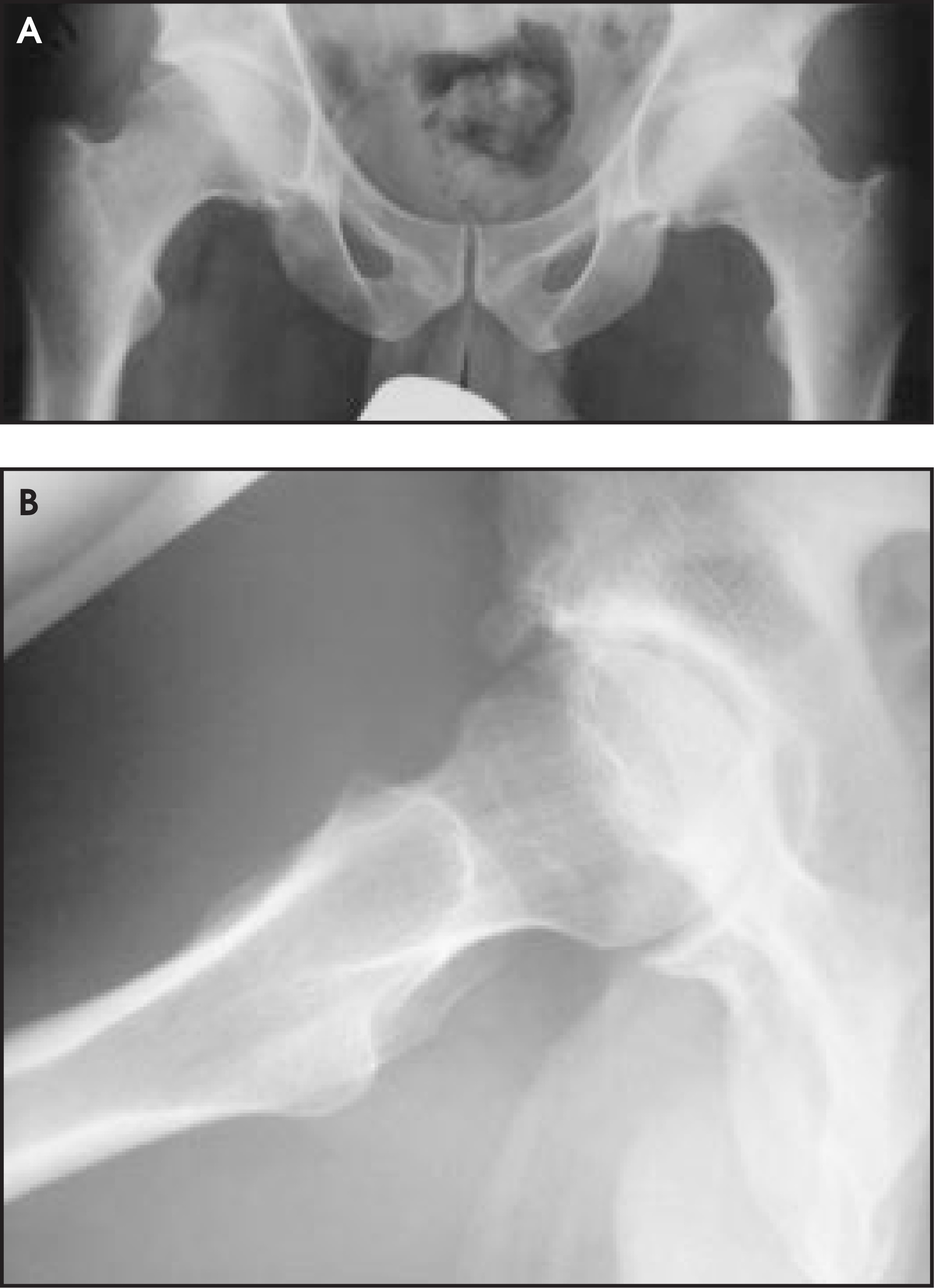
DIAGNOSIS
Intertrochanteric occult stress fracture in an avid Hacky Sack (WHAM-O, Inc., Emeryville, CA) player.
IMAGING FINDINGS
A plain radiograph of the right hip was unremarkable except for bilateral degenerative joint disease and the presence of os acetabulum at the right hip (Figure 1). A bone scan revealed augmented radionuclide uptake at the right hip, prominently at the intertrochanteric region (Figure 2). This area showed low signal intensity on Tl-weighted imaging (T1WI) (Figure 3A) and a band of bright signal on T2-weighted imaging (T2WI) (Figure 3B), consistent with a bone contusion or occult stress fracture. A CT-guided bone biopsy was performed, which revealed no tumor, infection, or other pathologic lesion. He underwent open reduction and internal fixation of the right hip with a Synthes dynamic screw (Synthes, Paoli, PA). The intertrochanteric region was scraped by curette and the bone specimen was sent to pathology. On microscopic examination, numerous small fragments of bone with empty lacunae, possibly due to necrosis or crush artifact, were noted. No evidence of tumor or osteomyelitis was noted.
DISCUSSION
Footbag is a growing sport. Although played as leisure for many years, it has existed as a competitive sport in several forms since the late 1970s and has a growing number of events each year. It is played mainly in the United States and Canada, but there are many footbag clubs and players around the world. During play, the bag, which is a small, hard or soft ball, may not contact the body except below the knee. Several forms of footbag sport include freestyle, footbag net, team freestyle, and footbag golf.
Bone contusions, so-called occult fractures, are microfractures of the cancellous bony trabeculae with edema and hemorrhage.1,2 These contusions were originally described by magnetic resonance (MR) as speckled or reticulated areas within cancellous bone revealing low signal intensity on T1WI and high signal intensity on T2WI. Occasionally, they would have band-like low-signal areas simulating stress fractures. They have been known by a variety of terms, such as bone bruises, occult fractures, osteochondral fractures, and occult osseous lesions. Accordingly, they may represent a spectrum of radiographically occult bone injury, ranging from simple trabecular hemorrhage, infarction, and edema; to microscopic compression fractures of cancellous bone; to osteochondral fractures that can be seen arthroscopically.2 Bone contusions are best imaged on fat-saturated or STIR MR imaging. When different MR techniques were examined, fast-spin-echo with fat saturation and fast-spin-echo short tau inversion recovery (STIR) sequences proved to be superior to conventional STIR sequences for the diagnosis of bone contusion.1
Femoral neck stress fracture (FNSF) is uncommon and accounts for 3% to 4% of stress fractures. It is generally seen in young, active endurance athletes and military recruits, or in the elderly with osteoporosis.3 Stress fractures are more common in the lower extremities, with 7.2% occurring in the femur, the fourth most common site in athletes after tibia (49%), tarsals (25.3%), and metatarsals (8.8%).4 However, the actual incidence of femoral stress fracture is difficult to assess. Stress fracture at the trochanteric region is very rare. Stress fractures are mostly transcervical at the femoral neck. Femoral neck stress fractures in younger individuals are often the result of athletic activities—such as long-distance running and ballet dancing—and are also seen in military recruits. They are especially associated with initiation of a new athletic activity or an increase in the frequency or intensity of a current activity.5
There are two primary types of FNSF: tension and compression stress fractures. The tension type is usually directed perpendicular to the line of force transmission in the femoral neck. It originates at the superior surface of the neck, is at increased risk for displacement, and is treated surgically. The compression type has radiologic changes (callus formation) on the inferior femoral neck without apparent cortical disruption and is usually managed nonsurgically.3 Stress fractures of the femoral neck are often (67%) not visible on plain radiographs. Unlike fractures in the cortical bone, which show radiolucent lines on X-ray, cancellous bone fractures are not visible on initial radiographic examination. They usually appear as a sclerotic line on follow-up study, with or without periosteal new bone formation.6 Rarely, a radiolucent line called “dreaded black line” may be the only subtle finding on the plain radiograph.7 Bone scan, although sensitive, is nonspecific for stress fractures. Magnetic resonance is considered the modality of choice for occult femoral neck fractures.4 Limited MRI with coronal TlWI spin-echo sequence is adequate for fast and inexpensive diagnosis of femoral neck fractures.8
Bone contusions in the distal femur are treated conservatively. Bone contusions or occult fractures in the femoral neck deserve careful consideration. Treatment is based upon the type of FNSF. Tension fractures are unstable and require operative stabilization with multiple screws or a sliding hip screw. The area of fracture can be curetted or reamed at surgery to induce biologic reaction and decrease the chance of nonunion.3 The compression type can be managed nonsurgically with several days of rest and protected weight bearing. Frequent radiographs may be needed to detect any changes or displacement. A displaced FNSF in a young adult is an orthopaedic emergency and requires open reduction and internal fixation.3
SUMMARY
This case reports the clinical and imaging findings of intertrochanteric fracture in an avid footbag player and discusses stress fractures of the femoral neck.
References
- Amdt W, Truax A, Bamett F. MR diagnosis of bone contusions of the knee: Comparison of coronal T2-weighted fast spin-echo with fat saturation and fast spin-echo STIR images with conventional STIR images.. AJR Am J Roentgenol. 1996;166:119-124.
- Zeiss J, PaIey K, Murray K. Comparison of bone contusion seen by MRI in partial and complete tears of the anterior cruciate ligament.. J Comput Assist Tomogr.. 1995;19:773-776.
- Egol K, Koval K, Kummer F. Stress fractures of the femoral neck.. Clin Orthop.. 1998;348:72-78.
- Boden B, Speer K. Femoral stress fractures.. Clin Sports Med.. 1997;16:307-317.
- Daffner R, Pavlov M. Stress fractures: Current concepts.. AJR Am J Roentgenol.. 1992;159:245-252.
- Pais M, Wang F, Tehranzadeh J, Serafini A, Pais M. Avulsion and Stress Injuries of the Musculoskeletal System.. 1989:65-98.
- Monteleone G. Stress fractures in the athlete.. Orthop Clin North Am.. 1995;26:423-432.
- Quinn S, McCarthy J. Prospective evaluation of patients with suspected hip fracture and indeterminate radiographs: Use of T1-weighted MR images.. Radiology.. 1993;187:469-471.
Citation
. RCOM RADIOLOGICAL CASE OF THE MONTH. Applied Radiology. 2002;31(11):63-65. doi:10.37549/AR1132.