RCOM RADIOLOGICAL CASE OF THE MONTH
Applied Radiology — Vol. 31 , Issue 12 , pp. 29 -30
DOI: 10.37549/AR1143
Published: December 1, 2002
Categories
CASE SUMMARY
A 60-year-old woman presented with a 2-month history of abdominal pain. Her laboratory values were normal, including amylase and lipase levels. An abdominal computed tomography (CT) scan was performed (Figure 1).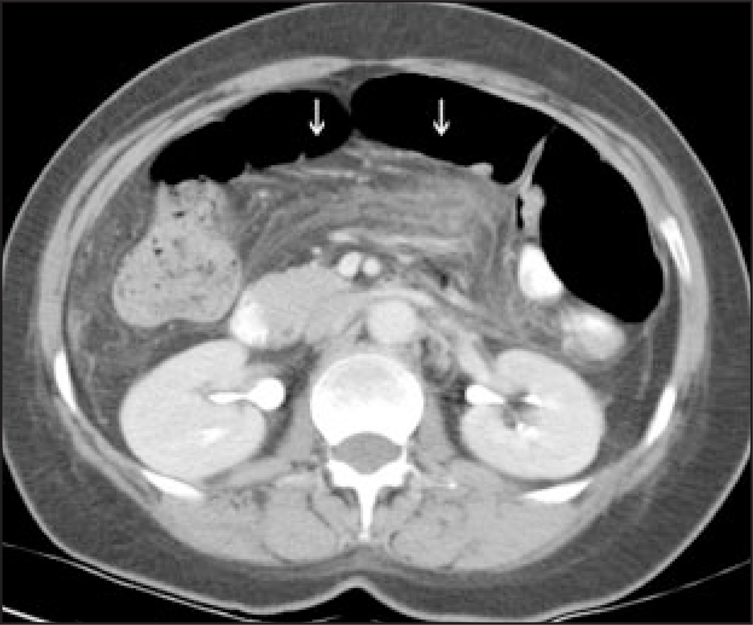
IMAGING FINDNGS
A contrast-enhanced CT scan of the abdomen demonstrates infiltration of the mesentery surrounding the mesenteric root (Figure 1). Because of recalcitrant symptoms, an exploratory laporatomy was eventually performed. Biopsies demonstrated vascular congestion with chronic inflammatory infiltrate and panniculitis. No other abnormality was discovered by imaging or at surgery.
DIAGNOSIS
Mesenteric panniculitis
DISCUSSION
Mesenteric panniculitis is a form of chronic inflammation of adipose tissue of the bowel mesentery. It is known by different names, including sclerosing mesenteritis, mesenteric lipodystrophy, and liposclerotic mesenteritis. It has been described as one stage of the three manifestations of mesenteric fat degeneration, which starts as mesenteric lipodystrophy with fatty degeneration, followed by mesenteric panniculitis with mesenteric inflammation, and ending in retractile mesenteritis with fibrosis. However, there is no evidence that this sequence occurs in all patients.1
Both pancreatitis and inflammatory bowel disease should be excluded before the diagnosis of mesenteric panniculitis is established. It has been associated with concurrent malignancy in up to 70% of cases, but a pathologic link has not been described. Additionally, up to 90% of patients with mesenteric panniculitis are asymptomatic. In fact, mesenteric panniculitis is frequently an incidental finding on abdominal CT scan. An autoimmune etiology has been conjectured as a cause for the inflammatory process, but this remains to be proven.2
Mesenteric panniculitis is seen on CT scans of the abdomen as infiltration of the mesentery secondary to inflammation, sometimes with small soft-tissue nodules and vessels surrounded by a hypodense fatty halo. It is more commonly seen on the left side of the abdomen along the orientation of the jejunal mesentery.2
Although mesenteric panniculitis is frequently concurrent with malignancy, it is a benign condition. It typically resolves without treatment. Steroids have been shown to have a possible benefit toward decreasing inflammation, but surgery is not indicated.3
CONCLUSION
Mesenteric panniculitis is a benign inflammatory condition of the mesentery, which is frequently asymptomatic and usually self-limiting.
References
- Patel N, Saleeb S, Teplick S. General case of the day: Mesenteric panniculitis. RadioGraphics. 1999;19:1083-1085.
- Daskalogiannaki M, Voloudaki A, Prassopoulos P. CT evaluation of mesenteric panniculitis: Prevalence and associated diseases. AJR Am J Roentgenol. 2000;174:427-431.
- Kirkiros C, Edis A. Mesenteric panniculitis resulting in bowel obstruction and response to steroids. Aust NZ J Surg. 1989;59:287-290.
Citation
. RCOM RADIOLOGICAL CASE OF THE MONTH. Applied Radiology. 2002;31(12):29-30. doi:10.37549/AR1143.