RCOM RADIOLOGICAL CASE OF THE MONTH
Applied Radiology — Vol. 31 , Issue 12 , pp. 31 -32
DOI: 10.37549/AR1147
Published: December 1, 2002
Categories
CASE SUMMARY
A 28-year-old woman presented with a chief complaint of dyspnea. Physical examination revealed an anxious woman with diffuse upper-airway wheezing. Her medical history included Nissen fundoplication for asthma symptoms attributed to gastroesophageal reflux. Over a 5-year period, she had been evaluated by numerous primary care and emergency medicine physicians and treated with inhalers, allergy medicines, and corticosteroids with no lasting relief of symptoms. Chest radiography was followed by high-resolution computed tomography (HRCT) of the chest, bronchoscopy, and thoracotomy.
DIAGNOSIS
Mucoepidermoid tumor of the lung
IMAGING FINDINGS
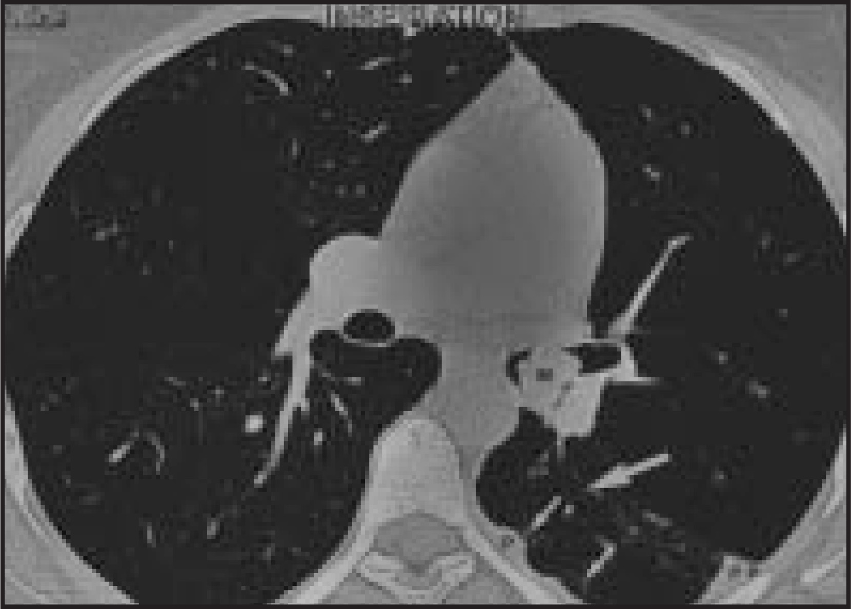
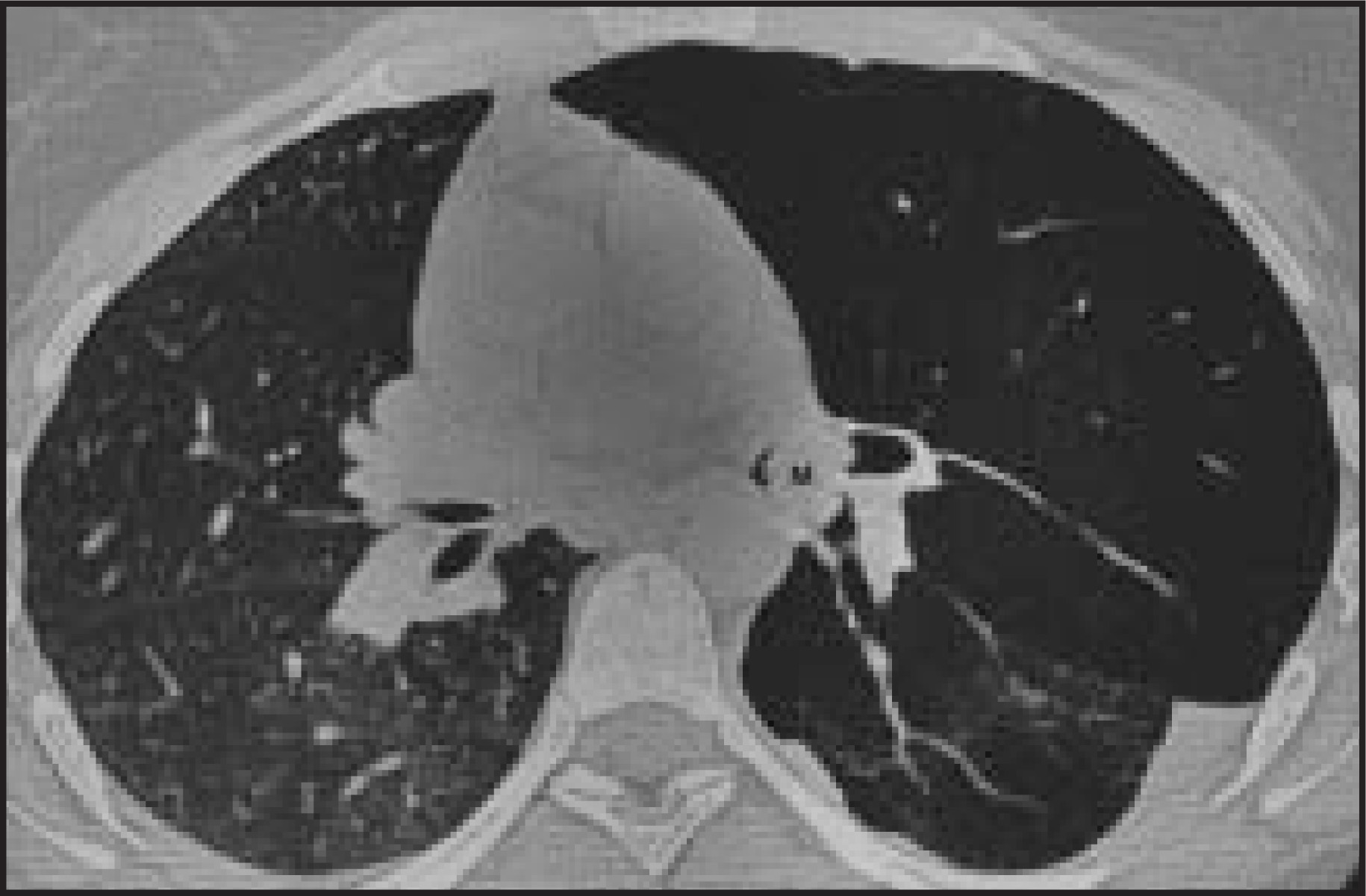
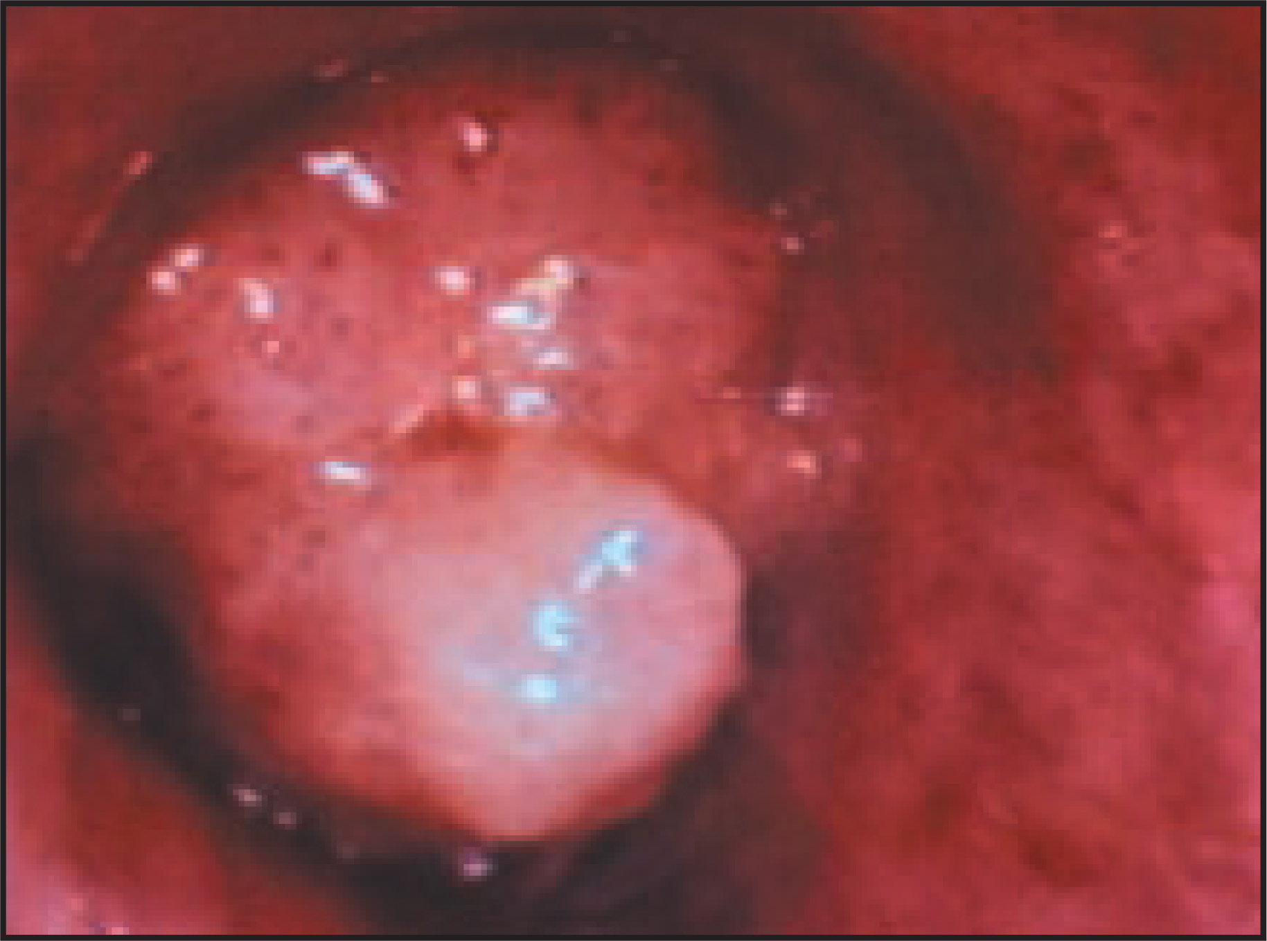
Chest radiograph at admission showed a hyperlucent left lung with diminutive vasculature (Figure 1). Inspiratory HRCT showed a lobulated soft-tissue mass in the left mainstem bronchus, extending into and straddling the left upper- and lower-lobe bronchi (Figure 2). The left upper lobe was hyperexpanded with diminutive vascularity, consistent with a ball-valve type obstruction and reflex vasoconstriction. In the left lower lobe there was volume loss, bronchial dilatation, small reticular opacities, and adjacent pleural thickening, attributed to previous episodes of obstructive pneumonia. Expiratory HRCT showed air trapping as a noncollapsing hyperlucent left lung, most markedly involving the left upper lobe (Figure 3), and contralateral mediastinal shift. Bronchoscopy revealed a smoothly marginated, polypoid “strawberry” lesion at the confluence of the left upper- and lower-lobe bronchi (Figure 4).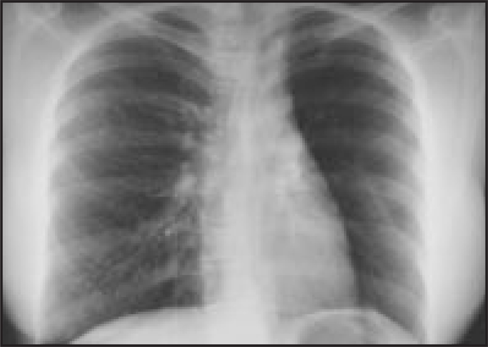
DISCUSSION
Mucoepidermoid tumors of the lung, first reported by Smetana et al,1 arise from tracheobronchial mucous glands and are similar in morphology to mucoepidermoid tumors arising from oropharyngeal salivary glands. They are one type of several tracheobronchial gland tumors that at one time were inaccurately referred to as bronchial adenomas.2,3 The diagnosis of mucoepidermoid tumor should be considered in anyone who has radiologic or endoscopic evidence of a polypoid intraluminal mass in the trachea or major bronchi. A history of adult-onset “asthma” that has increased in severity despite adequate therapy signals the possibility of a tracheobronchial lesion. The average duration of symptoms is between 8 and 18 months, but some individuals report symptoms for many years. The average age of patients is between 35 and 45 years, but patients as young as 6 years and as old as 78 years have been reported.4 Mucoepidermoid tumors occur most commonly in the main, lobar,4,5 and segmental bronchi.3 The chest radiograph may be interpreted as normal but usually shows signs of abnormal ventilation or perfusion to the affected lung, atelectasis, or pneumonia. A distinct endobronchial mass is often seen only on CT.3,6,7
CONCLUSION
A history of asthma in an adult should alert a radiologist to look for imaging signs of an endotracheal or endobronchial mass. These signs include hyperlucent lung, hyperexpanded lung, air trapping on expiration, lung/lobar collapse, recurrent pneumonia, and an endobronchial lesion.
References
- Smetana H, Iverson L, Swann L. Bronchogenic carcinoma, an analysis of 100 autopsy cases. Milit Surg. 1952;111:335-351.
- Heitmiller R, Mathisen D, Ferry J. Mucoepidermoid lung tumors. Ann Thorac Surg. 1989;47:394-399.
- Kim T, Lee K, Han J. Mucoepidermoid carcinoma of the tracheobronchial tree: Radiographic and CT findings in 12 patients. Radiology. 1999;212:643-648.
- Yousem S, Hochholzer L. Mucoepidermoid tumors of the lung. Cancer. 1987;60:1346-1352.
- Turnbull A, Huvos A, Goodner J, Foote F. Mucoepidermoid tumors of bronchial glands. Cancer. 1971;28:539-544.
- Noda S, Sundaresan S, Mendeloff E. Tracheal mucoepidermoid carcinoma in a 7-year-old child. Ann Thorac Surg. 1998;66:928-929.
- Fisher D, Mond D, Fuchs A, Khan A. Mucoepidermoid tumor of the lung: CT appearance. Comput Med Imaging Graphics. 1995;19:339-342.
Citation
. RCOM RADIOLOGICAL CASE OF THE MONTH. Applied Radiology. 2002;31(12):31-32. doi:10.37549/AR1147.