RCOM RADIOLOGICAL CASE OF THE MONTH
Applied Radiology — Vol. 32 , Issue 3 , pp. 38 -39
DOI: 10.37549/AR1170
Published: March 1, 2003
Categories
CASE SUMMARY
A 64-year-old man who presented with headaches underwent magnetic resonance imaging (MRI) of the brain, as an outpatient. He had no known neurologic deficits and no significant medical history. A gadolinium-enhanced T1-weighted axial brain MRI showed multiple brain nodules.
He underwent a quick MRI search for a primary malignancy. Two fast, coronal, large field-of-view sequences were performed: one centered over the chest and one centered over the abdomen and pelvis. It took 1 to 2 minutes to remove the head coil and center the patient in the body coil. Each of the two sequences took <30 seconds. Overall, this added only a few minutes to the exam time.
IMAGING FINDINGS
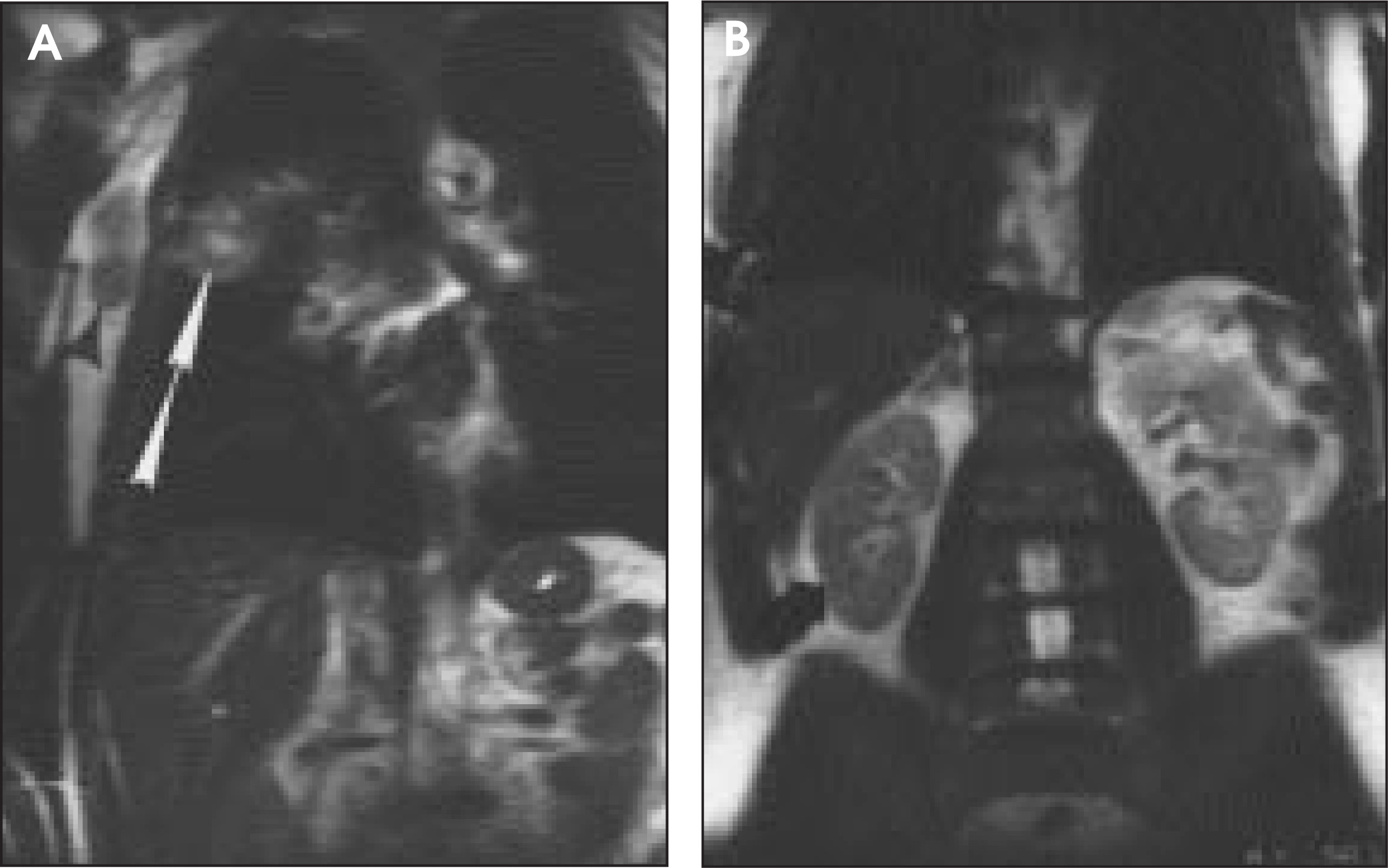
The preliminary MRI obviously shows multiple unsuspected brain metastases (Figure 1). The real question then is, “Where is the primary carcinoma?” A fast whole-body screening sequence identified a large lung mass in the right upper lobe with metastatic adenopathy in the right hilum and right axilla (Figure 2A). The abdomen and pelvis were grossly normal (Figure 2B).
With this information, a percu-taneous needle biopsy of the palpable right axillary mass was performed without the need for imaging guidance and without exposing the patient to the risk of pneumothorax. Pathology was positive for small cell carcinoma.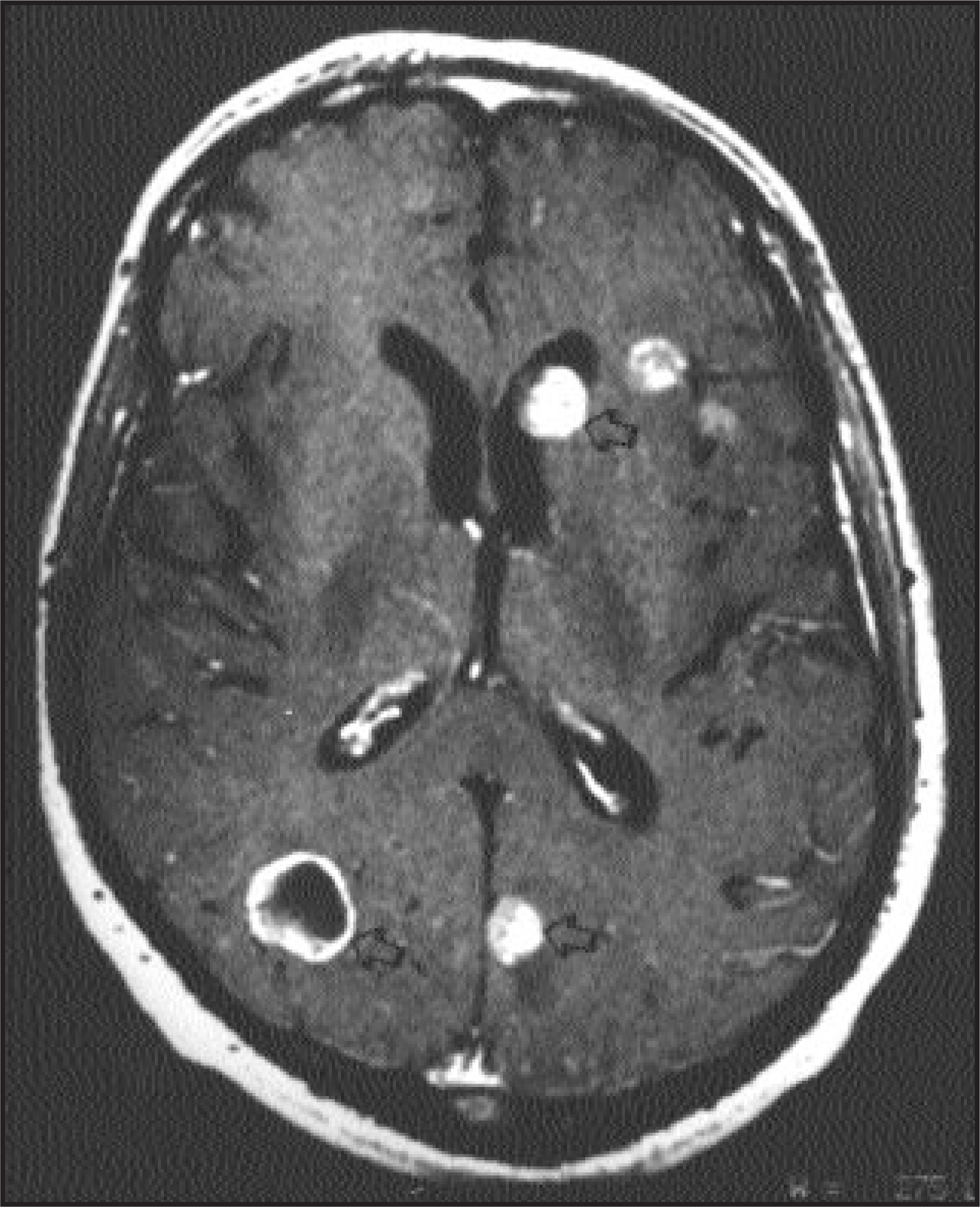
DIAGNOSIS
Small cell carcinoma
DISCUSSION
Occasionally, patients under-going brain or spine MRI are found unexpectedly to have MR findings suggestive of metastases. The MR technologists can usually recognize these findings. Our department has recently developed a fast whole-body MRI screening sequence that we run as a supplement to brain MRI of a patient who has been discovered to have brain metastases. This has proven to be very helpful and has added only a few minutes to the examination time.
In the case presented, we diagnosed the primary cancer, roughy staged the cancer, and found a readily accessible biopsy site. If this quick-search MR examination had been negative, we would have proceeded with the standard procedure for an unknown primary cancer, including chest X-ray, abdominal computed tomography (CT), pelvic CT, and, if required, chest CT, mammography, barium enema, and brain biopsy. In this case, fast whole-body MRI examination obviated the need for these additional exams and procedures.
CONCLUSION
This case illustrates the benefit of fast whole-body MRI sequences, which are helpful and can be performed easily and quickly on high-strength magnets.1-4
Further investigation of these fast MRI screening techniques should be considered to calculate the sensitivities and specificities of the whole-body MR screening examination. When brain or spine metastases are discovered unexpectedly, MR technologists and radiologists should consider adding this whole-body MRI screening sequence while the patient is still on the MR table.
References
- Walker R, Kessar P, Blanchard R. Turbo STIR magnetic resonance imaging as a whole-body screening tool for metastases in patients with breast carcinoma: A preliminary report. J Mag Reson Imaging. 2000;11:343-350.
- Mazumdar A, Siegel M, Narra V. Whole-body fast inversion recovery MR imaging of small cell neoplasms in pediatric patients: A pilot study. AJR Am J Roentgenol. 2002;179:1261-1266.
- Lauenstein T, Goehde S, Herborn C. Three-dimensional volumetric interpolated breath-hold MR imaging for whole-body tumor stapling in less than 15 minutes: A feasibility suidy. AJR Am J Roentgenol. 2002;179:445-449.
- Eustace S, Walker R, Blake M. Whole-body MR imaging. Practical issues, clinical applications, and future directions. Magn Reson Imaging Clin N Am. 1999;7:209-236.
Citation
. RCOM RADIOLOGICAL CASE OF THE MONTH. Applied Radiology. 2003;32(3):38-39. doi:10.37549/AR1170.