RCOM RADIOLOGICAL CASE OF THE MONTH
Applied Radiology — Vol. 32 , Issue 4 , pp. 45 -47
DOI: 10.37549/AR1176
Published: April 1, 2003
Categories
CASE SUMMARY
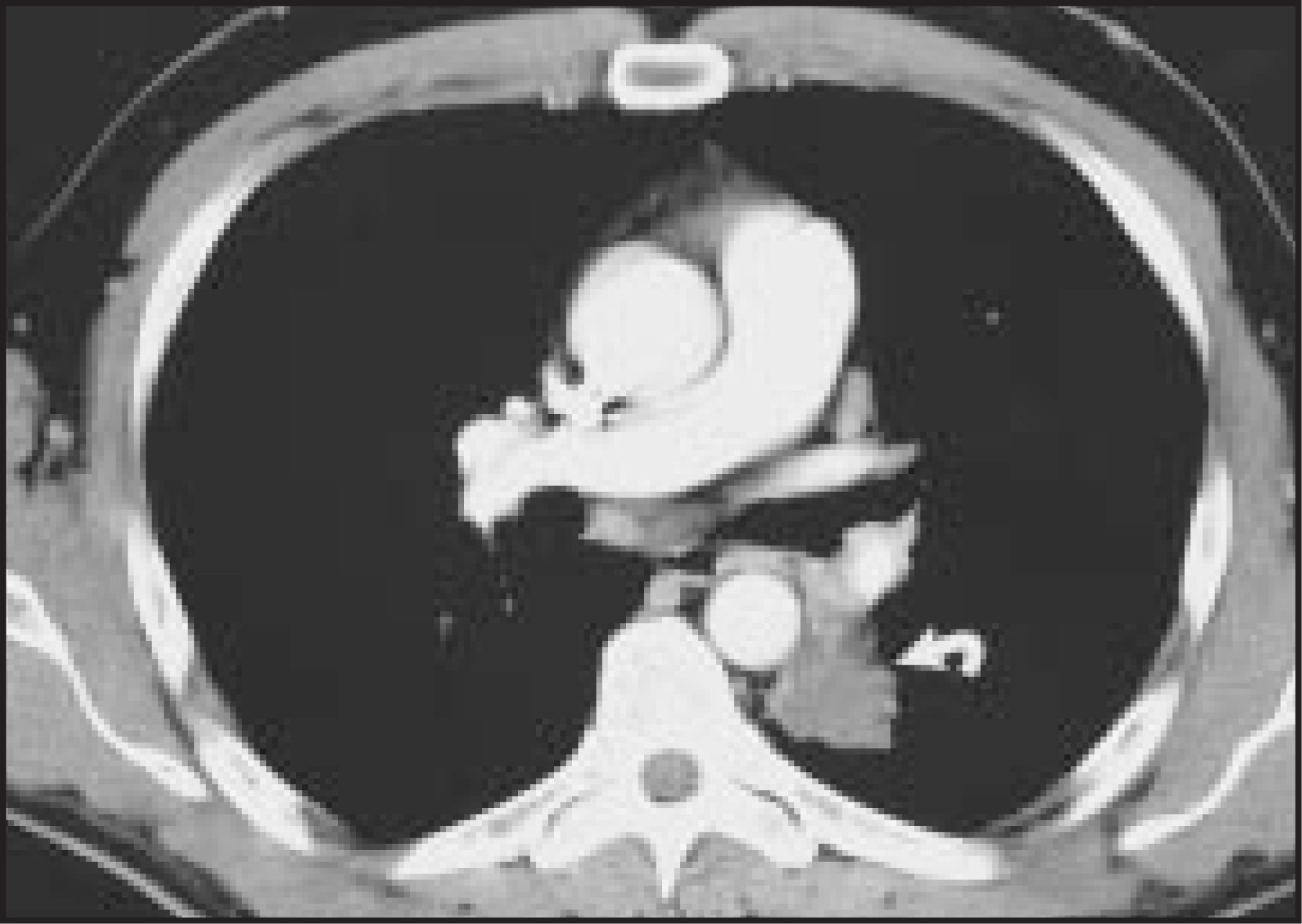
A 40-year-old male smoker presented to his primary care physician for persistent chest-wall pain following a mild sports injury. He also complained of worsening exertional dyspnea and night sweats. There was no report of significant surgical or medical history. The physical examination was unremarkable except for some minor chest-wall tenderness. Anteroposterior and lateral chest films were obtained (Figure 1). Based on these findings, a contrast-enhanced computed tomography (CT) study was performed (Figure 2).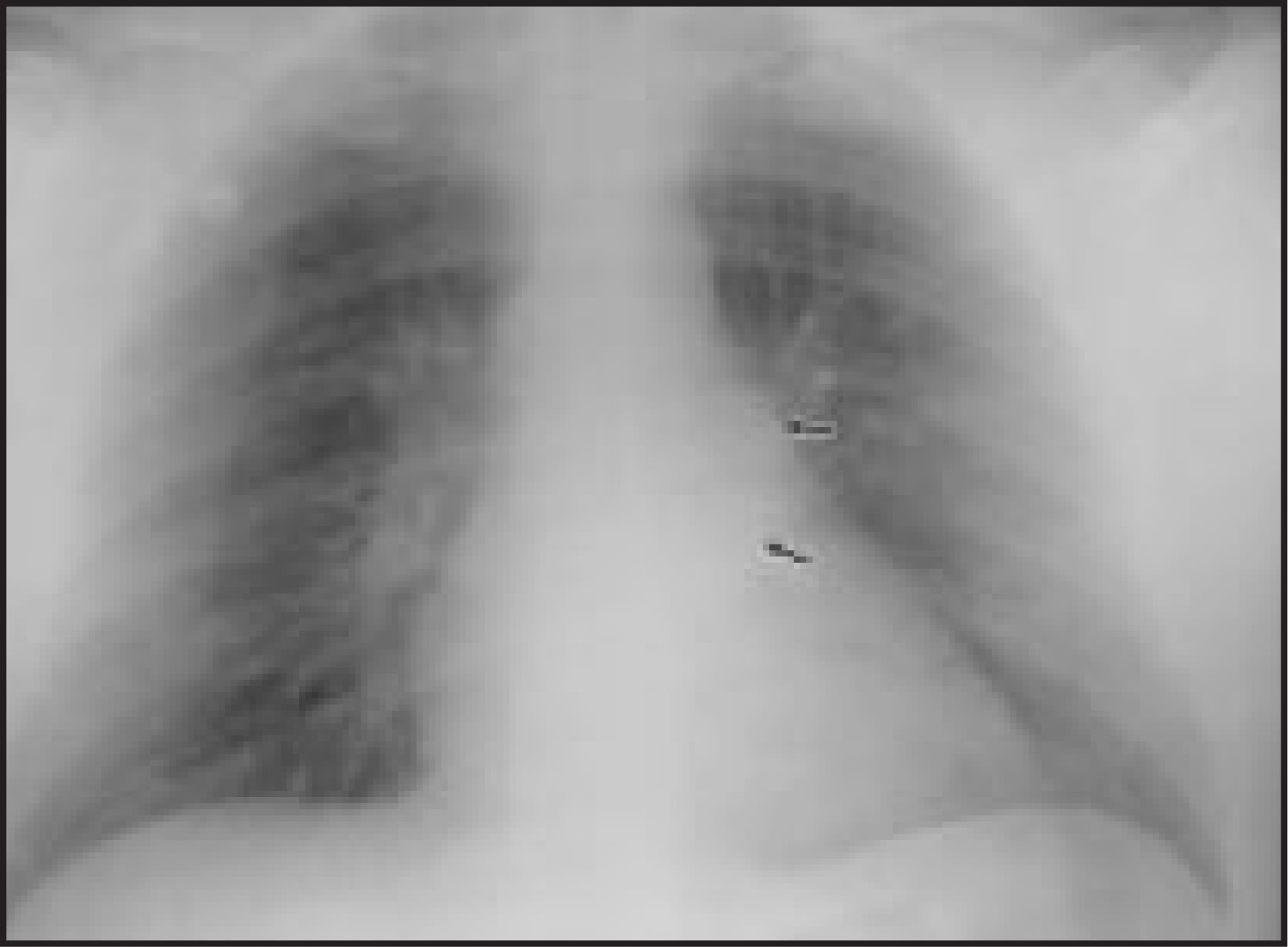
DIAGNOSIS
Typical bronchial carcinoid tumor
IMAGING FINDINGS
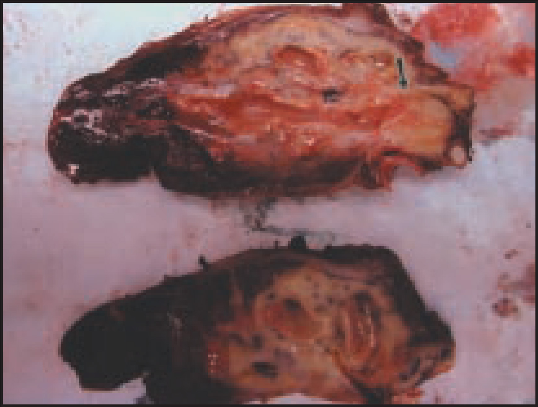
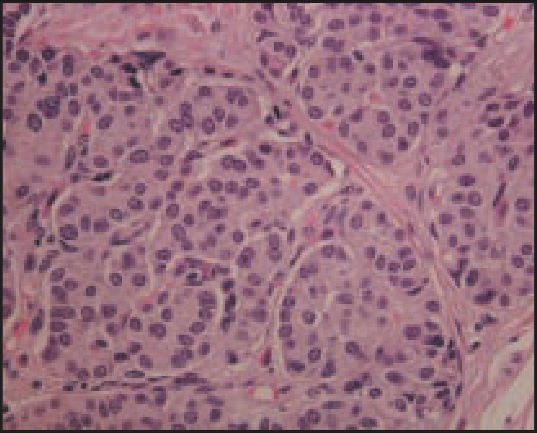
Anteroposterior (Figure 1) and lateral chest radiographs revealed a large (3- to 4-cm) mass at the posterior aspect of the left hilum. Contrast-enhanced CT scan of the chest revealed a large mass in the superior segment of the left lower lobe, with associated left hilar and subcarinal adenopathy (Figure 2). 18F-flourodeoxyglucose positron emission tomography (PET) revealed a nonavid mass, excluding the possibility of a bronchogenic carcinoma (not shown). Bronchoscopy revealed a mass within the superior segmental bronchus of the left lower lobe (Figure 3), from which a biopsy was obtained for pathologic identification (Figure 4). After the diagnosis was established, the patient underwent a successful left pneumonectomy. Histologic examination of the tumor was consistent with typical carcinoid tumor (Figure 5).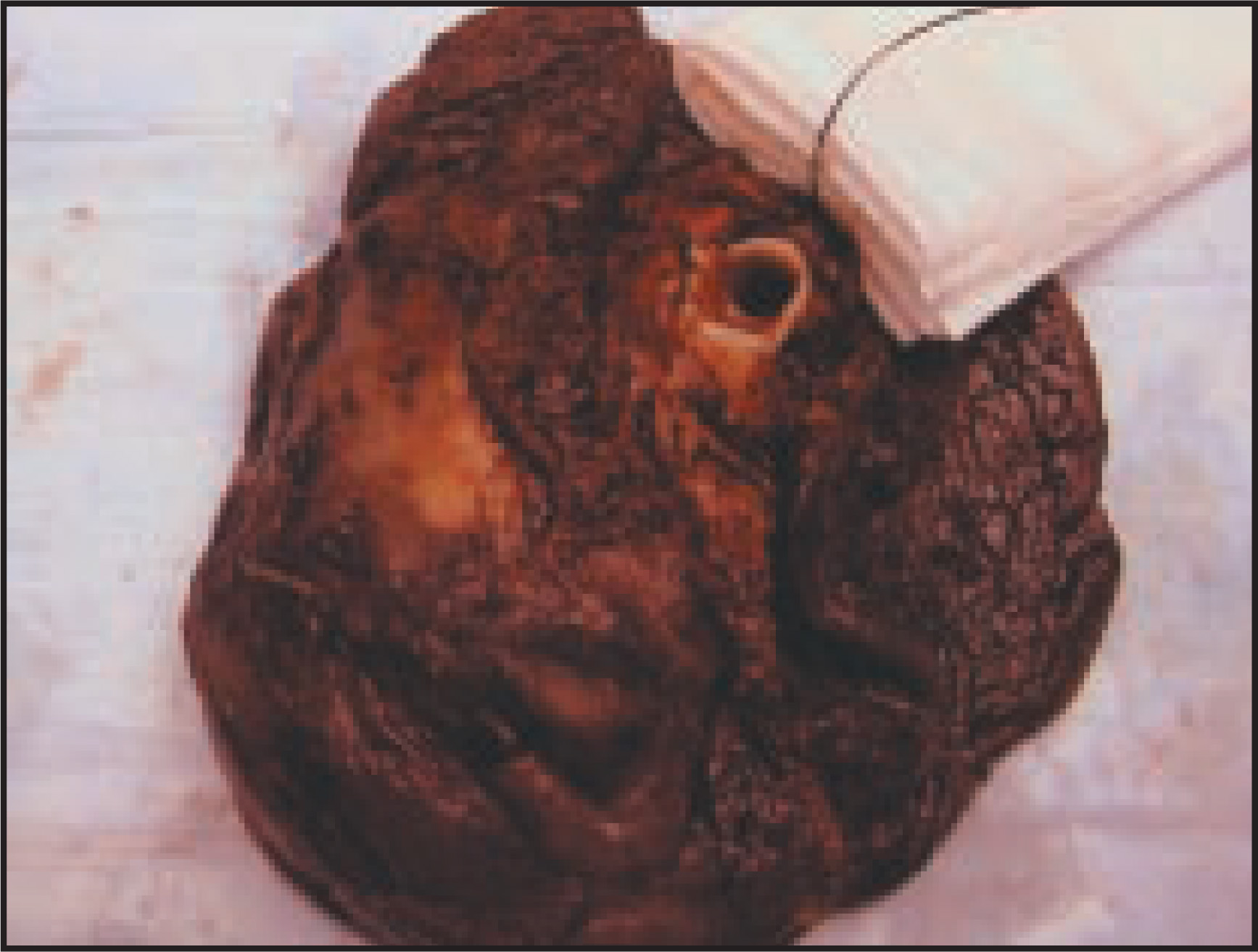
DISCUSSION
Carcinoid tumor is a serotonin-producing malignant tumor belonging to a class of neoplasms known as the neuroendocrine tumors. They are unique in that they can present with tumor-like symptoms or with a combination of symptoms related to endocrine dysregulation. Most cases of carcinoid are found incidentally in the appendix following an appendectomy. The prevalence of carcinoid in the general population is actually quite high, but >95% of these tumors occur in one of three sites: the appendix, rectum, or small intestine.1,2
Carcinoid tumors of the lung are much less common and represent 1% to 2% of all lung cancers.3 They arise from the neurosecretory cells of the bronchial mucosa. Two different types have been identified.4 The first and most common is referred to as typical carcinoid. Typical carcinoid tumors are low-grade tumors, with 10-year survival rates approaching 90%.5 They are capable of local invasion, including invasion of local lymph nodes, but rarely metastasize. The second type of carcinoid is referred to as atypical carcinoid. It is much more aggressive and carries a 5-year survival rate of 25% to 69%.6 Both subtypes tend to arise from the bronchial tree and spread by local invasion. Typical carcinoid tumors are more commonly found centrally within the major bronchi, whereas the atypical carcinoids tend to arise from the peripheral and central bronchi with equal frequency.7 The most frequently presenting symptoms of carcinoid are lobar obstruction, hemoptysis, dyspnea, cough, and lobar pneumonia secondary to obstruction. Some are found serendipitously on chest radiographs in asymptomatic patients; rarely, patients may present with endocrine abnormalities, such as Cushing’s syndrome.8 Both types of carcinoid tumors of the lung can occur in patients of any age, without gender preference. The role that smoking plays in the development of carcinoid is still under debate.5
Definitive diagnosis and classification is made by biopsy and pathologic examination of the tissue. Due to its predilection for arising from the luminal surface of the major bronchi, adequate tissue samples can often be obtained through endobronchial biopsy. Alternatively, tissue may be obtained through CT-guided fine-needle biopsy or thoracotomy.7
Helical chest CT is helpful in determining the size and position of the tumor but is not as accurate in assessing tumor extension as high-resolution CT (HRCT).9 Helical chest CT may also help in narrowing the differential diagnosis, by identifying characteristic findings of carcinoid. These findings include dense ossification, scattered calcifications, and intraluminal location.10 Since carcinoid tumors are metabolically sluggish relative to most other malignancies, PET imaging is most often negative. These results can be misleading, because it may falsely suggest that the lesion is benign.11
Carcinoid is treated surgically, with the approach dependent on the size, location, and tissue type. Treatment for intraluminal typical bronchial carcinoid may be attempted with bronchoscopic removal of the tumor. This technique combines direct bronchoscopic visualization with HRCT. Bronchoscopic removal of these tumors leads to complete eradication in the majority of patients.9 Tumors not meeting the criteria for endobronchial resection may be treated surgically with bronchial sleeve resection, segmentectomy, lobectomy, or pneumonectomy.8 Chemotherapy has not been demonstrated to be successful as a treatment option but, rather, is limited to a palliative role in the management of diffuse metastatic carcinoid; however, investigations involving chemotherapy are still ongoing.12-14
CONCLUSION
Bronchial carcinoid tumor is a low-grade malignant tumor with slow progression and is associated with a favorable prognosis. There are two subtypes, with the less aggressive type comprising approximately 90% of all bronchial carcinoids. The definitive diagnosis is made by histologic examination. Carcinoid tumors typically arise from the luminal surface of major bronchi, and definitive treatment involves surgical resection.
References
- Moertel C, Dockerty M, Judd E. Carcinoid tumors of the vermiform appendix. Cancer. 1968;21:270.
- Moertel C. Karnofsky memorial lecture. An odyssey in the land of small tumors. J Clin Oncol. 1994;5:1503.
- Davila D, Dunn W, Tazelaar H. Bronchial carcinoid tumors. Mayo Clin Proc. 1993;68:795-803.
- Arrigoni M, Woolner L, Bernatz P. Atypical carcinoid tumors of the lung. J Thorac Cardiovasc Surg. 1972;64:413-421.
- Filosso P, Rena O, Donati G. Bronchial carcinoid tumors: Surgical management and long-term outcome. J Thorac Cardiovasc Surg. 2002;123:303-309.
- Thomas C, Tazelaar H, Jett J. Typical and atypical pulmonary carcinoids: Outcome in patients presenting with regional lymph node involvement. Chest. 2001;119:1143-1150.
- Fink G, Krelbaum T, Yellin A. Pulmonary carcinoid: Presentation, diagnosis, and outcome in 142 cases in Israel and review of 640 cases from the literature. Chest. 2001;119:1647-1651.
- Davila D, Dunn W, Tazelaar H. Bronchial carcinoid tumors. Mayo Clin Proc. 1993;68:795-803.
- Van Boxem T, Golding R, Venmans B. High-resolution CT in patients with intraluminal typical bronchial carcinoid tumors treated with bronchoscopic therapy. Chest. 2000;117:125-128.
- Davis S, Zirn J, Govoni A. Peripheral carcinoid tumor of the lung: CT diagnosis. AJR Am J Roentgenol. 1990;155:1185-1187.
- Erasmus J, McAdams H, Patz E. Evaluation of primary pulmonary carcinoid tumors using FDG PET. AJR Am J Roentgenol. 1998;170:1369-1373.
- Moertel C, Hanley J. Combination chemotherapy trials in metastatic carcinoid tumor and the malignant carcinoid syndrome. Cancer Clin Trials. 1979;2:327.
- O’Byrne K, Schally A, Thomas A. Somatostatin, its receptors and analogs, in lung cancer. Chemotherapy. 2001;47(Suppl 2):78-108.
- Ruggieri M, Scocchera F, Genderini M. Therapeutic approach of carcinoid tumours of the lung. Eur Rev Med Pharmacol Sci. 2000;4(1-2):43-46.
Citation
. RCOM RADIOLOGICAL CASE OF THE MONTH. Applied Radiology. 2003;32(4):45-47. doi:10.37549/AR1176.