RCOM RADIOLOGICAL CASE OF THE MONTH
Applied Radiology — Vol. 32 , Issue 5 , pp. 44 -46
DOI: 10.37549/AR1181
Published: May 1, 2003
Categories
CASE SUMMARY
A 61-year-old white woman presented to the Urology clinic with left flank pain and a questionable history of weight loss. She has a medical history of nephrolithiasis and hypertension. Past surgeries included a cholecystectomy and hysterectomy. The patient has a history of long-term tobacco use. Urinanalysis on admission revealed microscopic hematuria. Hematologic workup was unremarkable.
DIAGNOSIS
Adrenal cortical adenoma with very atypical imaging features
IMAGING FINDINGS
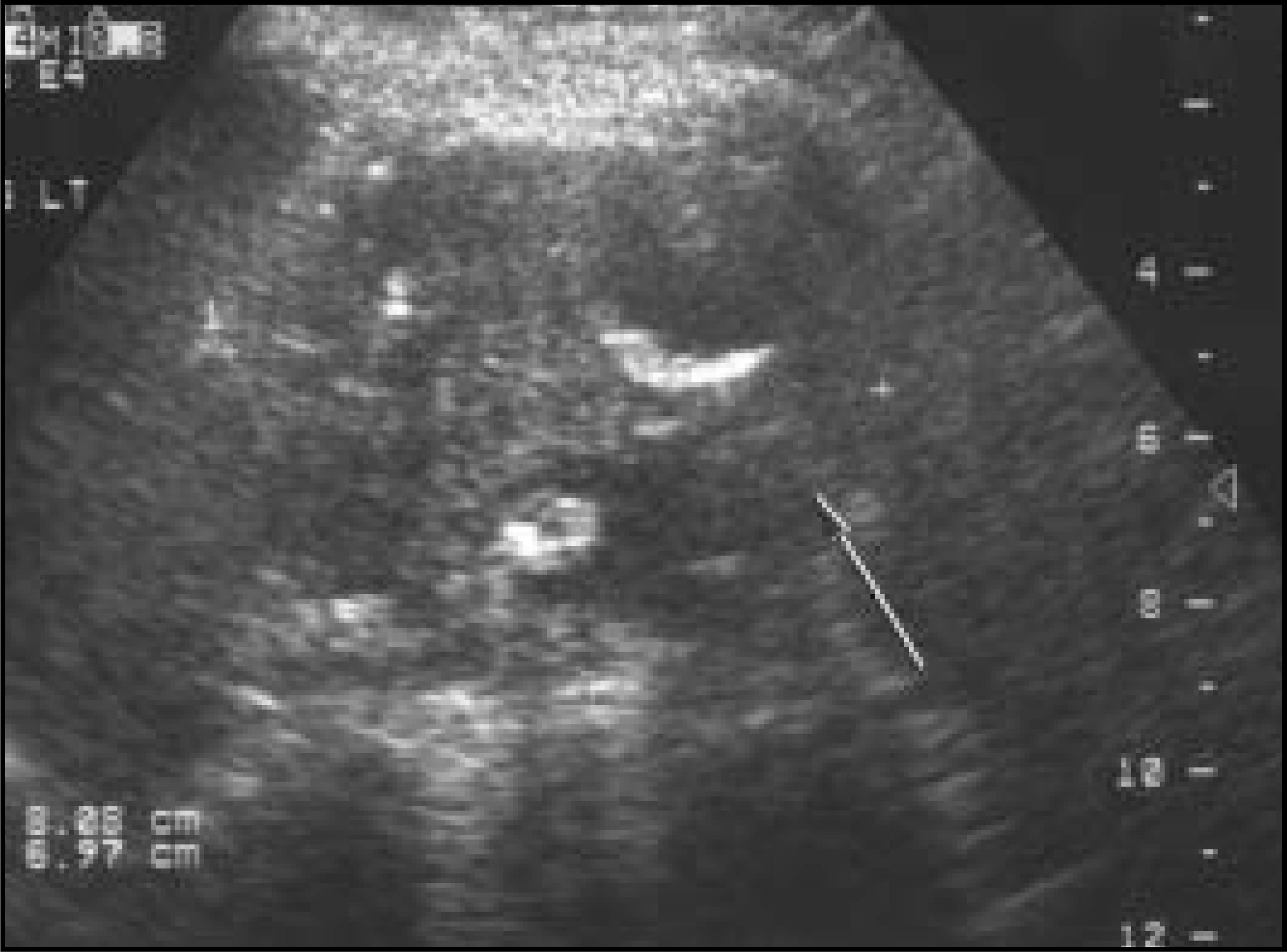
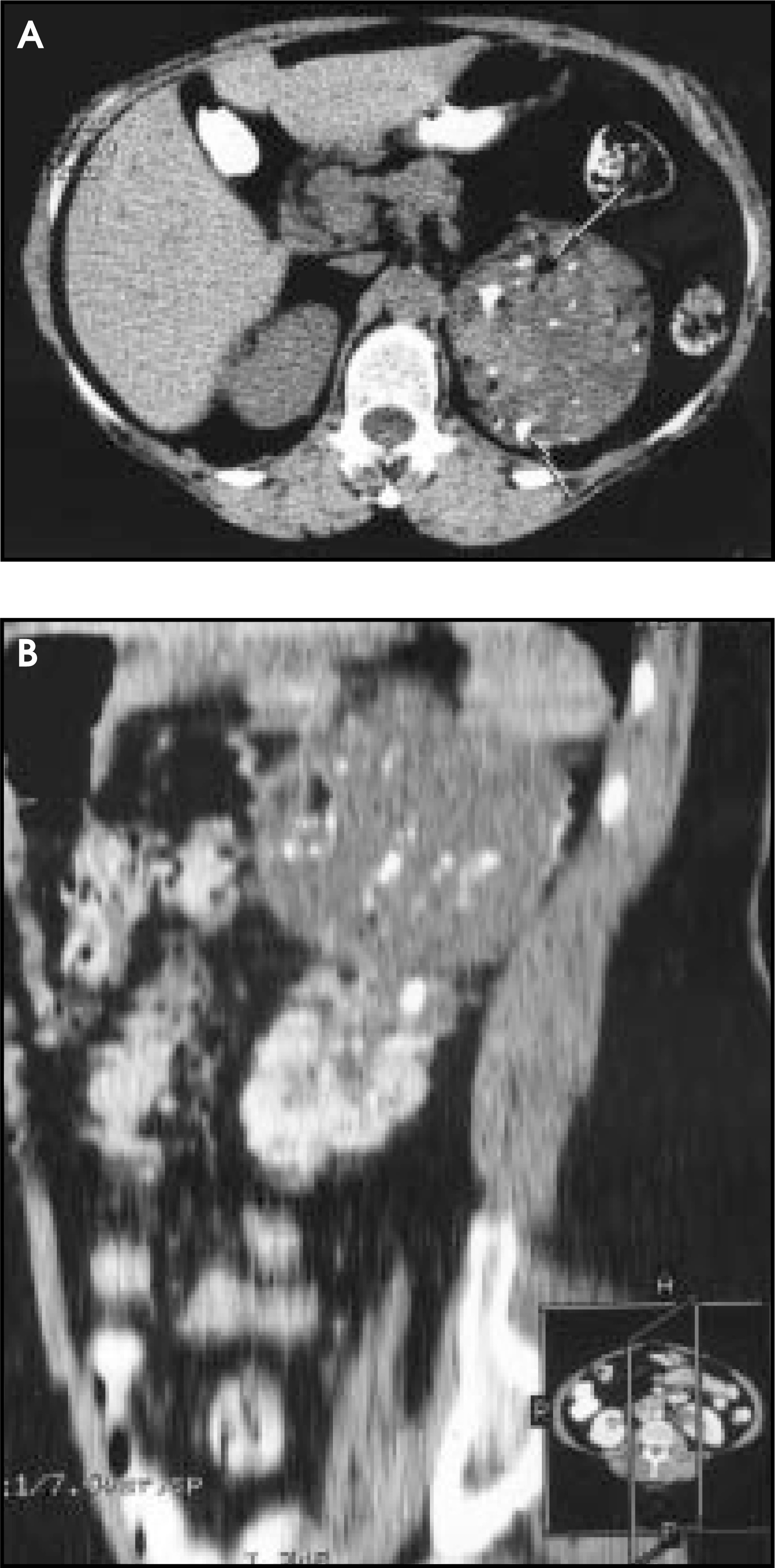
The initial radiologic examination, an abdominal X-ray of the kidney, ureter, and bladder (KUB), showed multiple amorphous calcifications in the left mid-flank region, possibly within an adrenal or upper renal pole mass (Figure 1). A follow-up intravenous urogram demonstrated inferior dis-placement of the left kidney with prominent areas of calcification superior to the left kidney. A renal ultrasound showed a heterogeneous echo-texture mass lesion in the expected location of the left adrenal gland, which appeared separate from a normal-sized left kidney (Figure 2). A noncontrast computed tomographic (CT) scan showed a heterogeneous attenuation 11-cm mass with large amorphous calcifications as well as islands of macroscopic fat, in the expected location of the left adrenal gland (Figure 3A). Contrast-enhanced CT showed minimal peripheral enhancement as well as prominent necrotic areas and a few thin enhancing septations (not shown). Sagittal CT reconstructions further substantiate the above findings (Figure 3B).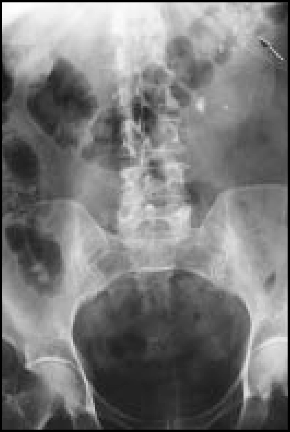
PATHOLOGIC CORRELATION
Grossly, the tumor mass measured 11 × 11 × 8 cm and weighed 480 g. It was adjacent to the kidney, but separated from it by fibrofatty tissue. On the cut surface, the tumor was yellow to gray and mostly smooth. There were large areas of hemorrhage and focal areas that appeared to be necrotic or degenerative. Scattered 0.5- to 1-cm cysts containing blood or necrotic debris were also present. On cutting, bony hard areas could be appreciated. Microscopically, the tumor mass contained uniform lipid-laden adrenal cortical cells. Most of the central part of the lesion was hemorrhagic with large areas of hyaline and myxoid degeneration. There were large bands of fibrous tissue traversing the areas of degeneration. Focal areas of fatty metaplasia and osseous metaplasia were present (Figure 4).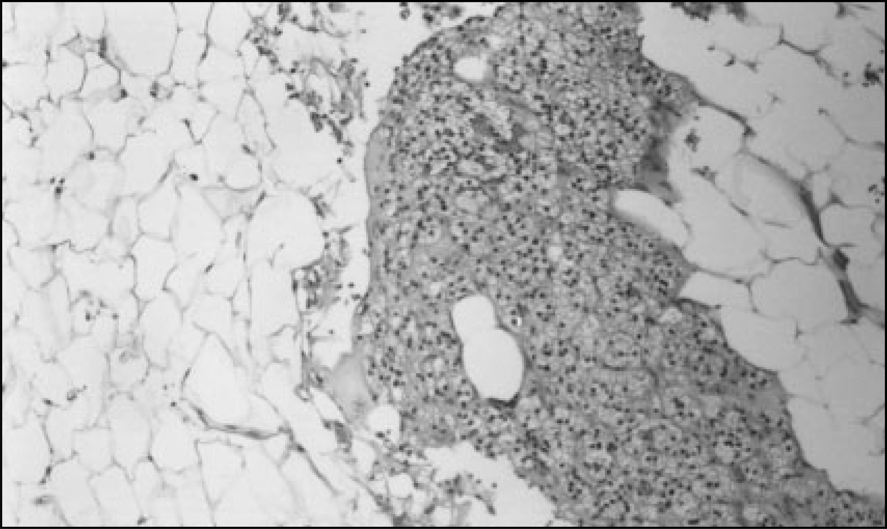
DISCUSSION
Characterization of adrenal masses is an important clinical problem and a focus of abdominal imaging research, because adrenal masses are frequently seen as the so-called “incidentaloma.” They appear in autopsy series with a reported prevalence of 2% to 9%.1,2 Although most incidentally discovered adrenal masses represent benign cortical adenomas, often imaging features cannot reliably differentiate an adenoma from more ominous lesions.3
On imaging, an adrenal adenoma is typically a small (usually <5 cm), well-circumscribed, homogeneous mass with near fluid attenuation values on precontrast CT and uniform contrast enhancement with rapid washout of iodinated or gadolinium-based contrast material. Adenomas frequently demonstrate the presence of intracellular lipid and almost never liquefy. Calcification is rare. They may occasionally undergo intratumoral hemorrhagic degeneration with the development of cystic internal spaces and subsequent fibrosis.4-6
The imaging findings in this case were not characteristic of a benign cortical adenoma. There were large areas of osseous metaplasia as well as scattered areas of necrosis. Additionally, prominent islands of adipose tissue were noted throughout the lesion. Furthermore, the size of the mass at 11 cm was, in itself, unusual for a benign adenoma.
Initial diagnostic considerations included a primary adrenal cortical malignancy, a metastatic lesion from an occult primary, and a myelolipoma. Pheochromocytoma and adenoma were believed to be less likely7,8 because of atypical imaging characteristics.
CONCLUSION
A subgroup of adrenal adenomas are large and heterogeneous. They calcify more frequently, and have areas of internal hemorrhage and necrotic components. They may show foci of fatty metaplasia. All of these characteristics are responsible for the bizarre imaging features. Imaging features alone frequently cannot differentiate these lesions from other large adrenal masses, such as adreno-cortical carcinoma and metastases. Surgical resection may be required for a definitive diagnosis.
References
- Glazer H, Weyman P, Sagel S. Nonfunctioning adrenal masses: Incidental discovery on computed tomography. AJR Am J Roentgenol. 1982;139:81-85.
- Mitnick J, Bosniak M, Megibow A, Naidich D. Non-functioning adrenal adenomas discovered incidentally on computed tomography. Radiology. 1983;148:495-499.
- Bernardino M, Walther M, Phillips V. CT-guided adrenal biopsy: Accuracy, safety, and indications. AJR Am J Roentgenol. 1985;144:67-69.
- Sato N, Watanabe Y, Saga T. Adrenocortical adenoma containing a fat component: CT and MR image evaluation. Abdom Imaging. 1995;20:489-490.
- Ichikawa T, Ohtomo K, Uchiyama G. Contrast-enhanced dynamic MRI of adrenal masses: Classification of characteristic enhancement patterns. Clin Radiol. 1995;50:295-300.
- Korobkin M, Lombardi T, Aisen A. Characterization of adrenal masses with chemical shift and gadolinium-enhanced MR imaging. Radiology. 1995;197:411-418.
- Newhouse J, Heffess C, Wagner B. Large degenerated adrenal adenomas: Radiologic-pathologic correlation. Radiology. 1999;210:385-391.
- Rosenthal D, Chew F. Radiology: MGH Clinical Review. 1994:140-144.
Citation
. RCOM RADIOLOGICAL CASE OF THE MONTH. Applied Radiology. 2003;32(5):44-46. doi:10.37549/AR1181.