RCOM RADIOLOGICAL CASE OF THE MONTH
Applied Radiology — Vol. 32 , Issue 6 , pp. 31 -33
DOI: 10.37549/AR1188
Published: June 1, 2003
Categories
CASE SUMMARY
A 20-year-old man presented to the clinic with a painful mass in his right knee. He recently had a plain radiographic knee study at another institution that was read as normal. At that time, the mass was noted to be smaller and less painful. A new radiographic knee study was obtained, followed by futher evaluation with magnetic resonance (MR) imaging.
DIAGNOSIS
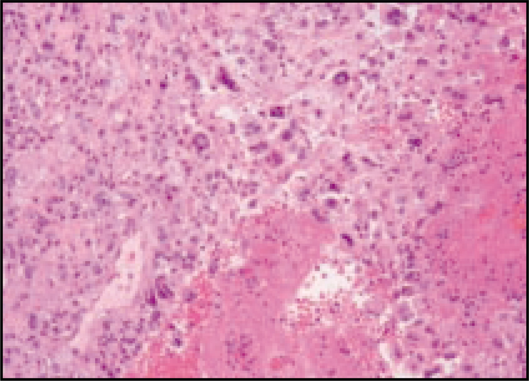
Telangiectatic osteosarcoma
IMAGING FINDINGS
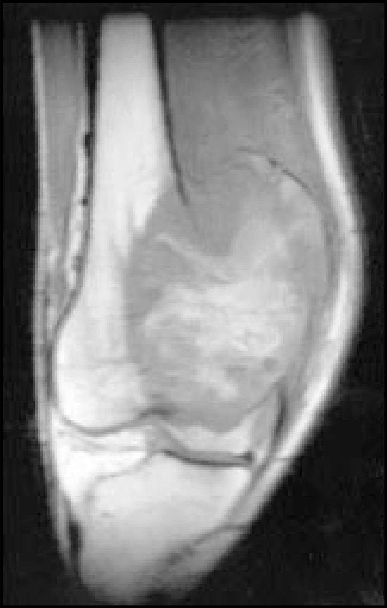
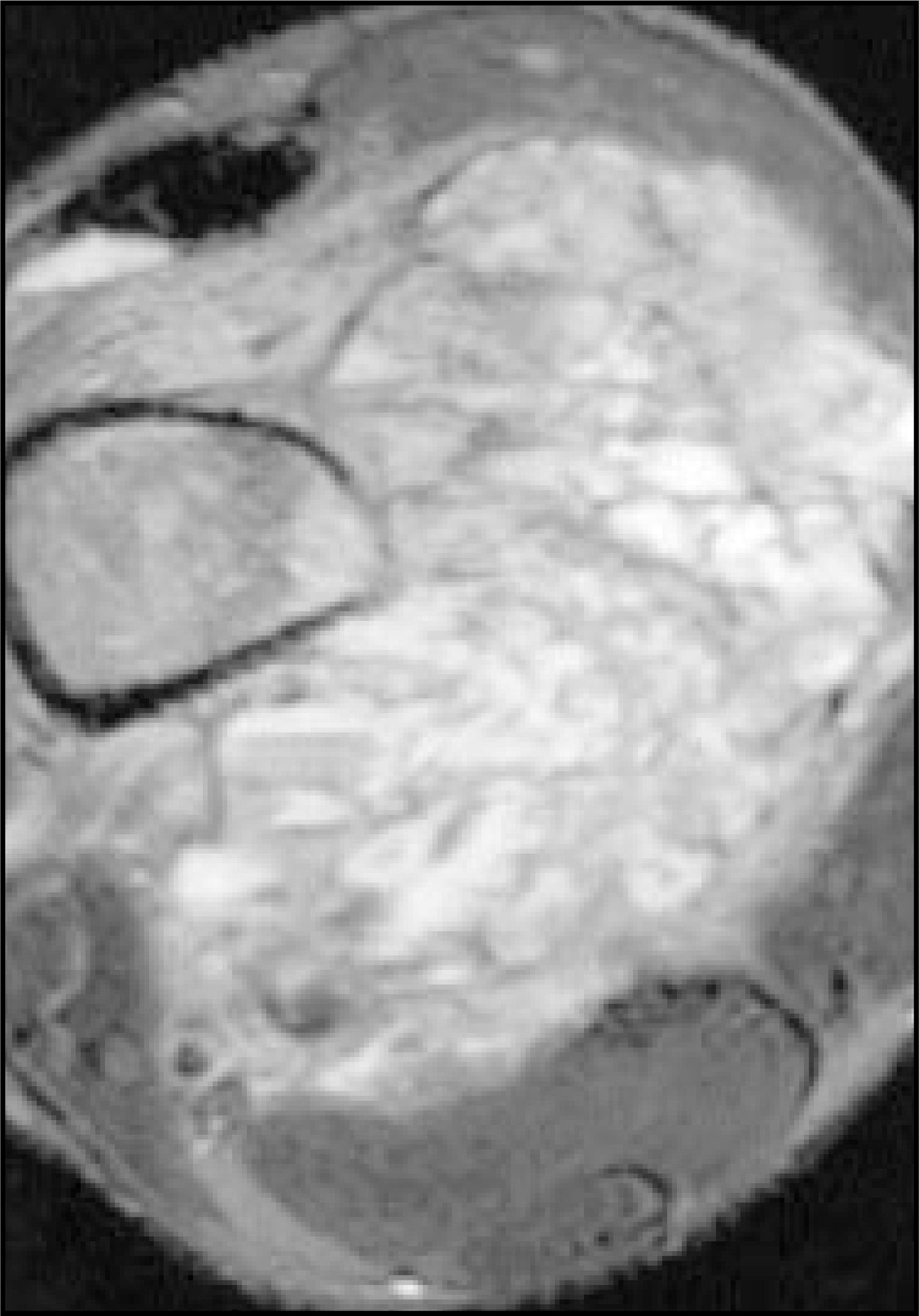
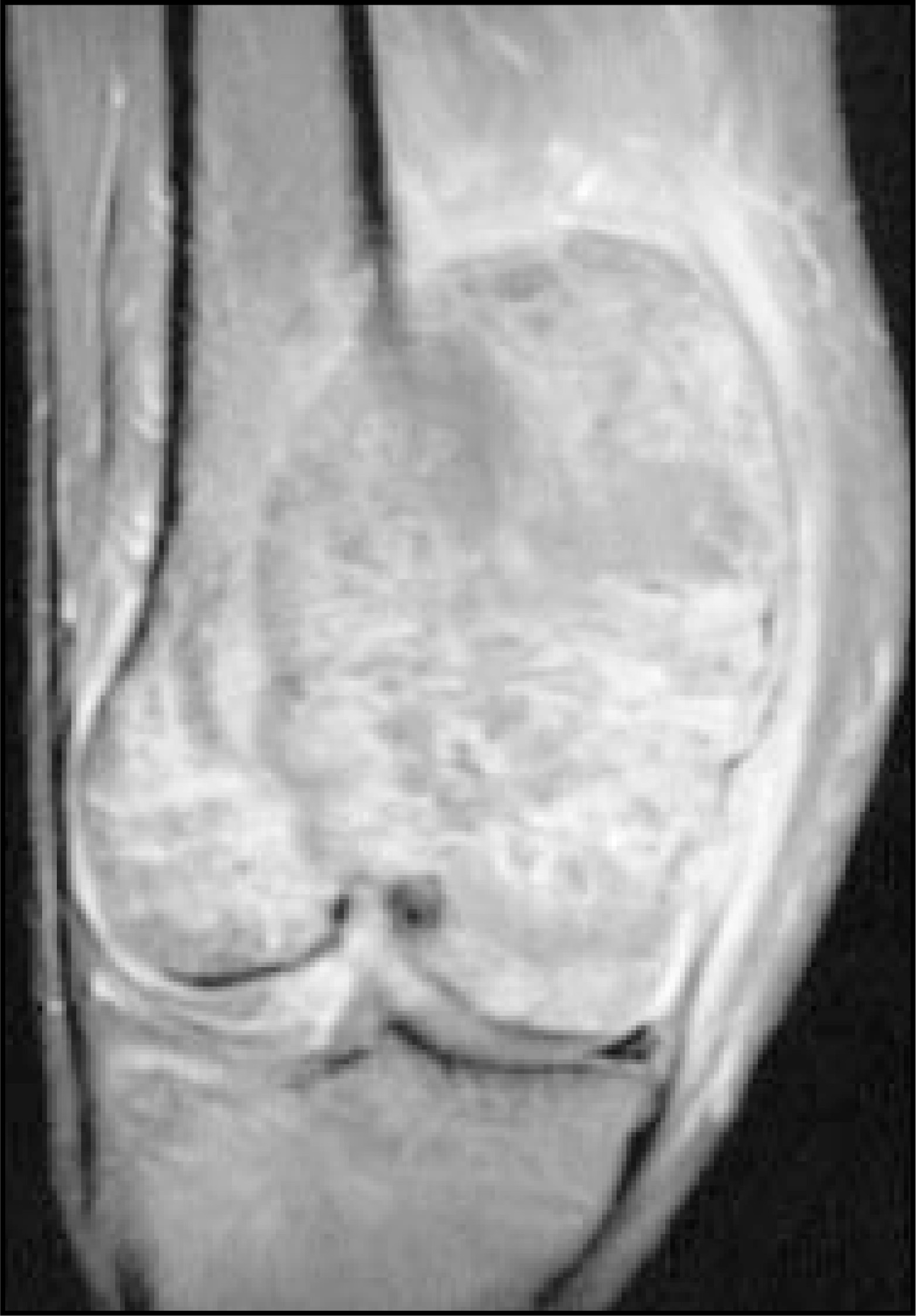
The anteroposterior radiograph of the right knee revealed obliteration of the cortex along the medial femoral metadiaphysis, associated with an ill-defined lucency involving the distal medial femur, as well as the epiphysis (Figure 1). Faint osseous matrix may be seen. No clear periostitis was identified, but there is an associated, large soft-tissue mass overlying and medial to the described osseous changes. The MR study confirms the presence of the large mass and associated cortical destruction (Figure 2). Additionally, multiple fluid-fluid levels are seen throughout the mass (Figure 3). There is extension into the intra-articular space and medial head of the gastrocnemius muscle, and displacement of the neurovascular bundle.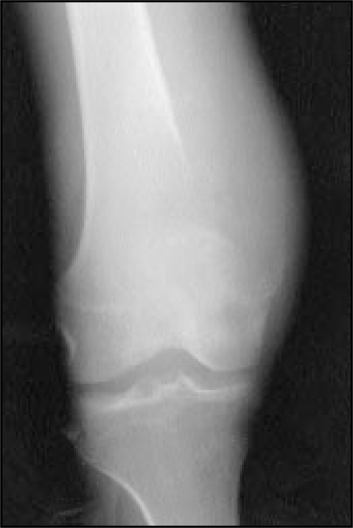
DISCUSSION
Osteosarcomas are the most common primary malignancies of bone, occurring predominantly in the population younger than age 30. They most frequently involve the ends of long bones, particularly the distal femur and proximal tibia.1 Common clinical presentations include pain, pathologic fracture, and soft-tissue mass. Telangiectatic osteosarcomas are an uncommon, yet aggressive, morphologic variant, comprising approximately 2% to 4% of all osteosarcomas.2 Radiographically, they typically appear destructive and radiolucent, often resembling the radiographic appearance of aneurysmal bone cysts.3 Although there is usually only a small amount of osseous matrix within this neoplasm, it is not unusual for there to be a significant associated soft-tissue component. Periosteal reaction in the form of a Codman’s triangle may be seen in association with a more rapid-growing tumor. Fluid-fluid levels on computed tomography (CT) and MR imaging, although nonspecific findings,4 can be seen in association with telangiectatic osteosarcomas and are thought to represent areas of hemorrhage into cystic spaces,5 which are characteristically lined by giant cells and tumor cells rather than the endothelial cells more typical of aneurysmal bone cysts. Osteosarcomas, in general, have been known to be metastatic, with the lungs being the most common site of invasion. The treatment generally involves wide local excision and, sometimes, limb amputation, depending on the extent of the disease. Some believe that there may be a role for chemotherapy.6 The prognosis is considered to be similar to that of conventional osteosarcomas.
CONCLUSION
The telangiectatic osteosarcoma is an uncommon type of osteosarcoma characterized by hemorrhage into dilated cystic spaces, which often results in fluid-fluid levels on CT and MR imaging. These patients typically present with severe pain, and care should be taken to distinguish this neoplastic process from aneurysmal bone cysts and other benign processes that may have a similar imaging appearance.
References
- Whitehead R, Melhem E, Kasznica J. Telangiectatic osteosarcoma of the skull base.. Am J Neuroradiol.. 1988;19:754-757.
- Klein M, Kenan S, Lewis M. Osteosarcoma: Clinical and pathological considerations.. Orthop Clin North Am.. 1989;20:327-345.
- Kyriakos M, Hardy D. Malignant transformation of aneurismal bone cyst, with an analysis of the literature.. Cancer.. 1991;68:1770-1780.
- Hudson T. Fluid levels in aneurysmal bone cysts: A CT feature.. AJR Am J Roentgenol.. 1984;142:1001-1004.
- Tsai J, Murray K, Fallon M. Fluid-fluid level: A nonspecific finding in tumors of bone and soft-tissue.. Radiology.. 1990;175:779-782.
- Bacci G, Ferrari S, Ruggieri P. Telangiectatic osteosarcoma of the extremity.. Acta Orthop Scand.. 2001;72:167-172.
Citation
. RCOM RADIOLOGICAL CASE OF THE MONTH. Applied Radiology. 2003;32(6):31-33. doi:10.37549/AR1188.