RCOM RADIOLOGICAL CASE OF THE MONTH
Applied Radiology — Vol. 32 , Issue 6 , pp. 34 -37
DOI: 10.37549/AR1191
Published: June 1, 2003
Categories
CASE SUMMARY
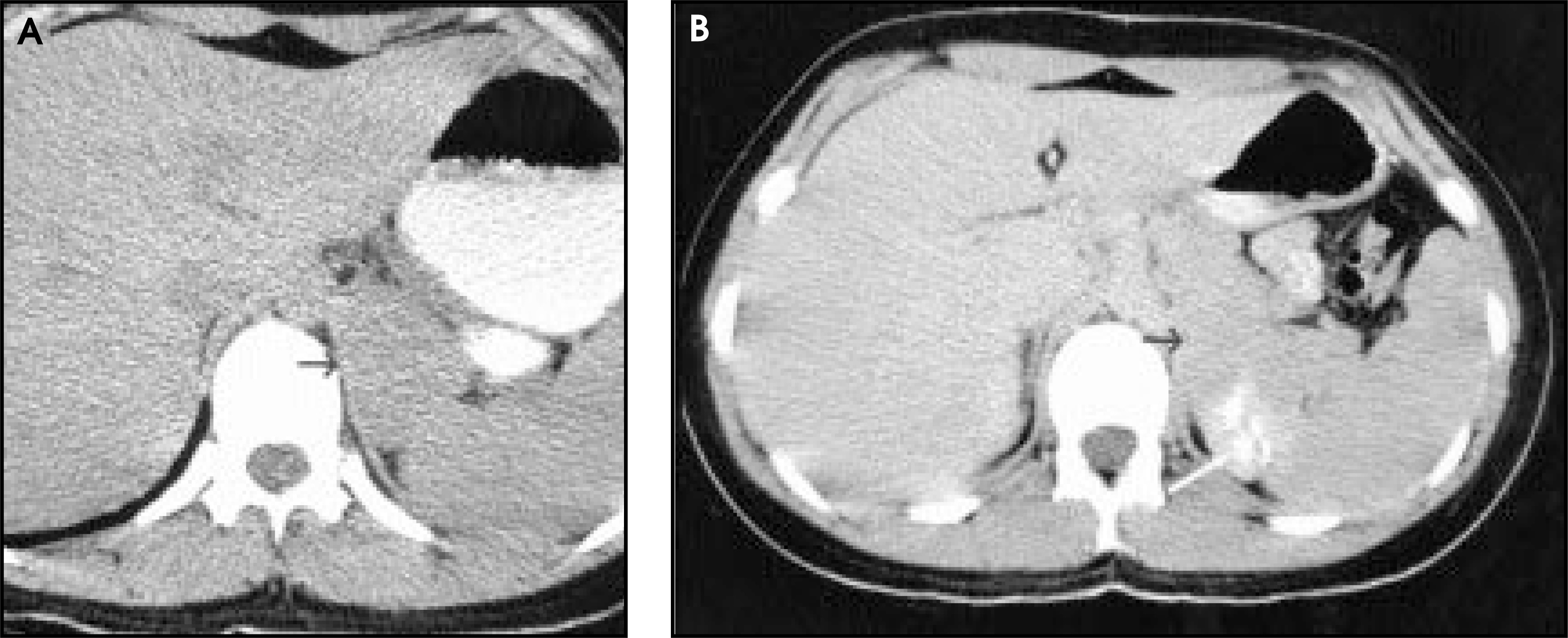
The patient is a 17-year-old girl, nonsmoker, who presented 1 year previously with a spontaneous pneumothorax. She failed conservative therapy and underwent pleurodesis procedures twice. Open-lung biopsy demonstrated pulmonary lymphangioleiomyomatosis (LAM), and a computed tomography (CT) study at another institution showed a left suprarenal mass. The histologic and immunohistochemical examination from a retroperitoneal biopsy were consistent with a lymphangiomyoma. No skin lesions were present and there was no family history of tuberous sclerosis, seizures, or mental deficiency.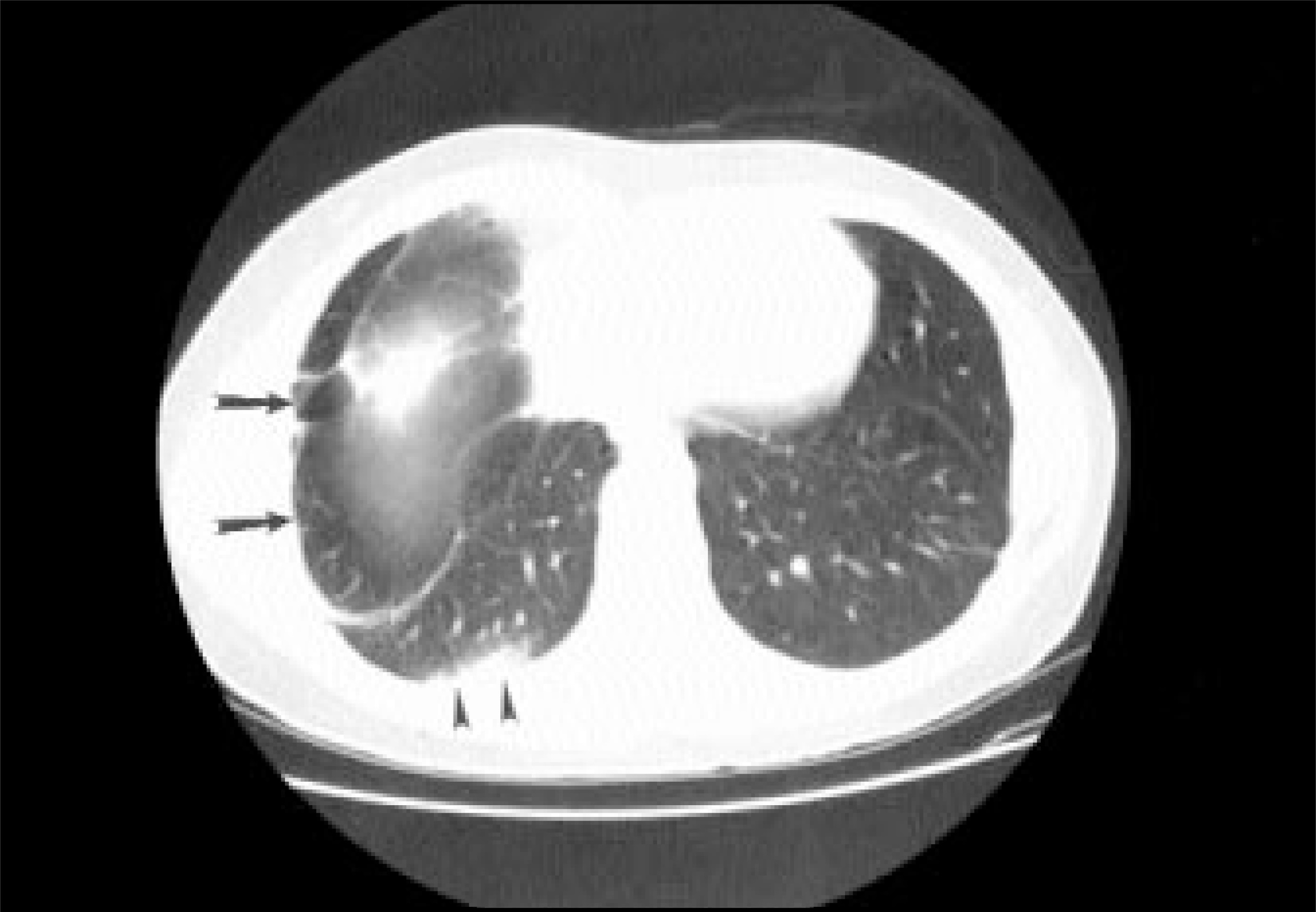
DIAGNOSIS
Pulmonary lymphangioleiomyomatosis with retro-peritoneal lymphangiomyoma.
DISCUSSION
Lymphangioleiomyomatosis is a rare disease that occurs almost exclusively in women of reproductive age. Although the etiology of the disease is unknown, it is hypothesized that it may be related to the amplifying effect on mutant proteins by exogenous estrogen or pregnancy.1 The main feature of the disease is the abnormal proliferation of immature smooth muscle at the level of the distal airway, small blood vessels, and lung lymphatic system, including the mediastinal and retroperitoneal lymphatic system2 and thoracic duct.3 The most common clinical symptoms at initial presentation are thoracic in nature and include exertional dyspnea, spontaneous pneumothorax, and chylothorax.4 The characteristic clinical findings are probably due to the proliferation of the atypical smooth muscle cells that obstruct bronchioles, lymphatics, and venules5 and cystic dilatation caused by matrix metalloproteinases produced by LAM.6 Many patients do not live more than 10 years from the initial diagnosis, and treatment is controversial, although hormone therapy (directed at progesterone receptors on spindle cells), oophorectomy, and lung transplantation may offer some benefit.7
Chest radiographs may be normal initially. The earliest findings usually consist of an interstitial infiltrate with several different appearances that has been described as lymphangitic; soft, reticular, or accentuated at the bases; fine nodular; and septal.8 Eventually, an interstitial infiltrate associated with airflow obstruction, air trapping, and hyperinflation is usually seen. A study of 39 women with biopsy-proven LAM found a decreased FEV1, FEV1/FVC, and DLCO.9 Cysts and bullae are common and eventually honeycomb lung develops.10
In recent years many authors have been exploring the benefit of CT in the diagnosis of LAM. In a study of 69 patients, Urban et al5 found that all the patients had characteristic bilateral thin-walled cysts. After radiographic chest evaluation of 39 women with LAM, Avila et al9 found additional cysts on 92% of thin-section CT scans. Kirchner et al11 supported the characteristic appearance of LAM on high-resolution CT. In 11 women with open-biopsy–proven LAM they universally found multiple, thin-walled cysts uniformly distributed throughout the lung.11 Although they also found interstitial changes in most of their patients, they concluded that the cystic changes described are almost pathognomonic for LAM. In a separate study of 80 women with biopsy-proven LAM, Avila et al12 found that 76% of the patients also had positive findings on abdominal CT or ultrasound. The most common findings were renal angiomyolipomas (AMLs), enlarged abdominal lymph nodes, chylous ascites, and lymphangiomyomas.12 In a study of 22 women with known extrapulmonary involvement of LAM, Matsui et al6 found that 68% of them were AMLs. Other studies have reported AMLs in up to two thirds of patients with LAM at initial presentation.13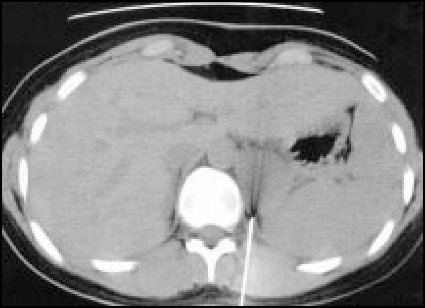
A single paper describes ultrasound findings in diffuse retroperitoneal lymphangiomyomatosis. They found multiple anechoic cystic structures, irregular in shape, thin walled, with intraluminal septa, and associated enlarged nodes. These findings, in diffuse lymphangiomyomatosis, represent multiple ectatic lymphatic vessels.14
There is a varied spectrum to the disease depending on the degree of intra-and extrapulmonary involvement. This led one group of authors15 to divide the extrapulmonary LAM into three groups: 1) pulmonary parenchymal LAM with diffuse mediastinal and or retroperitoneal involvement; 2) localized mediastinal or retroperitoneal LAM without pulmonary parenchymal involvement; and 3) pulmonary parenchymal and lymph node disease in patients with stigmata of tuberous sclerosis.8 The first two categories of involvement are extremely rare, each consisting of approximately four reported case studies. The case described here reports pulmonary involvement with localized retroperitoneal involvement.
In the English literature, LAM has been strongly associated with renal angiomyolipomas. Angiomyolipoma can also occur as a component of the tuberous sclerosis complex. Lymphangioleiomyomatosis has long been suspected to be related to angiomyolipoma and tuberous sclerosis because of clinical associations. Many cytological features, morphological characteristics, and immunophenotypic profiles all support a close relationship between LAM and angiomyolipoma, which probably represent different morphological manifestations of hamartomatous proliferation of a particular form of HMB-45 positive smooth muscle.16 The pathology of the lung in LAM and tuberous sclerosis appears identical histologically. Since renal angiomyolipomas and extrapulmonary lymphangiomas have been found in both, some authors have suggested that LAM is a “form fruste” of tuberous sclerosis.8 The largest study of LAM evaluated 80 women without tuberous sclerosis from 1995 to 1998 using thin-section CT and ultrasound. Only four women were found to have a localized retroperitoneal lymphangiomyoma as seen in this case.12
CONCLUSION
It is clear that LAM is highly associated with a variety of benign extrapulmonary tumors, especially AMLs, lymphangiolipomas, and enlarged lymph nodes. A classic presentation on CT or MRI and a high index of suspicion for these lesions in a woman with known LAM could avoid the risks of unnecessary surgery for a suspected malignancy. In a presentation such as this case of a retroperitoneal lymphangiomyoma, methods of acquiring information that are less invasive than surgical biopsy should be considered. These lesions are often amenable to image-guided biopsy if the etiology of an extrapulmonary mass in a patient with LAM is uncertain. In this case, a lymphangiomyoma was the suspected diagnosis but because of patient concern and the lack of a well-defined relationship between LAM and benign retroperitoneal masses, a needle-guided biopsy was performed.
Several studies in the last several years have included women with LAM who were not diagnosed by pulmonary biopsy. These studies relied on imaging techniques, biopsy of extrapulmonary lesions, and clinical presentation to make the diagnosis of pulmonary LAM in a minority of their patients. Specifically, they looked for the characteristic thin-walled cysts on chest CT and at least one of the following: unexplained recurrent pneumothoraces or hemoptysis, biopsy-proven AML or lymphangiomyoma, persistent chylothorax, or transbronchial biopsy that was suspicious for but not diagnostic of LAM. Given the recent information, which has further established the relationship between LAM and benign mediastinal and retroperitoneal masses, it may now be possible to establish an imaging diagnosis of a retroperitoneal AML in a patient with biopsy-proven LAM and thereby avoid exposing the patient to the inherent risks of an invasive procedure. Provided that the appropriate imaging characteristics are present, it may be time that retroperitoneal masses in patients with LAM could be diagnosed as benign with a high degree of certainty without exposing the patient to any potential morbidity associated with a biopsy of the mass.
References
- Shen A, Iseman M, Waldron J, King T. Exacerbation of pulmonary lymphangiomyomatosis by exogenous estrogens.. Chest.. 1987;91:782-785.
- Johnson S, Davey D, Cibull M. Lymphangioleiomyomatosis.. Am Surg.. 1993;59:395-399.
- Villegas Fernandez F. Pulmonary lymphangioleiomyomatosis associated with renal angiomyolipoma. Presentation of a case and review of the literature.. Revista Clinica Expaniola.. 1992;191:144-147.
- Zompatori M. Pulmonary lymphangioleiomyomatosis. Radiologic aspects and the diagnostic role of computed tomography.. Radiol Med.. 1990;80:286-292.
- Urban T, Lazor R, Lacronique J. Pulmonary lymphangioleiomyomatosis: A study of 69 patients.. Medicine.. 1999;78:321-337.
- Matsui K, Tatsuguchi A, Valencia J. Extrapulmonary lymphangioleiomyomatosis (LAM): Clinicopathologic features in 22 cases.. Hum Pathol.. 2000;31:1242-1248.
- Matsui K, Takeda K, Yu Z. Role for activation of matrix metalloproteinases in the pathogenesis of pulmonary lymphangioleiomyomatosis.. Arch Pathol Lab Med.. 2000;124:267-275.
- Pear B. Pulmonary and retroperitoneal lymphangiomyomatosis.. AJR Am J Roentgenol.. 1980;134:1288-1289.
- Avila N, Chen C, Chu S. Pulmonary lymphangioleiomyomatosis: Correlation of ventilation-perfusion scintigraphy, chest radiography, and CT with pulmonary function tests.. Radiology.. 2000;214:441-446.
- Vodoz V, Leuenberger P. Pulmonary lymphangiomyomatosis and renal angiomyolipoma: Report of 2 cases.. J Suisse Med.. 1987;117:958-963.
- Kirchner J, Stein A, Viel K. Pulmonary lymphangioleiomyomatosis: High-resolution CT findings.. Eur Radiol. 1999;9:49-54.
- Avila N, Kelly J, Chu S. Lymphangioleiomyomatosis: Abdominopelvic CT and US findings.. Radiology. 2000;216:147-153.
- Chu S, Horiba K, Usuki J. Comprehensive evaluation of 35 patients with lymphangioleiomyomatosis (LAM).. Chest.. 1999;115:1041-1052.
- Di Matteo A, Luisetti M, Brunetti E, Filice C. Ultrasound findings in retroperitoneal lymphangiomyomatosis.. Bildgebung.. 1990;57:88-89.
- Silverstein E, Ellis K, Wolff M. Pulmonary lymphangiomyomatosis.. AJR Am J Roentgenol.. 1974;120:832-850.
- Chan J, Tsang W, Pau M. Lymphangiomyomatosis and angiomyolipoma: Closely related entities characterized by hamartomatous proliferation of HMB-45-positive smooth muscle.. Histopathology. 1993;22:445-455.
Citation
. RCOM RADIOLOGICAL CASE OF THE MONTH. Applied Radiology. 2003;32(6):34-37. doi:10.37549/AR1191.