RCOM RADIOLOGICAL CASE OF THE MONTH
Applied Radiology — Vol. 32 , Issue 7 , pp. 45 -47
DOI: 10.37549/AR1201
Published: July 1, 2003
Categories
CASE SUMMARY
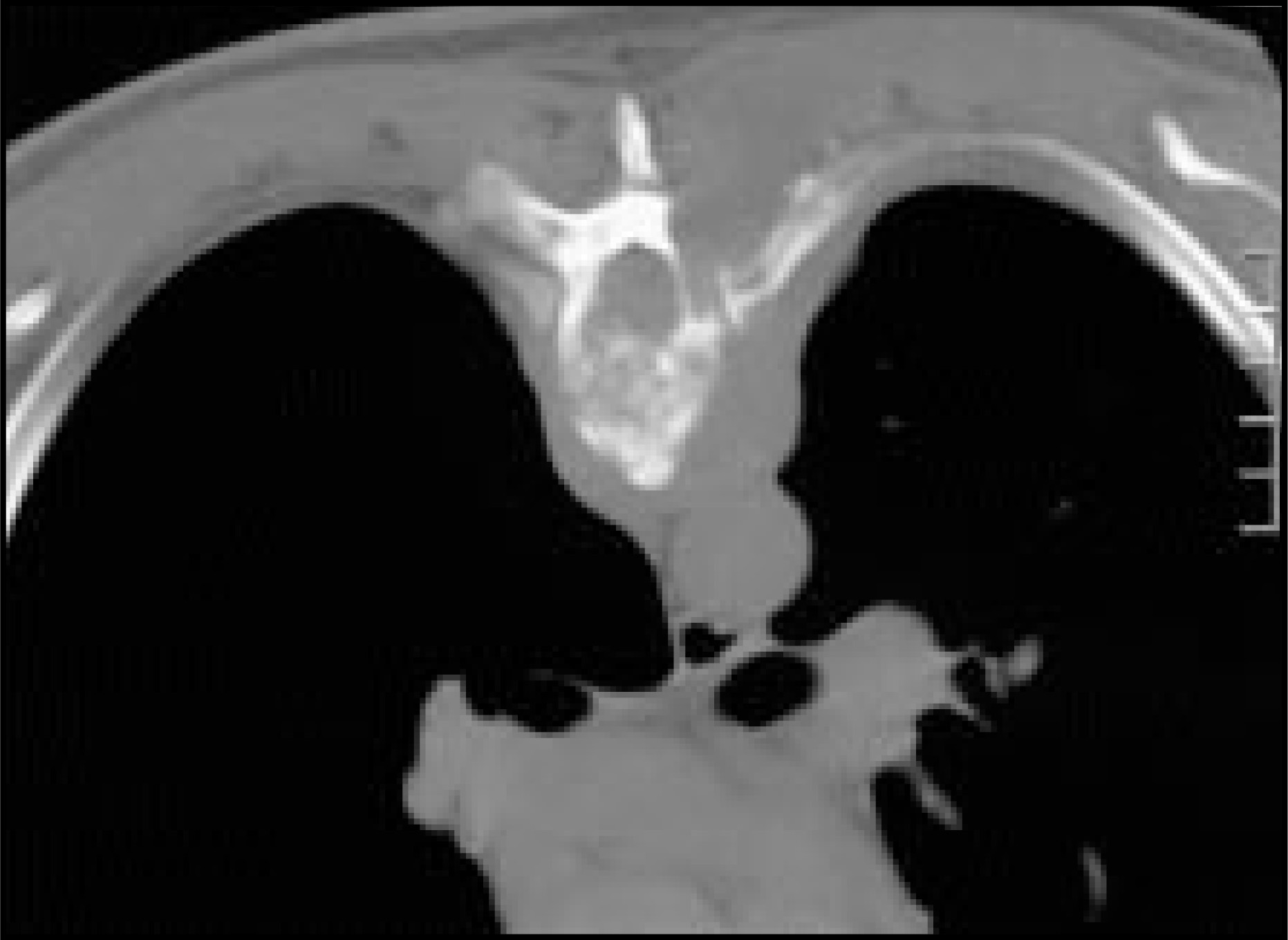
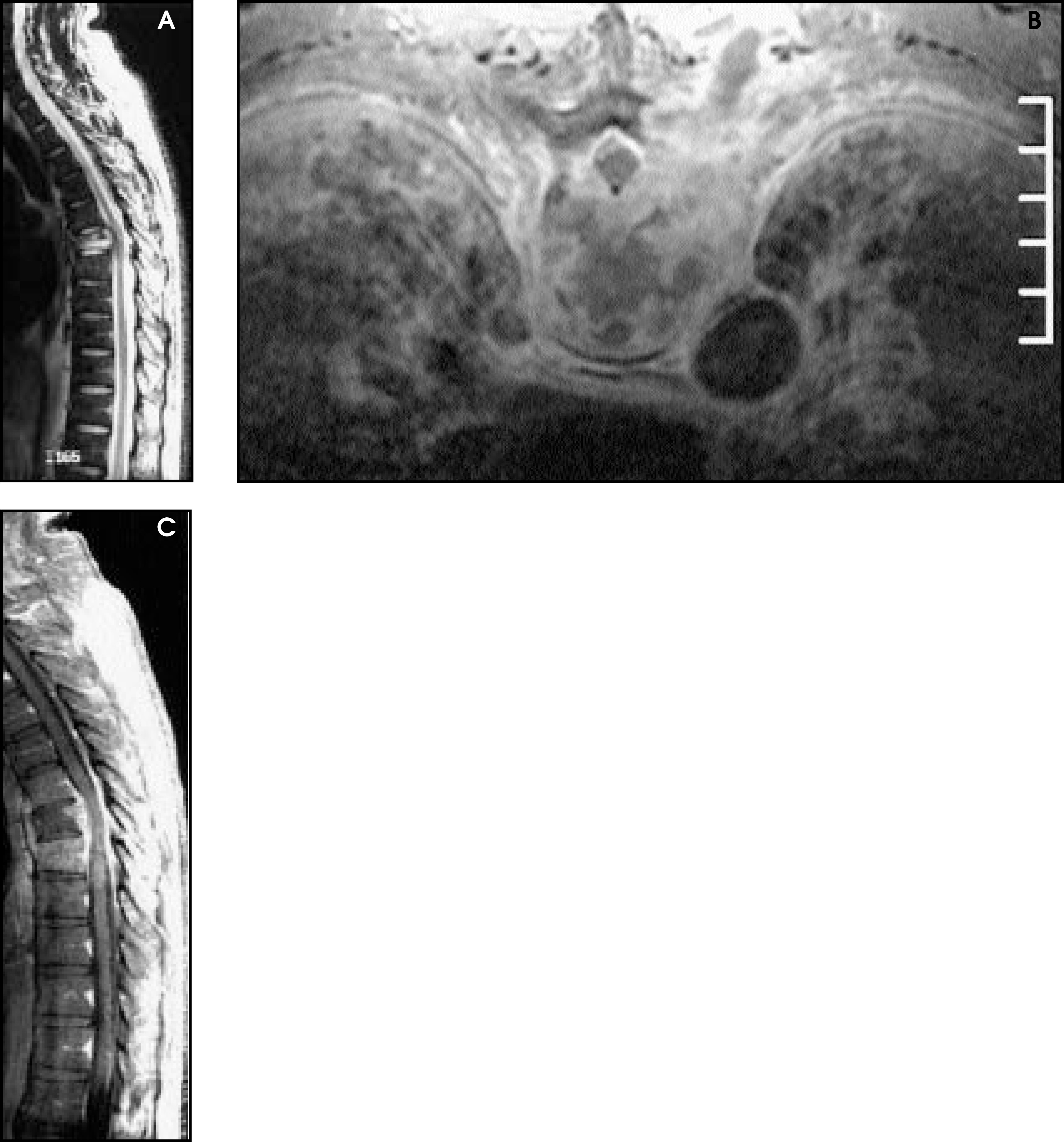
A 51-year-old man presented to the emergency department with chest pain radiating to the right shoulder. The pain had been present for several months but had become refractory to analgesics. Past medical history was remarkable for recently diagnosed diabetes mellitus with negative cardiac and gastrointestinal workups. Physical examination revealed the patient had a low-grade fever and pain localized over the midthoracic spine. A radiograph of the thoracic spine (Figure 1) prompted subsequent computed tomography (CT; Figure 2) and magnetic resonance (MR; Figure 3) examinations.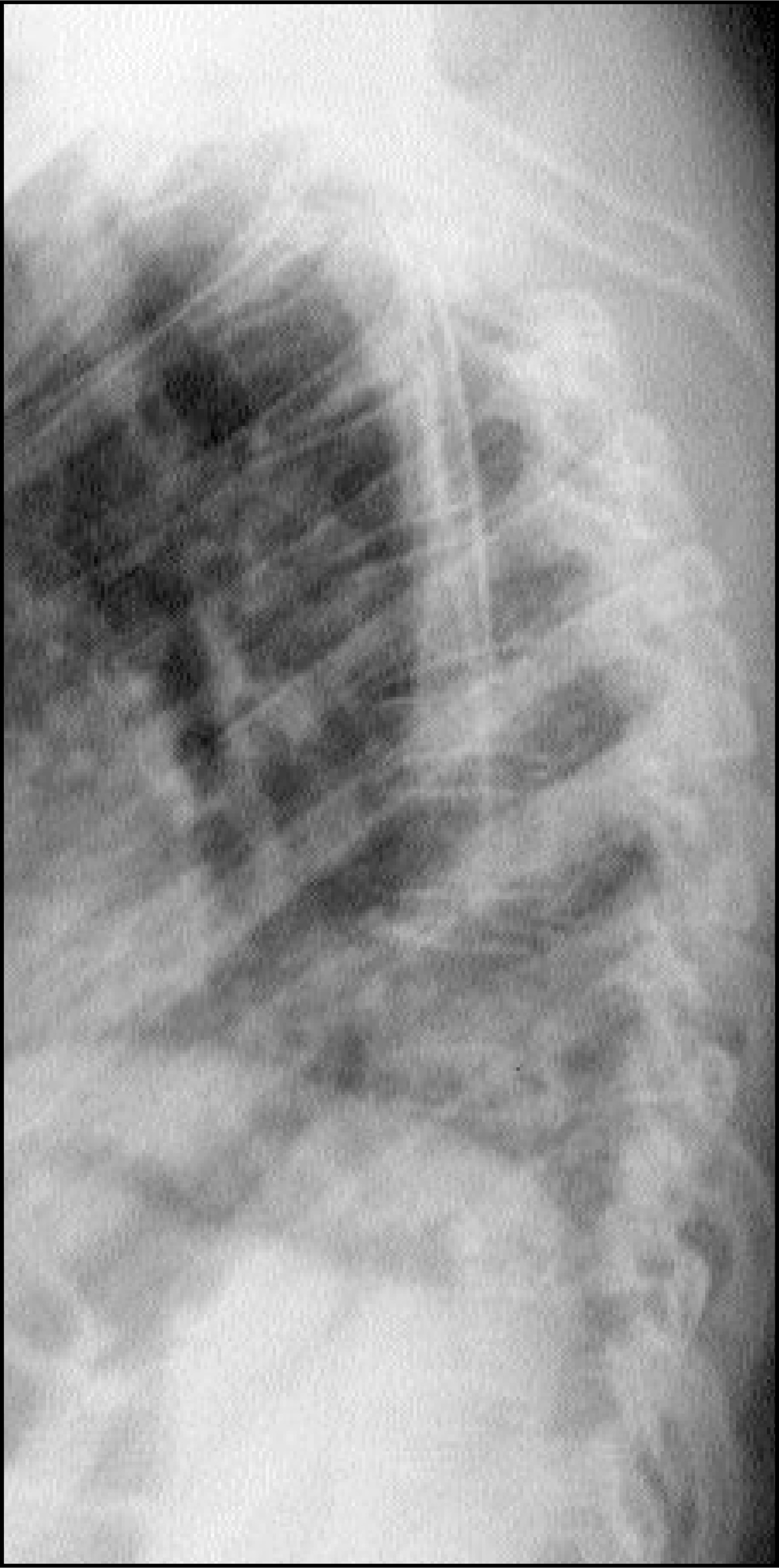
DIAGNOSIS
Cryptococcal osteomyelitis
IMAGING FINDINGS
Radiography of the thoracic spine demonstrated compression fracture of T6 (Figure 1). CT demonstrated a paravertebral soft-tissue mass with destruction of T6 vertebral bodies, posterior elements, and posterior left rib (Figure 2). MR imaging revealed abnormal enhancement within the T6 vertebral body with sparing of the adjacent intervertebral discs (Figure 3).
DISCUSSION
Cryptococcus neoformans is a ubiquitous yeast (Figure 4) that grows abundantly in soil containing bird (especially pigeon) droppings. Disseminated infection of man and animals occurs mainly in the immunocompromised, with the lungs and central nervous system most commonly affected. Approximately 50% of patients with cryptococcal meningitis, however, show no evidence of immunosuppression. Osteomyelitis occurs in 5% to 10% of disseminated cases, but isolated bone involvement is rare. Crypto-coccal meningitis is common in HIV-infected patients; however, no case of cryptococcal osteomyelitis has been reported in this population.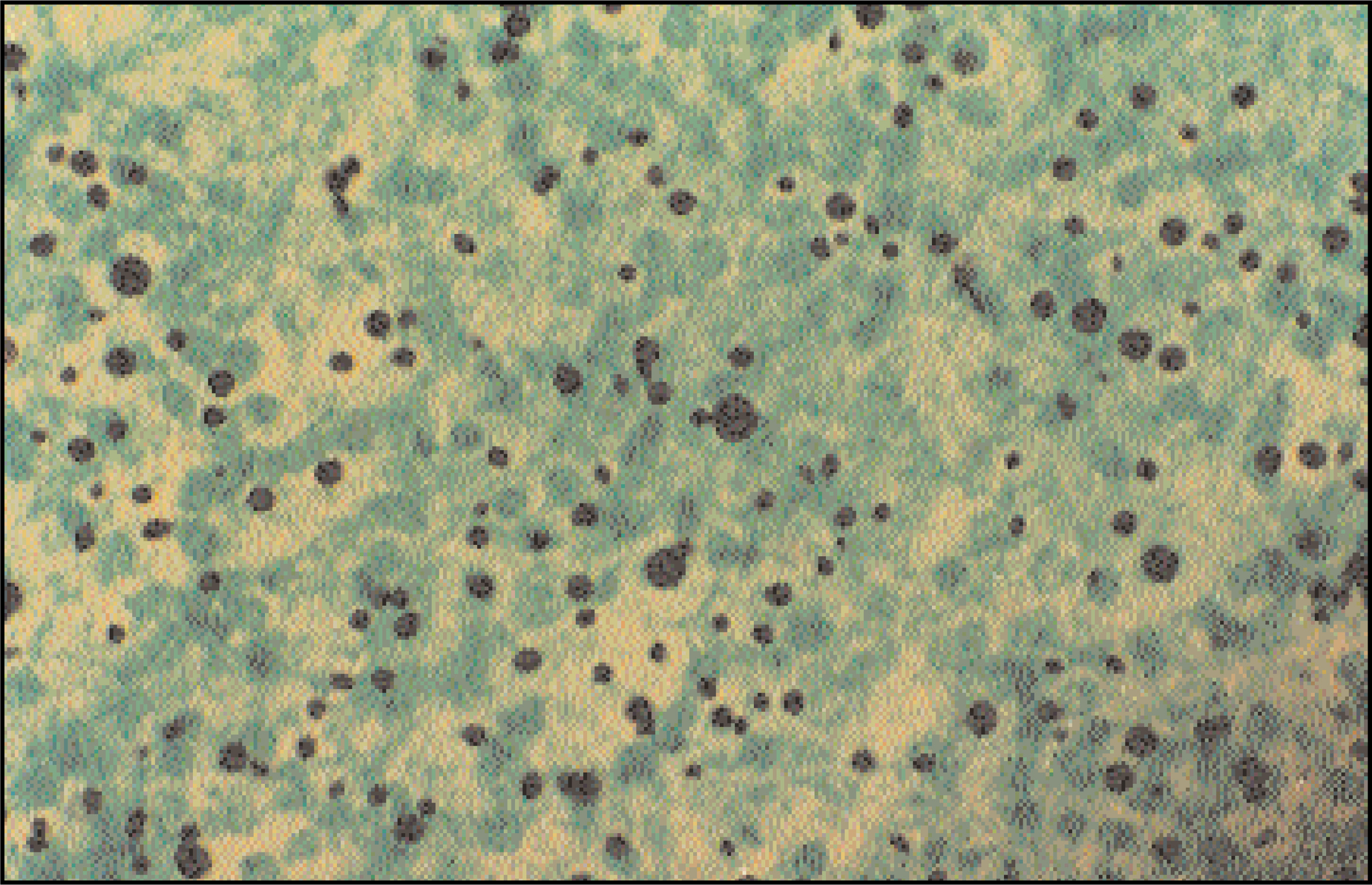
A recent review of 40 reported cases found most cases involved one site. The most common site was the spine, followed by the femur, tibia, humerus, and ribs. The most common comorbidity was sarcoidosis, followed by tuberculosis, steroid therapy, and, as in this case, diabetes mellitus.
Differential diagnostic considerations include tuberculosis, plasmacytoma, metastatic disease, and lymphoma.
References
- Liu P. Cryptococcal osteomyelitis: Case report and review.. Diagn Microbiol Infect Dis.. 1998;30:33-35.
- Resnick D. Diagnosis of Bone and Joint Disorders.. 1995:2505-2506.
Citation
. RCOM RADIOLOGICAL CASE OF THE MONTH. Applied Radiology. 2003;32(7):45-47. doi:10.37549/AR1201.