RCOM RADIOLOGICAL CASE OF THE MONTH
Applied Radiology — Vol. 33 , Issue 1 , pp. 40 -42
DOI: 10.37549/AR1211
Published: January 1, 2004
Categories
CASE SUMMARY
A 42-year-old woman presented with a 5-month history of abdominal pain, nausea, and vomiting. Physical examination revealed a palpable epigastric mass. The patient had undergone an elective transabdominal hysterectomy for uterine leiomyomata 5 months earlier. Her medical history and laboratory values were unremarkable. An abdominal computed tomography (CT) scan was performed (Figure 1).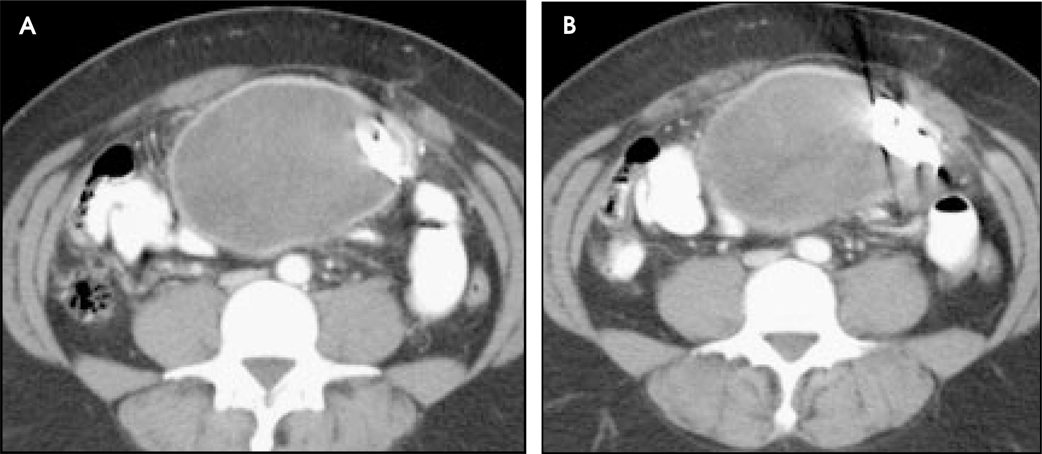
DIAGNOSIS
Retained laparotomy sponge (gossypiboma)
IMAGING FINDINGS
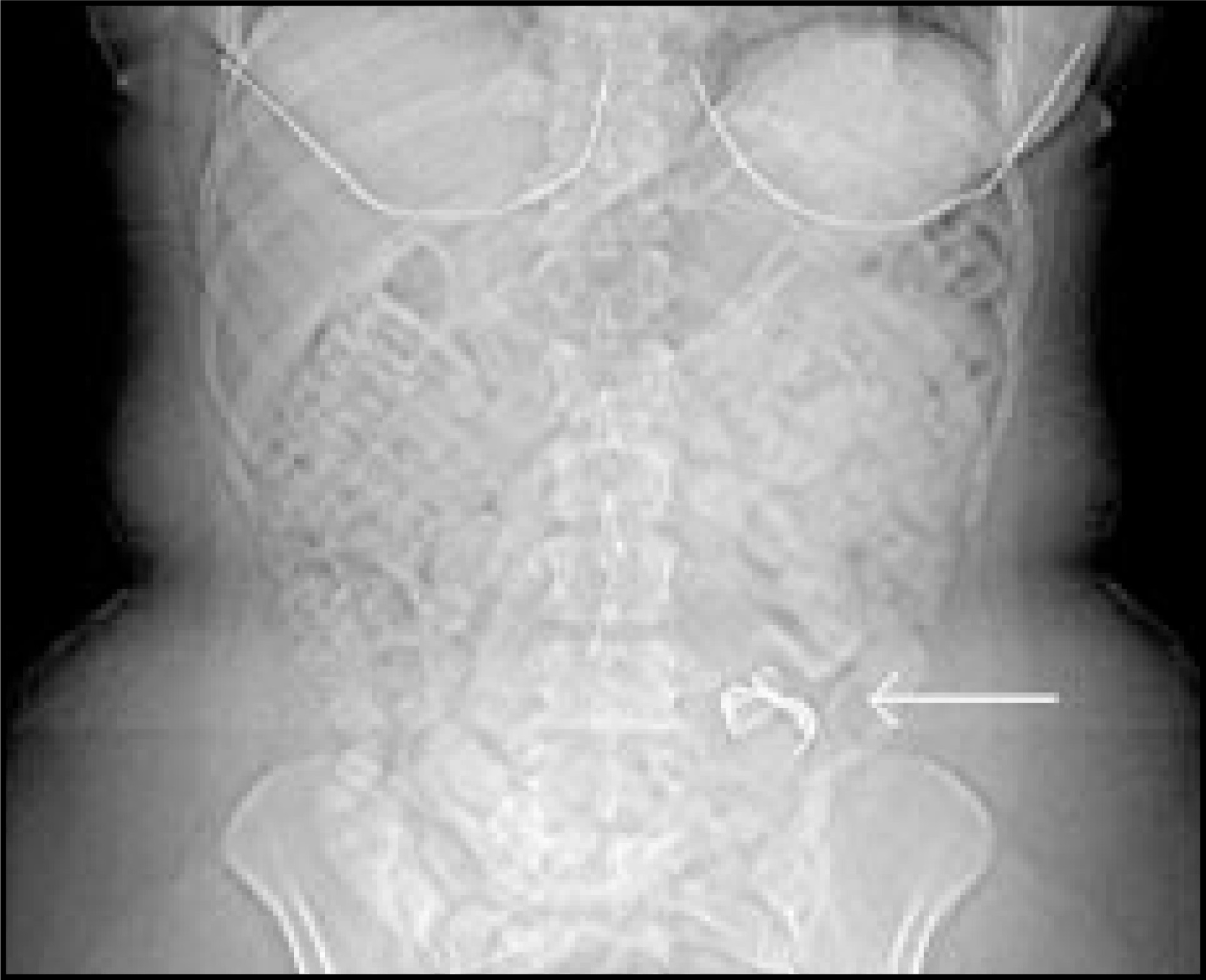
A contrast-enhanced CT scan of the abdomen reveals a well-circumscribed rim enhancing fluid collection within the anterior abdomen. A curvilinear metallic density is seen in the inferior aspect of the collection (Figure 1). There are relative hypoattenuating areas in the upper aspects of the fluid collection (Figure 2). Review of the scout image from the CT shows a density consistent with a laparotomy sponge in the left lower quadrant of the abdomen (Figure 3).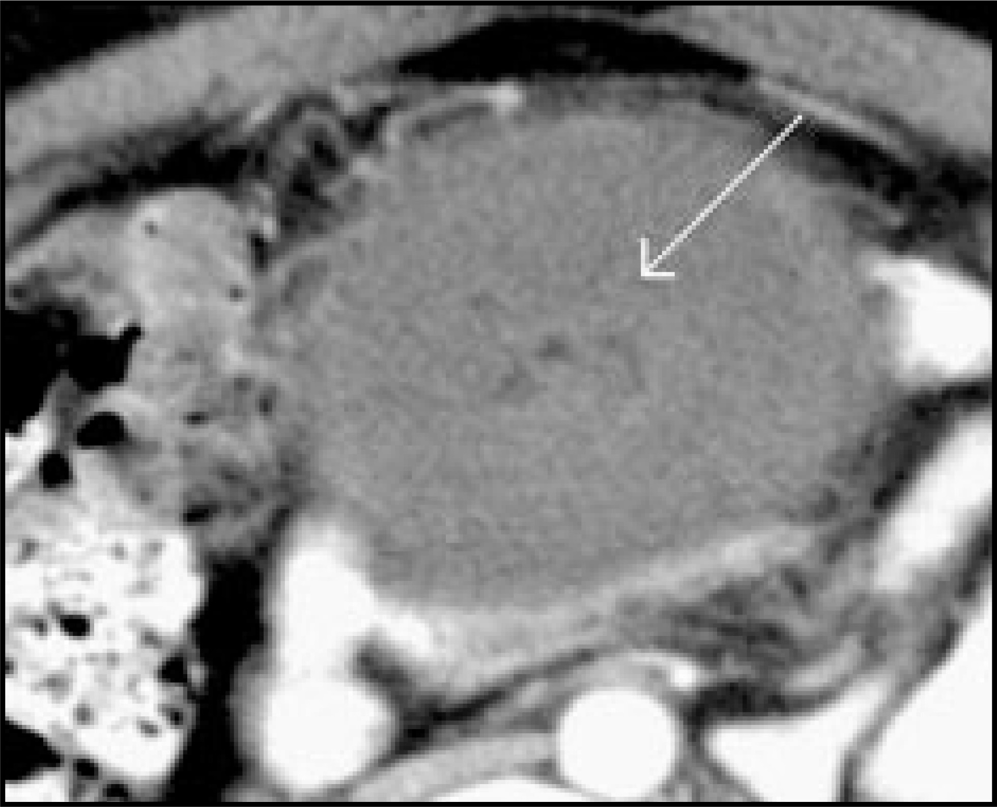
DISCUSSION
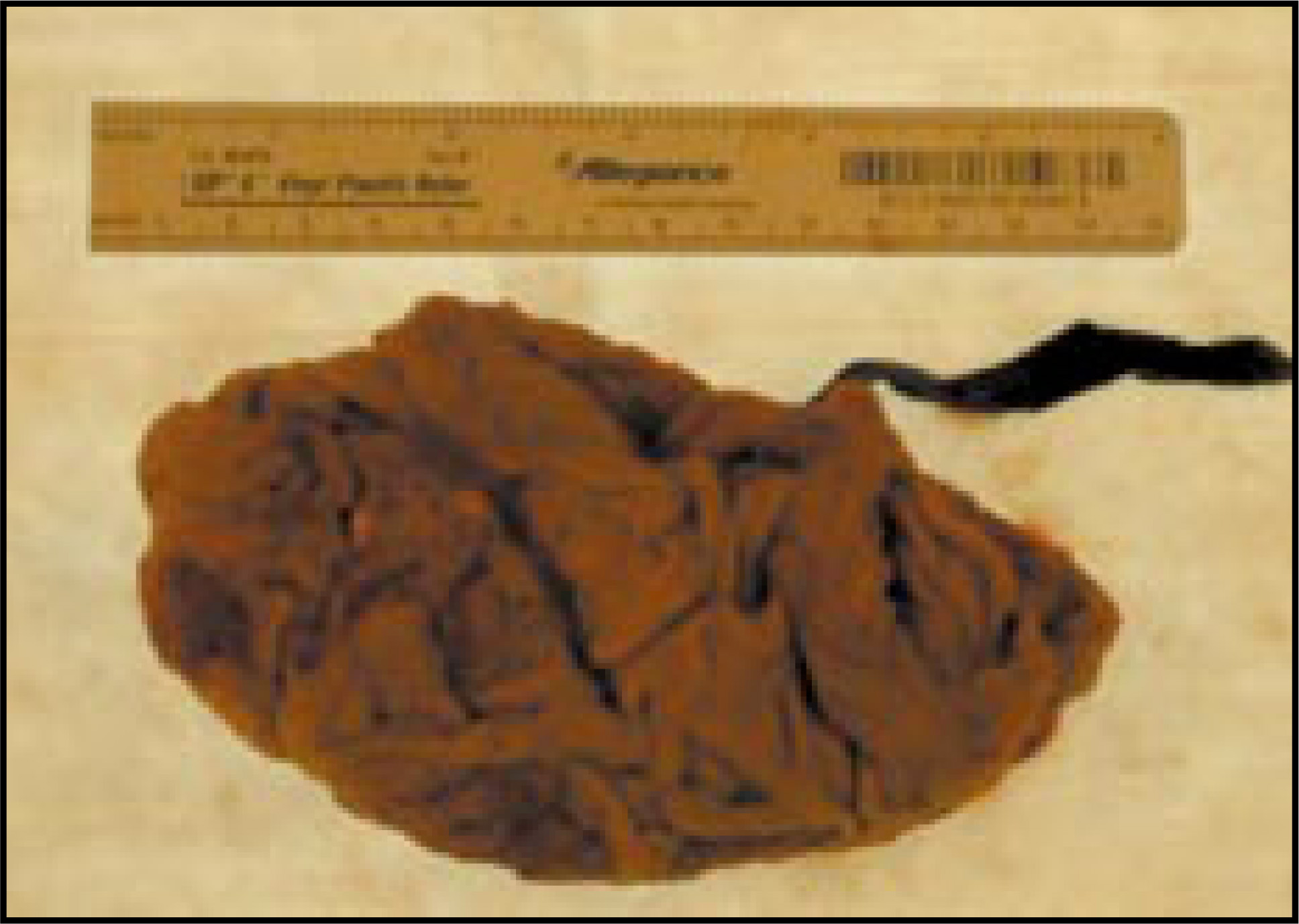
A gossypiboma, or retained surgical sponge (Gossypium [Latin]: cotton, Boma [Kiswahili]: place of concealment), is an uncommon surgical complication that has gained recent attention in the press. A study by The New England Journal of Medicine (NEJM) estimates that 1500 cases of retained surgical foreign objects occur annually in the United States, of which the majority are retained surgical sponges.1 These retained sponges were first referred to as “textilomas,“ but were renamed “gossypiboma” in 1978.2,3
Most retained foreign bodies are detected soon after surgery; the NEJM study reported a mean of 21 days to detection. However, 26% remain undetected for ≥60 days.1 More acute presentations are generally related to sepsis and abscess formation, while more delayed presentations usually occur with a palpable mass or increasing mass effect on adjacent structures.
Imaging plays a vital role in identifying retained foreign objects. Barium-impregnated threads are woven into one side of all surgical sponges available in the operating room, and these are visible on plain films as curvilinear densities (Figure 3). It is vital for the radiologist to recognize sponge markers as well as the radiographic appearance of all objects used in the operating room. Surgical sponges should never be used as a topical dressing, and a sponge visualized on a film should prompt immediate notification of the attending physician. Sponge fibers and any retained gas bubbles are easily visualized by ultrasound, but care should be taken not to misinterpret the areas of increased echogenicity and posterior acoustic shadowing as markers for a more malignant process.4
CT is the most useful modality to identify a retained sponge and its complications. There is often an associated fluid collection or inflammatory process, and the barium-impregnated threads of the sponge are easily seen. Gas lucencies may be seen in the fluid collection even in the absence of infection. The gas trapped in the interwoven threads of the surgical sponge may persist for weeks or months postoperatively. Similarly, calcification and enhancement of the wall of the fluid collection may also occur in the absence of infection.2,5 In this case, the patient had no symptoms of infection, even though gas lucencies were visible on the CT (Figure 2).
Emergency procedures, unexpected changes in the operation, and large body habitus are the factors most closely associated with an increased risk of retained foreign body.1 While removal of the sponge is safely performed in the majority of the cases, prevention is obviously the treatment of choice.
CONCLUSION
Gossypiboma is a term used to describe a retained surgical sponge, which is a relatively rare occurrence. Radiologists should be familiar with the imaging characteristics of these foreign bodies by multiple modalities, thus decreasing further patient pain and suffering and expediting the correct diagnosis.
References
- Gawande A, Studdert D, Orav E. Risk factors for retained instruments and sponges after surgery. N Engl J Med. 2003;348:229-235.
- Mackenzie D. Am Scientist. 1997.
- Williams R, Bragg D, Nelson J. Gossypiboma—the problem of the retained surgical sponge. Radiology. 1978;129:323-326.
- Zbar A, Agrawal A, Saeed I, Utidjian M. Gossypiboma revisited: A case report and review of the literature. J Roy Col Surg Edinburgh. 1998;43:417-418.
- Kopka L, Fischer U, Gross A. CT of retained surgical sponges (textilomas): Pitfalls in detection and evaluation. J Comput Assist Tomogr. 1996;20:919-923.
Citation
. RCOM RADIOLOGICAL CASE OF THE MONTH. Applied Radiology. 2004;33(1):40-42. doi:10.37549/AR1211.