RCOM RADIOLOGICAL CASE OF THE MONTH
Applied Radiology — Vol. 33 , Issue 1 , pp. 34 -37
DOI: 10.37549/AR1216
Published: January 1, 2004
Categories
CASE SUMMARY
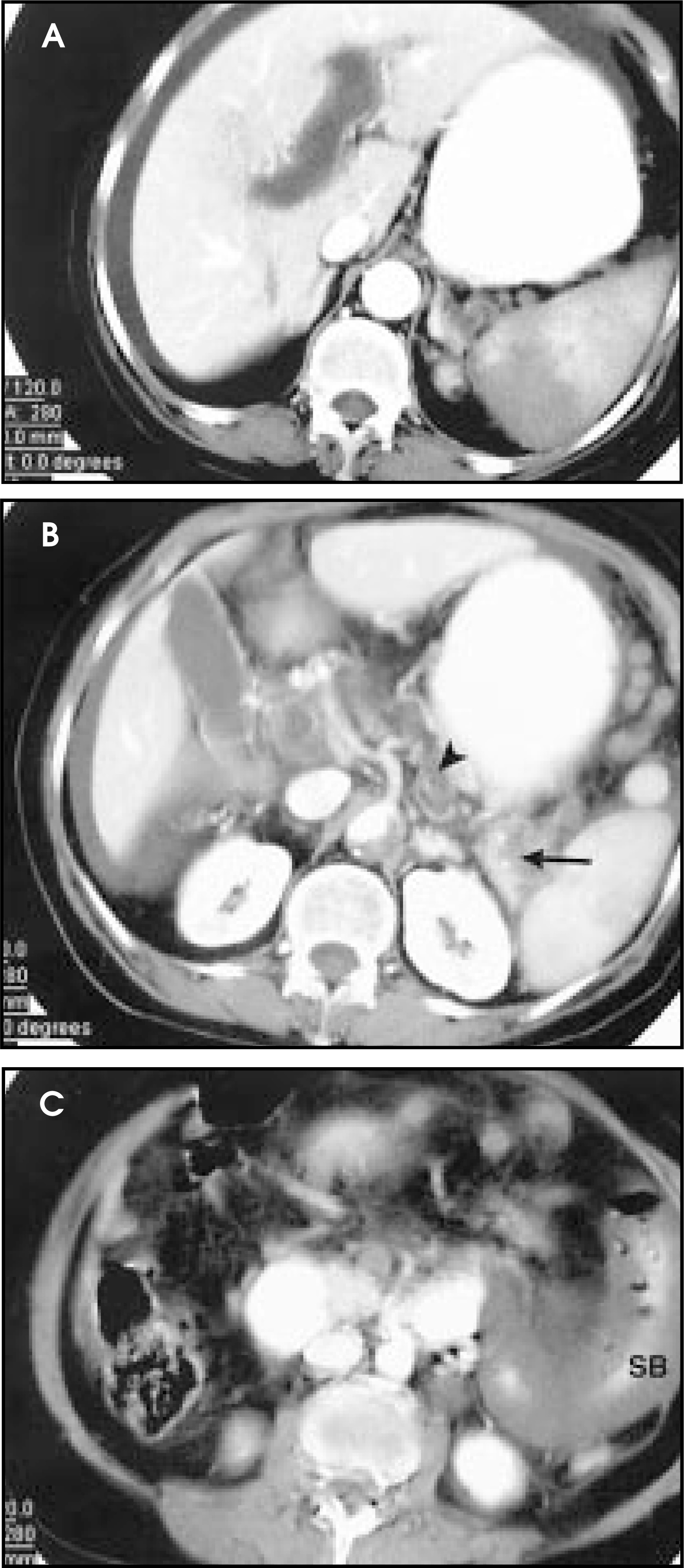
A 70-year-old Hispanic woman with a medical history of hypertension presented to the emergency department with 2 weeks of progressive abdominal pain, 4 days of constipation, and 2 days of vomiting. Physical examination was significant for moderate dehydration, abdominal distention, diffuse abdominal tenderness, and guarding suggestive of an acute abdomen.Abdominal ultrasonography (Figure 1) and contrast-enhanced computed tomography (CECT) of the abdomen and pelvis (Figure 2) were performed consecutively, which revealed portal vein thrombosis. Exploratory laparotomy revealed small-bowel gangrene, which was successfully resected. Further work-up to find the cause of the thrombosis indicated protein C deficiency. The patient recovered uneventfully.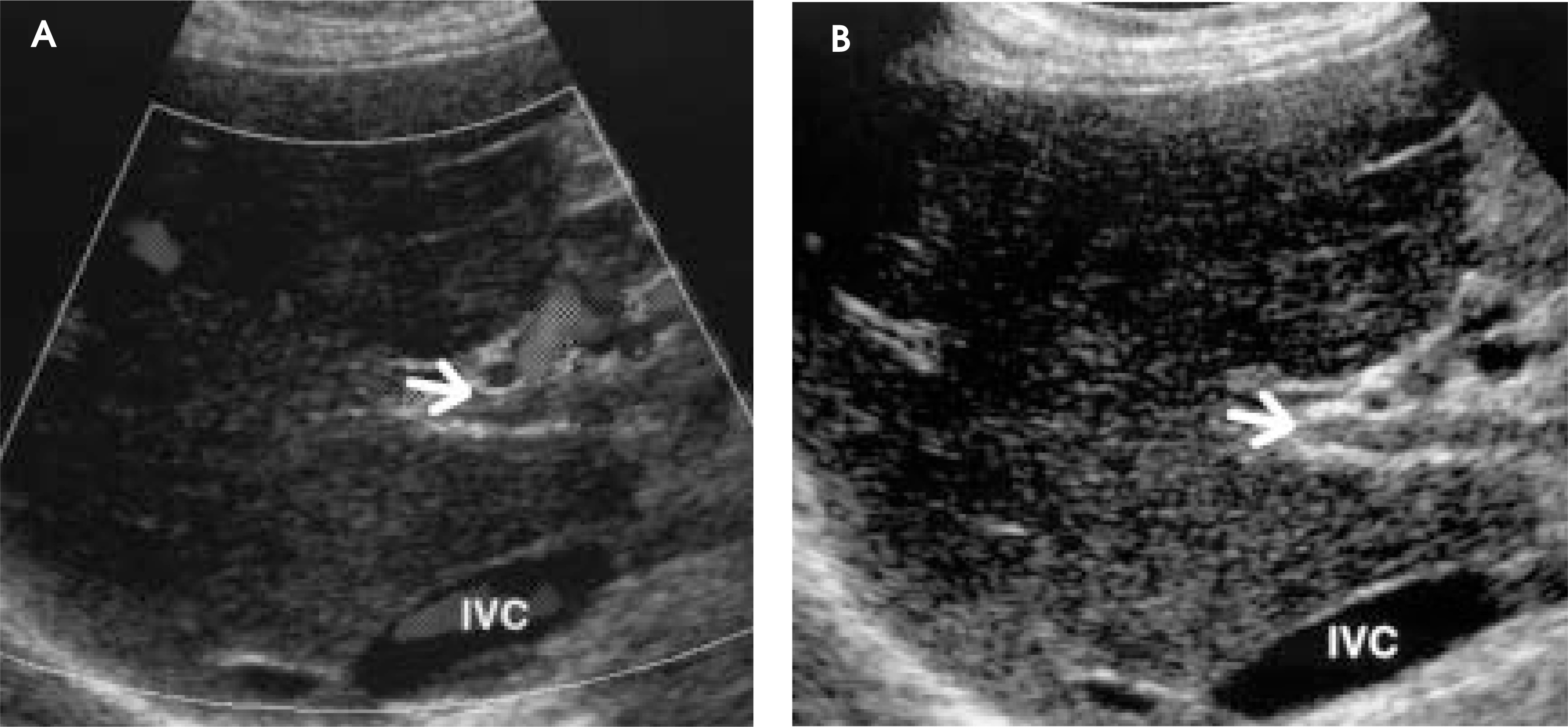
DIAGNOSIS
Portal vein thrombosis (PVT) with small-bowel gangrene
IMAGING FINDINGS
Ultrasonographic images of the portal vein are depicted in Figure 1. The color Doppler sonography at the level of the porta hepatis shows the longitudinal image of the portal vein with the lumen filled with hyperechogenic material (Figure 1A), which is consistent with thrombus.This is further demonstrated in Figure 1B. Normally, the lumen of the portal vein is hypoechoic, similar to the inferior vena cava (IVC). In this case, the lumen of the portal vein is echogenic because of intraluminal thrombosis. The portal vein can be distinguished from the hepatic vein by the imaging characteristics of presence of fat around it,which is represented by the highly hyperechoic wall.
Contrast-enhanced images of the abdomen are shown in Figure 2. Figure 2A (which was taken approximately 5 cm below the right dome of the diaphragm and at the level of the IVC and caudate lobe of the liver) demonstrates hypodensity, dilatation, and nonopacification of the portal vein with contrast, consistent with portal vein thrombosis. The same image shows nonopacification of the splenic vein and wedge-like low attenuation of the inferior portion of the spleen, representing splenic vein thrombosis and spleen infarction, respectively. Figure 2B clearly shows filling defects in the portal vein, as well as the superior mesenteric and splenic veins, representing thromboses in these veins. The aorta shows large semilunar filling defects in its posterior half, consistent with plaque.
However, there were no symptoms referable to aortic thrombus, no renal involvement, and no evidence of limb ischemia; therefore no further treatment was instituted. Figure 2C demonstrates marked wall edema and wall thickening of the segment of the small-bowel loop. There is also evidence of air tracking on the wall of the small bowel, which is consistent with pneumatosis. These constellations of findings strongly suggest small-bowel ischemia.
The patient underwent surgical resection of a gangrenous small-bowel segment, which mainly involved the jejunum. Histologic examination confirmed small-bowel infarction affecting a 45-cm segment of the jejunum, and thrombosis of the mesenteric veins.
DISCUSSION
We present a case of PVT with small-bowel gangrene secondary to protein C deficiency. Portal vein thrombosis is the most common cause of prehepatic portal hypertesion and occurs in equal frequency in children and adults, representing approximately 50% of cases.1 The peak age of incidence is 6 years for children and 40 years for adults.2
From the pathophysiologic standpoint,occlusion of the portal vein leads to buildup of pressure within the portal system. When the pressure rises above the normal of 8 mm Hg, the portosystemic collateralization is stimulated in order to restore the portal perfusion. This process is called cavernous transformation of the portal vein. The portosystemic collaterals develop at the esophagogastric, retroperitoneal, anorectal, and umbilical regions and may result in the development of gastroesophageal varices, hemorrhoids, and caput medusae, respectively. The portal vein accounts for 75%of blood supply to the liver. With PVT, the hepatic arterial flow generally increases to maintain hepatic perfusion and prevent possible hepatic infarction; this is called the buffer response.3
While the liver may be able to withstand the effects of PVT, the small bowel may show variable changes from normal, dilation, to infarction. The portal vein, which is formed from the confluence of the splenic and superior mesenteric veins, serves as the common venous drainage of the small and large bowel. When venous drainage from a segment of the bowel is compromised, stasis and back flow develop. This leads to congestion of the bowel wall, then dilation, which progressively cuts off the arterial supply, resulting in gangrene,4 which presents clinically as an acute abdomen requiring surgical intervention.
Causes of PVT can be broadly categorized into neoplastic, infective, inflammatory, hypercoagulable states, and miscellaneous. Neoplasm is the most common cause and includes hepatocellular carcinoma, cholangiocarcinoma, gastric, and pancreatic carcinomas.2,5,6
Infective processes include hepatobiliary infections and intra-abdominal sepsis.2 Hypercoagulable states (namely, protein C and S deficiency) can be caused by thrombocytopenia, antiphospholipid syndrome, oral contraceptives, and myeloproliferative disorders.2,7,8 Miscellaneous causes include pregnancy, postprocedure complications (eg, umbilical vein catheterization, chemoembolization), hypotension, cirrhosis, and, rarely, inflammatory bowel diseases.2,5,9,10
In children, umbilical vein catheterization and sepsis are the most common causes of PVT.2,5
However, a review of children from developing countries who developed PVT implicated protein C deficiency or markedly elevated antiphospholipid antibody in about 70% to 80% of cases.8 The patient in the case we report here had a protein C level of <0.5 mg/L (normal: 2.5–5.5mg/L), all of the other tests were within normal limits.
Clinical presentation of PVT depends on the underlying or precipitating factors,acuteness or chronicity of the condition, and presence or absence of portal hypertension. Generally, acute onset typically presents with features of acute abdomen: diffuse abdominal pain with tenderness, guarding, and rebound.4,5 In the chronic form, hemetemesis from variceal bleeding due to portal hypertension occurs.5,11 Our patient presented with the signs and symptoms of acute abdomen with gangrene of small bowel (involving part of duodenum and jejunum).
Ultrasonography (US), CECT, and magnetic resonance imaging (MRI) provide important diagnostic information. In acute settings, US is the best imaging modality for screening and it is simple, inexpensive, and noninvasive. It depicts PVT as a hyperechogenic focus, and power Doppler demonstrates hepatofugal flow, especially in the context of portal hypertension. Portal vein thrombus is hyperdense in noncontrast CT, and appears as hypodense filling defect in CECT.12 MRI may show areas of signal void in portal vein collaterals, and the thrombus appears hyperintense on T1-weighted images. However, intraluminal thrombi of <5 weeks duration appear markedly hyperintense relative to liver and muscle in both T1- and T2-weighted images, while older thrombi tend to be hyperintense in only T2-weighted images.13 While both US and CECT can evaluate the pancreatic and hepatic malignant lesions that may be responsible for PVT, CECT is particularly helpful in evaluating the mesenteric vessels, bowel, and other intra-abdominal structures to provide a global picture for adequate and effective therapeutic intervention.
In our patient, while US provided the initial diagnostic probability of PVT (Figure 1), CECT clearly confirmed the diagnosis and also showed segmental small-bowel dilation and moderate wall edema/thickening and pneumatosis (Figure 2) necessitating surgical intervention that ultimately saved the patient’s life.
Complications of PVT include portal hypertension, variceal bleeding, hepatic infarction, mesenteric vein thrombosis, and bowel ischemia.11,12
Bowel ischemia is a major complication of acute PVT with gangrenous development necessitating resection. Our patient had small-bowel gangrene, which was elucidated with CECT and subsequently resected successfully. Variceal bleeding complicating PVT can be treated with sclerotherapy; however, an interventional radiologist can provide transjugular intrahepatic portasystemic shunts (TIPS) when other palliative measures fail.
CONCLUSION
In this case, PVT caused by protein C deficiency resulting in small-bowel gangrene was accurately diagnosed by US and CECT. Contrast-enhanced CT (using normal intravenous contrast protocol) is a powerful tool in elucidating the possible causes and complications of PVT and should be utilized before deciding on any surgical intervention.
References
- Rikkers L, Townsend C, Beauchamp D, Evers M. Sabiston Textbook of Surgery: The Biological Basis of Modern Surgical Practice. 2001:1061-1075.
- Ferri F. Ferri’s Clinical Advisor 2002: Instant Diagnosis and Treatment. 2002:561.
- Lautt W. The 1995 Ciba-Geigy Award Lecture. Intrinsic regulation of hepatic flow. Canad J Physiol Pharmacol. 1996;74:223-233.
- Martin K, Balfe D, Lee J. Computed tomography of portal vein thromosis: Unusual appearances and pitfalls in diagnosis. J Comput Assist Tomogr. 1989;13:811-816.
- Dahnert W. Radiology Review Manual. 1999:610.
- Janssen H, Wijnhoud A, Haagsma E. Extrahepatic portal vein thrombosis: Etiology and determinants of survival. Gut. 2001;49:720-724.
- Hirohata Y, Murata A, Abe S, Otsuki M. Portal vein thrombosis associated with antiphospholipid syndrome. J Gastroenterol. 2001;36:574-578.
- Yachha S, Aggarwal R, Sharma B. Functional protein C and anti-cardiolipin antibody in children with deep vein thrombosis. Indian J Gastroenterol. 2001;20:47-49.
- Hagimoto T, Seo M, Okada M. Portal vein thrombosis successfully treated with a colectomy in active ulcerative colitis: Report of a case. Dis Colon Rectum. 2001;44:587-590.
- Baccarani U, Gasparini D, Risaliti A. Percutaneous mechanical fragmentation and stent placement for the treatment of early post-transplantation portal vein thrombosis. Transplantation. 2001;72:1572-1582.
- Weissleder R, Rieumont M, Wittenberg J. Primer of Diagnostic Imaging. 1997:631.
- Mori H, Hayashi K, Uetani M. High-attenuation recent thrombosis of the portal vein: CT demonstration and clinical significance. Radiology. 1987;163:353-356.
- Zirinsky K, Markisz J, Rubenstein W. MR imaging of portal venous thrombosis: Correlation with CT and sonography. AJR Am J Roentgenol. 1988;50:283-238.
Citation
. RCOM RADIOLOGICAL CASE OF THE MONTH. Applied Radiology. 2004;33(1):34-37. doi:10.37549/AR1216.