RCOM RADIOLOGICAL CASE OF THE MONTH
Applied Radiology — Vol. 33 , Issue 2 , pp. 9 -25
DOI: 10.37549/AR1220
Published: February 1, 2004
Categories
CASE SUMMARY
A previously healthy 2-year-old girl was seen by her pediatrician with a history of increasingly poor appetite and decreased energy and activity over the prior 2 months. She had occasional vomiting and foul-smelling stools. On physical examination, the child appeared pale and emaciated. Her weight had dropped from the 60th percentile at 19 months to below the 25th. She was lethargic, but cooperative. Her abdomen was protuberant, soft, and nontender. There was no organomegaly. An abdominal ultrasound was performed (Figure 1). A subsequent computed tomography (CT) scan demonstrated no abnormality (not shown).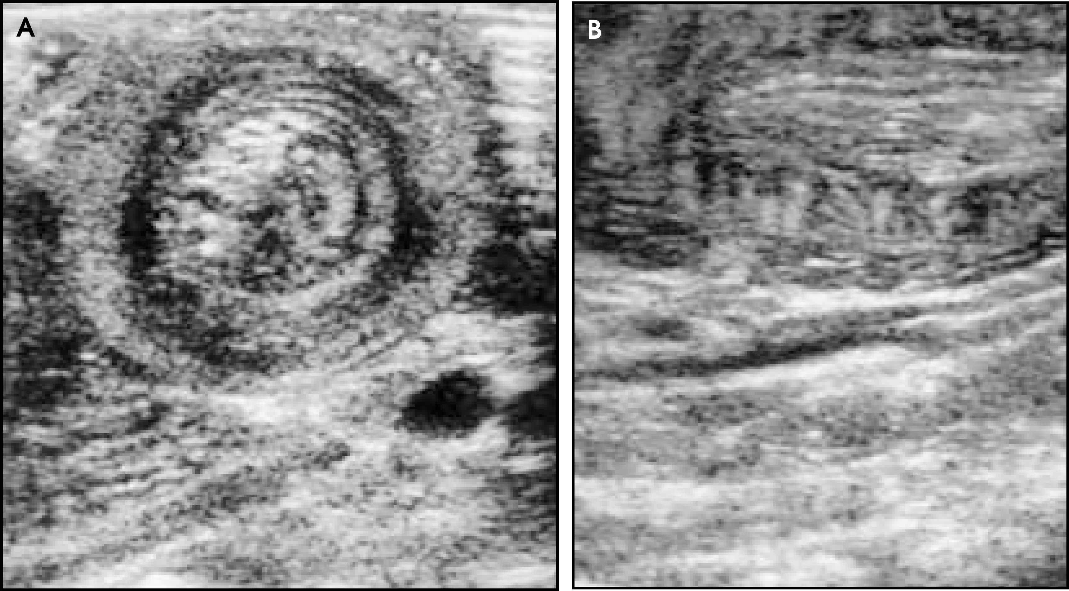
DIAGNOSIS
Small bowel intussusception associated with celiac sprue
IMAGING FINDINGS
An ultrasound of the abdomen was normal except for a target appearance to the bowel in the mid-abdomen (Figure 1). A focal segment of bowel showed a hypoechoic rim of thickened bowel wall measuring 6 to 7 mm and heterogeneous echogenic structures within the lumen, which appeared to be small bowel loops. These findings are classically compatible with intussusception.1 A repeat examination (not shown) performed 45 minutes later revealed no abnormality, suggesting intermittent intussusception. There was no tenderness, ascites, or fluid-filled bowel.
CASE FOLLOW-UP
A gluten-free diet was started empirically (on the mother’s initiative following an Internet search). The patient improved within 2 days: vomiting ceased, appetite improved, and she returned to normal activities. Laboratory studies were performed for endomysial antibodies IgA and anti-gliadin antibodies; IgG and IgA were strongly positive with endomysial IgA titer 1:10,240, gliadin IgG 108, and gliadin IgA >500. At follow-up, physical examination showed a normal, active 2-year-old child with a 9-oz weight gain. A biopsy, the diagnostic test of choice for celiac sprue, was not performed at the mother’s request because of her child’s dramatic improvement on dietary therapy. Because the laboratory results were strongly suggestive of a diagnosis of celiac sprue, this was the presumptive diagnosis, and the patient's progress was followed on a gluten-free diet with continuing good health.
DISCUSSION
Intussusception is easily diagnosed by ultrasound. Sonographic appearance can be variable in detail but most characteristically shows a target or donut configuration of bowel with a hypoechoic rim and a dense central echogenic core and no central movement in the donut.1 This pattern is known as the crescent-in-donut sign.1 Studies have shown this to be a sensitive and specific sign of intussusception.2
Intussusception in adults is much less common than in children, and it is more often caused by a specific gastrointestinal (GI) abnormality, usually a neoplasm.3 Adult intussusception is not an uncommon complication of sprue, but it is asymptomatic.4 In the pre-CT and ultrasound GI radiology literature, it was considered a specific sign of celiac disease.5
Celiac sprue, or gluten-sensitive enteropathy, is an autoimmune reaction to the gliadin fraction of gluten protein found in wheat, rye, oats, and barley. It does not present until gluten-containing foodstuffs have been introduced into the diet. Typically this means the disorder presents between 6 months and 2 years, the same age range as idiopathic intussusception, but it can manifest at any age. It affects more than 1 in 10,000 live births. The incidence appears to be much higher in Europe than in the United States. It is a malabsorption syndrome whose pathophysiology is the obliteration of intestinal villi and drastic reduction in absorptive surface area. Common signs and symptoms include diarrhea, failure to thrive, vomiting, anorexia, abdominal distension, and foul stools. Abdominal pain is rare. Affected children are often irritable and unhappy. There is no cure. The only treatment is a gluten-free diet that is a lifetime commitment that restricts patients to corn and rice.6
Until recently, diagnosis of sprue has depended on serial small-bowel biopsy that shows the characteristic histologic changes and interval improvement with cessation of gluten consumption. Antigliadin antibody tests, the first serologic tests, were not very reliable. Newer IgA-endomysial antibody tests have approached a sensitivity and specificity of 100%. Biopsy is still the definitive test, in particular if a repeat biopsy returns to normal following a gluten-free diet. The serologic tests normalize over 6 to 12 months following institution of a gluten-free diet, and this has been accepted as an alternative way to follow the resolution of sprue, instead of repeat biopsy.7
The case of a childhood small-bowel intussusception apparently caused by sprue has been rarely reported and is usually reported from Europe.8,9,10 There is one U.S. based report.11 One study using ultrasound in 17 pediatric sprue patients did not mention observing intus-susception.12 The largest prospective study of ultrasound for suspected intussusception showed many cases of clinically suspected intussusception that were not confirmed by radiologic study.2 The fraction of these cases that represent intermittent or transient cases with spontaneous resolution is unknown.
CONCLUSION
Infantile intussusception is considered a life-threatening emergency that requires immediate radiological and possible surgical intervention.1,13 This is a case of a child with an unexpected finding of transient intussusception on ultrasound apparently associated with active celiac sprue.
References
- Del-Pozo G, Albillos J, Tejedor D. Intussusception in children: Current concepts in diagnosis and enema reduction. RadioGraphics. 1999;19:299-319.
- Verschelden P, Filiatrault D, Garel L. Intussusception in children: Reliability of US diagnosis—a prospective study. Radiology. 1992;184:741-744.
- Gayer G, Apter S, Hofmann S. Intussusception in adults: CT diagnosis. Clin Radiol. 1998;53:53-57.
- Cohen M, Lintott D. Transient small bowel intussusception in adult coeliac disease. Clin Radiol. 1978;29:529-534.
- Masterson J, Sweeney E, Path M. The role of small bowel follow-through examination in the diagnosis of coeliac disease. Br J Radiol. 1976;49:660-664.
- Hyams J. Celiac disease: New thoughts on an old problem. Curr Opinions Pediatr. 1997;9:487-489.
- Miara S, Arnent M. Diagnosis of celiac sprue in 1994. Gastroenterol Clin North Am. 1995;24:133-143.
- Germann R, Kuch M, Prinz K. Celiac disease: An uncommon cause of recurrent intussusception. J Pediatr Gastroenterol Nutr. 1997;25:415-416.
- Gonzalez J, Gonzalez J, Crespo M, Sancho C. Acute gallbladder distension and recurrent small bowel intussusception in a child with celiac disease. J Gastroenterol Nutr. 1998;27:444-445.
- Mushtaq N, Marven S, Walker J. Small bowel intussusception in celiac disease. J Pediatr Surg. 1999;34:1833-1835.
- Martinez G, Israel N, White J. Celiac disease presenting as entero-enteral intussusception. Pediatr Surg Int. 2001;17:68-70.
- Riccabona M, Rossipal E. Sonographic findings in coeliac disease. J Pediatr Gastroenterol Nutr. 1993;17:198-200.
- DiFiore J. Intussusception. Semin Pediatr Surg. 1999;8:214-220.
Citation
. RCOM RADIOLOGICAL CASE OF THE MONTH. Applied Radiology. 2004;33(2):9-25. doi:10.37549/AR1220.