RCOM RADIOLOGICAL CASE OF THE MONTH
Applied Radiology — Vol. 33 , Issue 8 , pp. 38 -40
DOI: 10.37549/AR1267
Published: August 1, 2004
Categories
CASE SUMMARY
A 55-year-old woman with a history of radiation enteritis and biliary colic, resulting in laparoscopic cholecystectomy 10 days prior to admission, presented to the emergency department (ED) after 7 days of progressive epigastric pain, abdominal distention, nausea, and vomiting. Physical examination revealed diffuse abdominal tenderness, which was greatest in the epigastric region and right upper quadrant, without palpable mass. Abdominal radiography showed a dilated loop of small bowel, scattered air-fluid levels, and decompressed colon (Figure 1). An emergent right upper quadrant ultrasound request from the ED was considered, but computed tomography (CT) scanning was recommended, as it would be able to assess the small-bowel obstruction and possible complications of recent cholecystectomy.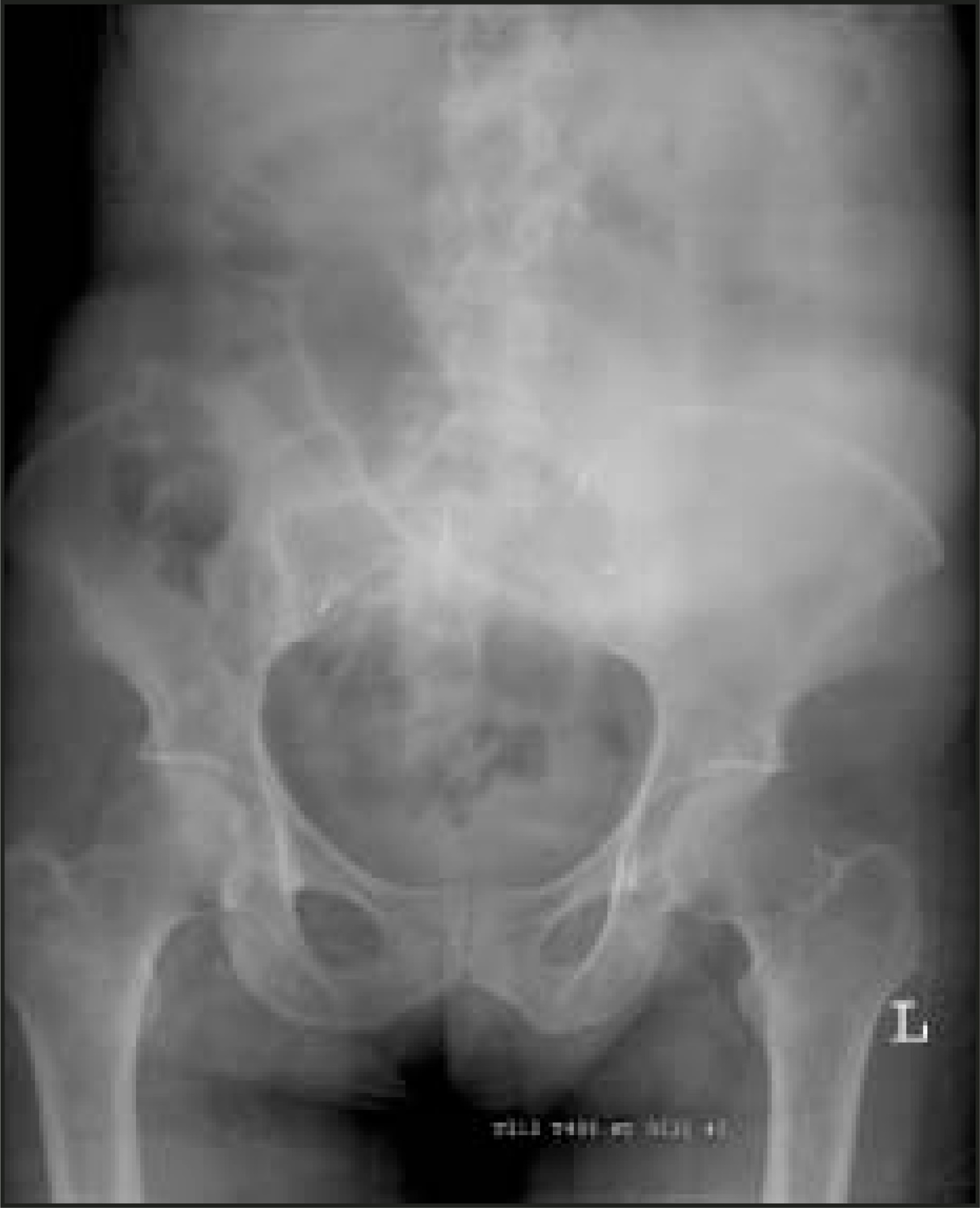
IMAGING FINDINGS
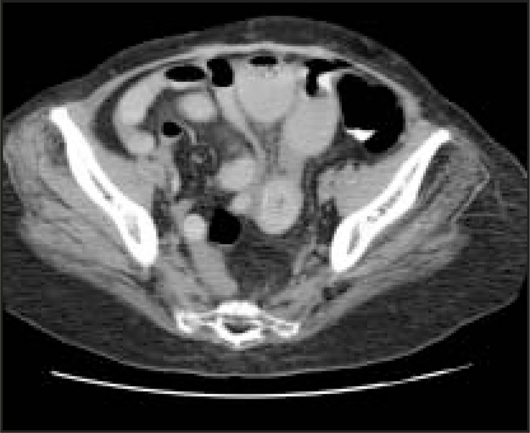
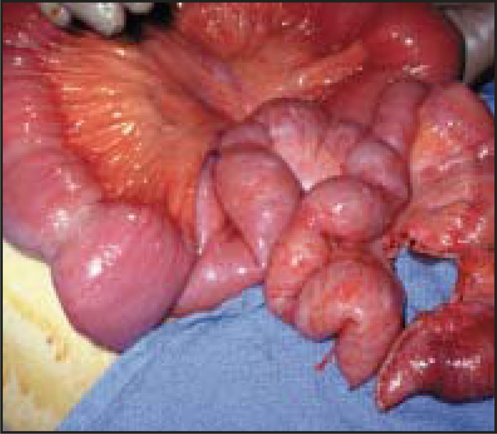
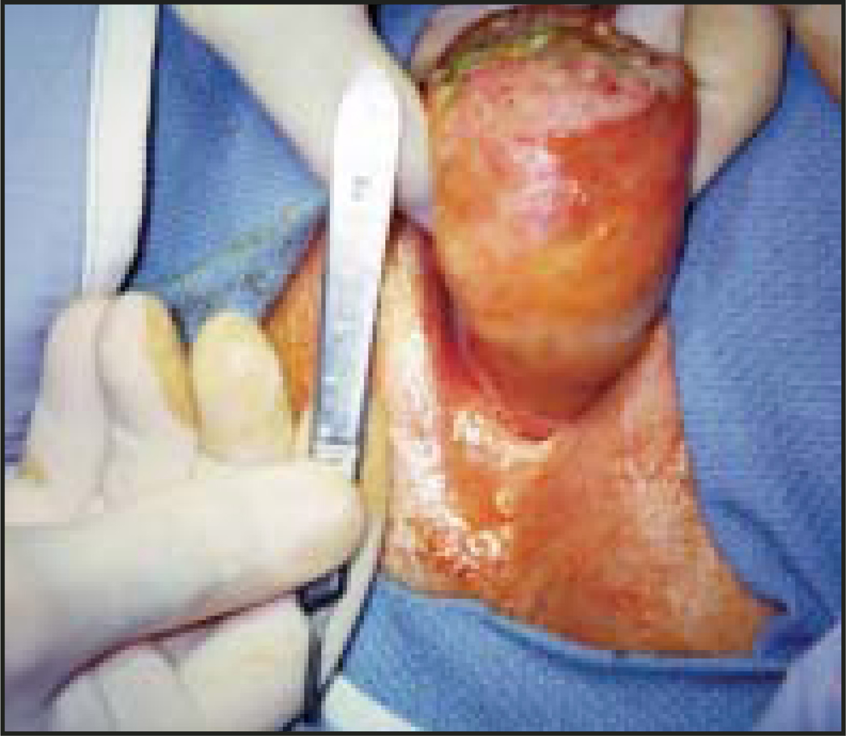
CT evaluation showed several dilated and fluid-filled loops of small bowel with a single lamellated, intra-luminal stone, measuring 2.5 cm in diameter, with a calcified rim (Figure 2). Repeat CT imaging performed 4 hours later revealed the changing position of the calculi, suggesting mobility (Figure 3). A large obstructive stone was surgically removed from the mid-jejunum (Figures 4 and 5) and was found by laboratory analysis to be composed of bilirubin and cholesterol crystals.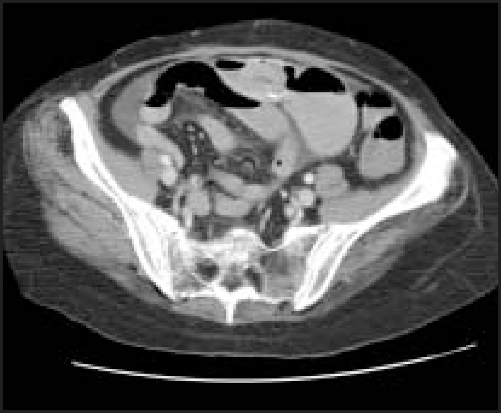
DIAGNOSIS
Gallstone ileus
DISCUSSION
Mechanical obstruction of the gastrointestinal (GI) tract by a gallstone is an uncommon but important complication of biliary stone disease. It is primarily a disease of the elderly, occurring in 25% of all small-bowel obstructions in patients older than 65 years.1 Concomitant illnesses, such as diabetes and cardio-vascular disease, in this population contribute to the significant morbidity and mortality associated with gallstone ileus. Because the radiographic triad of intestinal obstruction, pneumobilia, and aberrantly located gallstone described by Rigler et al2 in 1941 is present in less than half of patients, early use of CT, particularly in the elderly with radiographic findings suggesting small-bowel obstruction, plays a vital role in early and accurate detection.3
Gallstone disease affects 10% to 15% of people in the United States and produces symptoms in 25% to 30% of cases. Well-known complications include acute cholecystitis, biliary colic, acute pancreatitis, ascending cholangitis, gangrenous gallbladder, Mirizzi syndrome, and gallstone ileus. Complete or partial small-bowel obstruction occurs when one or more gallstones erode through the gall-bladder or biliary duct into the GI tract, creating a cholecystoenteric fistula. Common fistula sites include the duodenum, stomach, colon, and jejunum. Once in the GI tract, stones may pass spontaneously or become impacted, causing mechanical obstruction. Common sites of obstruction include the duodenum (Bouveret’s syndrome), terminal ileum, ileocecal valve, jejunum, then stomach and colon.4 Obstruction is influenced mainly by segment motility, size and number of calculi, and luminal diameter of bowel, as in the terminal ileum and ileocecal valve, both common sites of obstruction.5 Many believe small calculi increase in size as layers of bowel sediment deposit on stone surfaces, thereby increasing the likelihood of obstruction over time.6
Symptoms of gallstone obstruction are vague. Patients often present with partial or complete small-bowel obstruction, complaining of abdominal distension, nausea, and vomiting. Patients may be dehydrated, and many report anorexia and weight loss. Mean duration of symptoms before admission is reported as 5 to 14 days.7After admission, average reported delay in diagnosis ranges from 2 to 19 days, with an average of 3 days. Medical history is often unreliable, as 35% to 72% of patients suffering from gallstone ileus report antecedent biliary disease.8 Therefore, proper imaging plays an important role in establishing an accurate and timely diagnosis.
The abdominal radiograph is the mainstay of imaging in small-bowel obstruction and can be important in establishing a diagnosis of gallstone ileus. As reported by Rigler et al2 in 1941, the most frequent findings are small-bowel obstruction, pneumobilia, ectopic gallstone, and change in location of a stone on serial examinations. Roughly half of patients with confirmed gallstone ileus display two of the first three findings.9 In most cases, the most common finding by radiography is evidence of small-bowel obstruction, though little information is generally discovered concerning etiology. As a result, CT scanning is increasingly used in the assessment of the acute abdomen, particularly when small-bowel obstruction is suspected.
The use of CT is well-established in the evaluation of small-bowel obstruction and in the evaluation of the acute abdomen.10 Numerous authors have reported cases of gallstone ileus presenting with acute or subacute abdomen diagnosed by CT. Common findings, in order of frequency, include small-bowel obstruction with transition point, ectopic intraluminal calculi, gas-fluid levels in gallbladder fossa, free abdominal fluid, cholecystoduodenal fistula, pneumobilia, and thickened duodenum.11 Importantly, CT also assesses for strangulation, provides accurate alternative diagnoses in patients presenting with acute abdomen, and provides valuable information as to whether early laparotomy is warranted or whether nonoperative management should be considered.12
CONCLUSION
Mechanical obstruction from gallstone is an uncommon but important and detectable complication of gallstone disease, often presenting as partial or complete small-bowel obstruction in the elderly patient with an acute abdomen. Though classic radiographic findings are often absent, delays in diagnosis are avoidable and further complications can be mitigated if well-established criteria for the use of CT in small-bowel obstruction are applied in the emergency setting.
References
- Reisner R, Cohen J. Gallstone ileus: A review of 1001 reported cases. ;60:441-446.
- Rigler L, Borman C, Noble J. Gallstone obstruction. Pathogenesis and roentgen manifestations. JAMA. 1941;117:1753-1759.
- Seal E, Creagh M, Finch P. Gallstone ileus: A new role for abdominal computed tomography. Post Graduate Med J. 1995;71:331-315.
- Abou-Saif A, Al-Kawas F. Complications of gallstone disease: Mirizzi syndrome, cholecystocholedochal fistula, and gallstone ileus. Am J Gastroenterol. 2002;97:249-254.
- Van Hillo M, van der Vliet J, Wiggers T. Gallstone obstruction of the intestine: An analysis of ten patients and review of the literature. Surgery. 1987;101:273-276.
- Wittman D, Eggert A. Zur pathogenese des gallensteinileus. Chirurg. 1977;48:678-680.
- Kurtz R, Heimann T, Beck A, Kurtz A. Patterns of treatment of gallstone ileus over a forty-five year period. Am J Gastroenterol. 1985;80:95-98.
- Svartholm E, Andren-Sandberg A, Evander A. Diagnosis and treatment of gallstone ileus. Acta Chir Scand. 1982;148:435-438.
- Balthazar E, Schechter L. Air in gallbladder: A frequent finding in gallstone ileus. AJR Am J Roentgenol. 1978;131:219-222.
- Taourel P, Fabre J, Pradel J. Value of CT in the diagnosis and management of patients with suspected acute small bowel obstruction. AJR Am J Roentgenol. 1995;165:1187-1192.
- Swift S, Spencer J. Gallstone ileus: CTfindings. Clin Radiol. 1998;53:451-454.
- Maglinte D, Balthazar E, Kelvin F, Megibow A. The role of radiology in the diagnosis of small bowel obstruction. AJR Am J Roentgenol. 1997;168:1171-1180.
Citation
. RCOM RADIOLOGICAL CASE OF THE MONTH. Applied Radiology. 2004;33(8):38-40. doi:10.37549/AR1267.