RCOM RADIOLOGICAL CASE OF THE MONTH
Applied Radiology — Vol. 33 , Issue 8 , pp. 34 -37
DOI: 10.37549/AR1269
Published: August 1, 2004
Categories
CASE SUMMARY
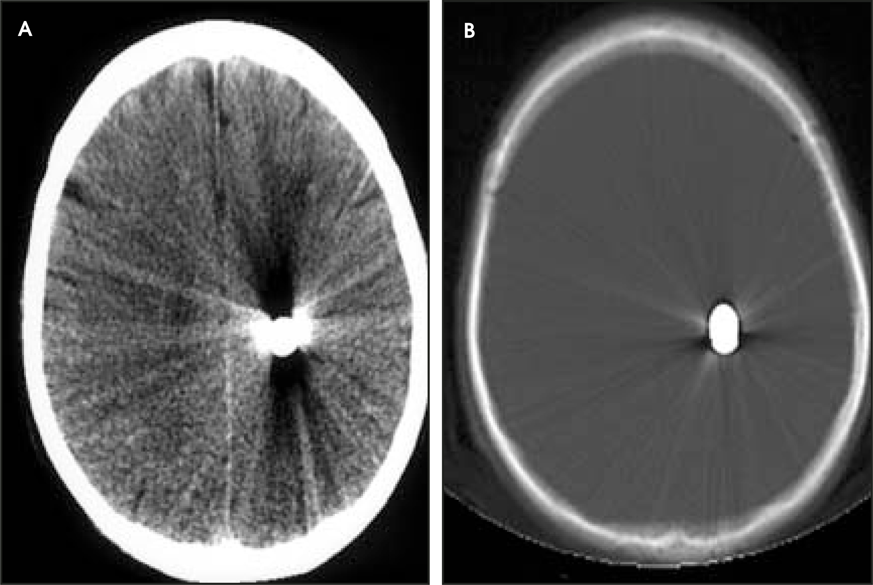
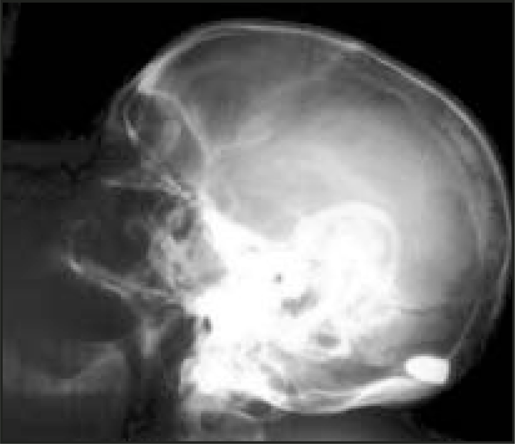
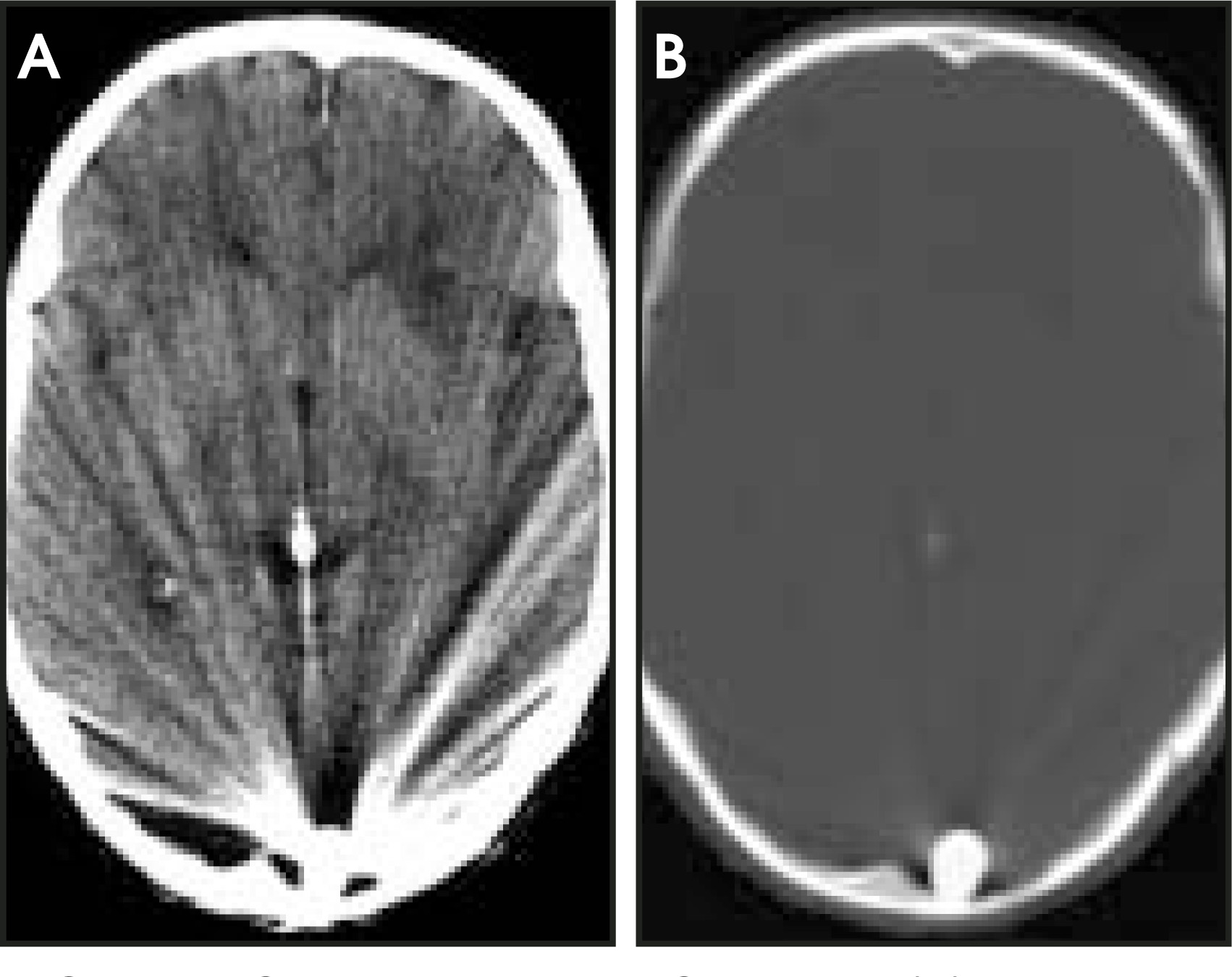
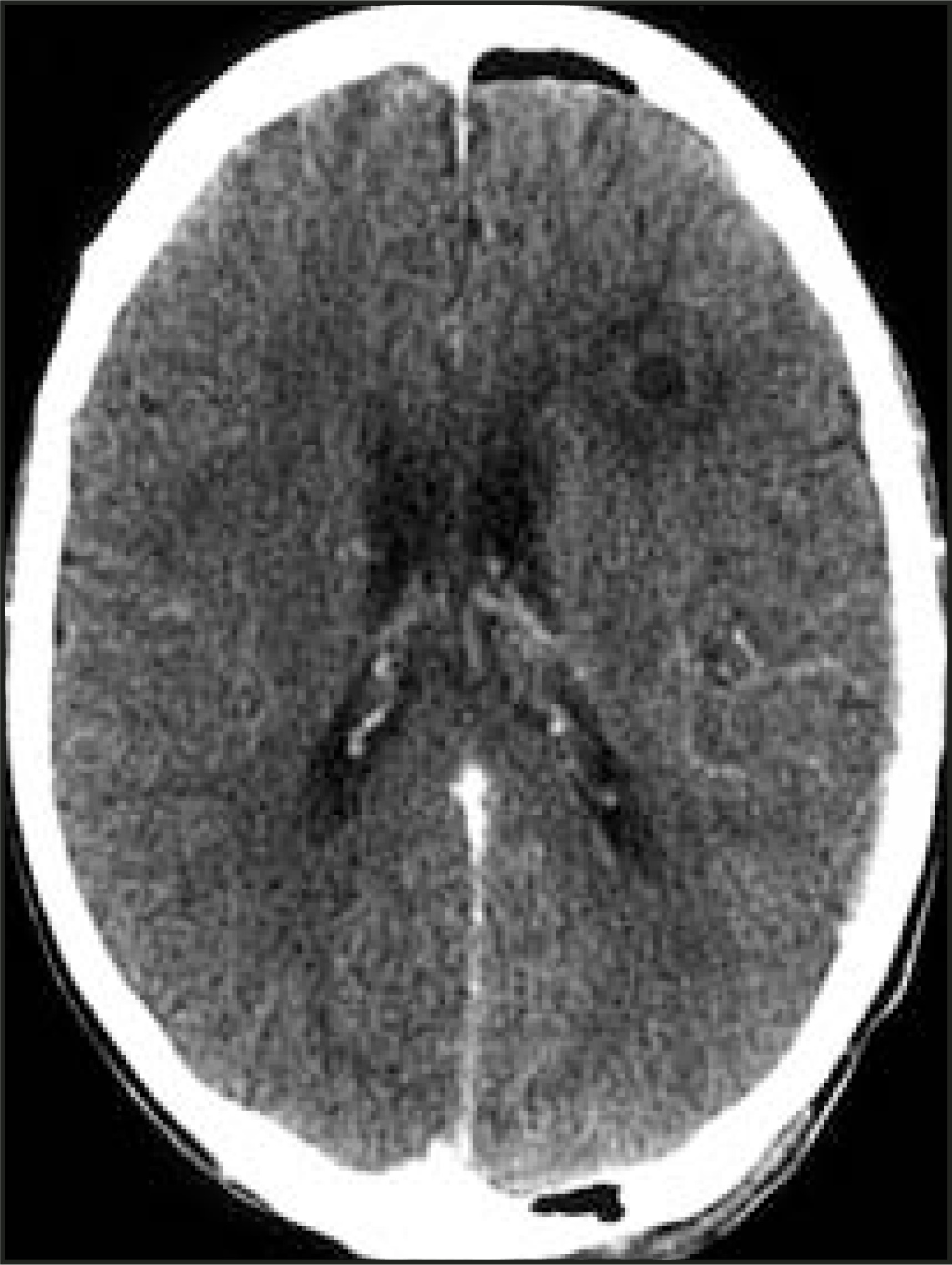
A 54-year-old man was admitted to the emergency department after being shot with a handgun. Physical examination revealed a single entry wound in the upper jaw near the left second premolar tooth with no observed exit wound. There was periorbital edema and ecchymosis on the left. Neurologic examination revealed normal findings except for a mild right hemiparesis. Lateral cranial roentgenography (Figure 1) and computed tomography (CT) (Figure 2) were performed. On the third day after admission, control roentgenogram (Figure 3) and CT (Figure 4) examinations were repeated. Ten days later, a cranial CT (Figure 5) was performed.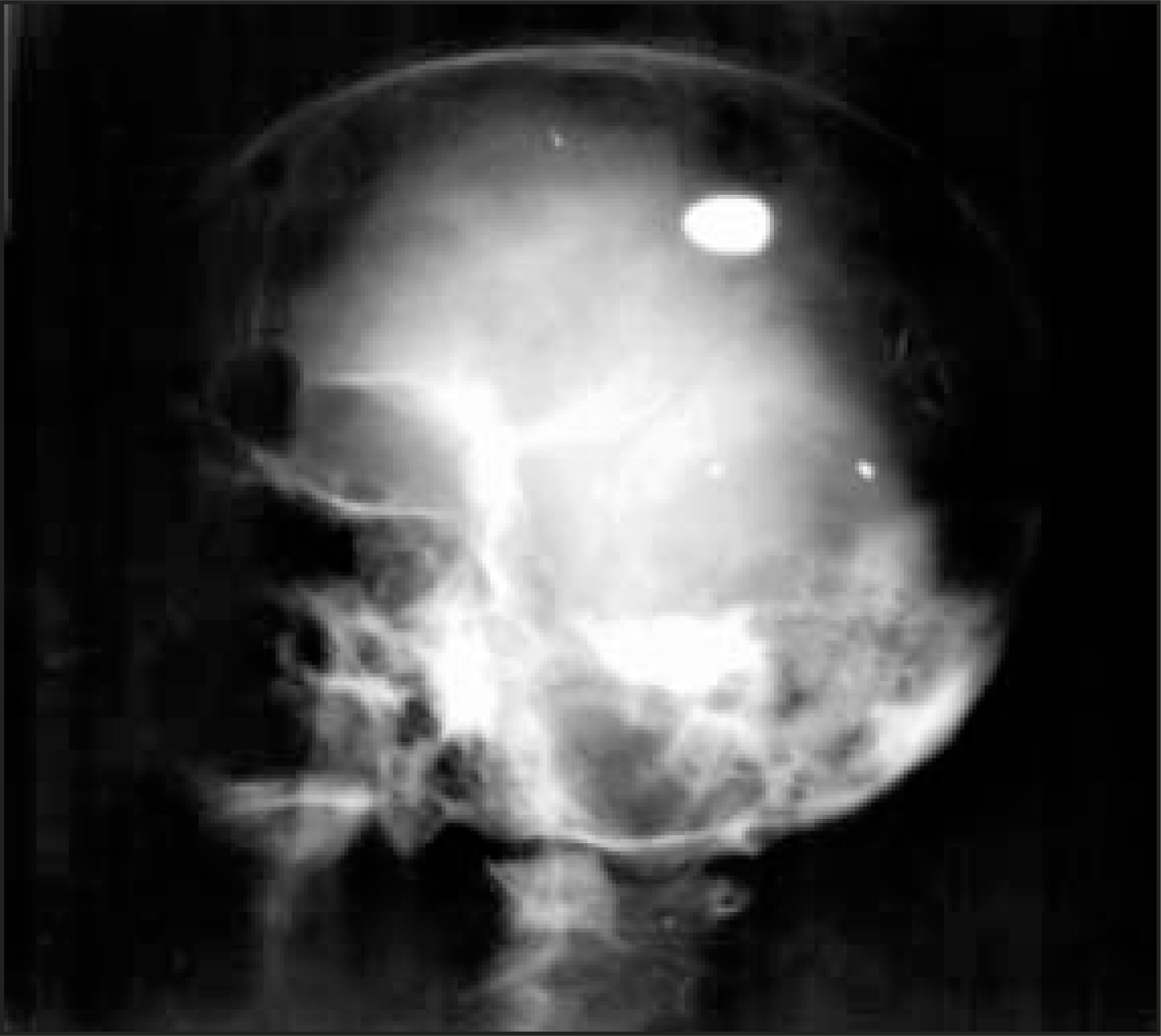
DIAGNOSIS
Intracranial bullet migration causing brain abscess
IMAGING FINDINGS
Lateral cranial roentgenogram revealed a bullet fragment lodged in the parietal region (Figure 1). Cranial CT examination showed an hyperdense appearance in the left deep parietal region with metallic artifact (Figure 2). Coronal paranasal sinus CT showed soft-tissue densities in the left maxillary sinus and in the left ethmoid cells. The neurosurgery team decided to treat the patient conservatively; and intravenous antibiotics were administered for prophylaxis of brain abscess. Three days after admission, control roentgenograms revealed that the bullet had migrated to the posterior part of the left occipital lobe (Figure 3). The final location of the bullet was the post-erobasal part of the left occipital lobe just above the tentorium, lateral to the torcular Herophili (Figure 4). On follow-up, it was discovered that the patient dev-eloped brain abscess (Figure 5), and surgery was performed. There was no complication in the postoperative period.
DISCUSSION
Cranial gunshot wounds are an important problem of neurosurgery in both military and civil practice. In such cases, brain tissue becomes damaged due to skull fracture, disruption of white matter integrity, and fluid percussion waves resulting from energy transferred by the bullet.1 The retention of the bullet and its fragments inside the cranium after cranial gunshot wounds is a commonly encountered phenomenon that leads to a treatment dilemma in neurosurgical practice. Although the bullet or its fragments could be left in situ if they are away from the bullet tract, the retained bullet fragments might lead to secondary events in the late period, such as increased infection risk due to foreign body reaction and migration.2–5
This complication was first described by Wilvandre and Morgan in 1916.6 Gravity, pulsation of the brain, and the loss of tissue resistance due to injury are among the possible mechanisms for migration.1,7,8 Most of the migration cases were reported to occur within the same compartment. In rare cases, transcompartmental migration could be seen. The bullet in this case most likely migrated from the frontoparietal region to the occipital lobe due to the effect of gravity, decreased tissue resistance causing softening, and pulsations from neighboring vascular structures. This migration was stopped at the torcular Herophili.
The determination of the exact location of the bullet in deeply located cases is an important surgical problem. In such cases, intraoperative ultrasound is a useful choice to decrease morbidity. Since the bullet was located just below the dura in this case, intraoperative ultrasound was not required. The ex-traction of the bullet from valuable neural locations, such as the motor cortex, might lead to greater damage. In such cases, conservative therapy should be preferred.
CONCLUSION
Retained bullet fragments could migrate to unexpected locations in the cranium. In these cases, serial radiological imaging should be performed to determine the bullet location before any surgical attempt.
References
- Rengachary S, Carey M, Templer J. The sinking bullet. Neurosurgery. 1992;30:291-295.
- Maltby G. Penetrating craniocerebral injuries. Evaluation of late results in a group of 200 consecutive penetrating cranial war wounds. J Neurosurg. 1946;3:239-249.
- Raimondi A, Samuelson G. Craniocerebral gunshot wounds in civilian practice. J Neurosurg. 1970;32:647-653.
- Martin J, Campbell E. Early complication following penetrating wounds of the skull. J Neurosurg. 1946;3:58-73.
- Wood E. The diagnostic significance of change in position of metallic foreign bodies in brain abscess. AJR Am J Roentgenol. 1948;59:52-58.
- Wilvandre G, Morgan J. Movements of the foreign bodies in the brain. Arch Radiol Electrother. 1916;21:22-27.
- Drew J, Fager C. Delayed brain abscess in relation to retained intracranial foreign bodies. J Neurosurg. 1954;11:386-391.
- Milhorat T, Elowitz E, Johnson R, Miller J. Spontaneous movement of bullets in the brain. Neurosurgery. 1993;32:140-143.
Citation
. RCOM RADIOLOGICAL CASE OF THE MONTH. Applied Radiology. 2004;33(8):34-37. doi:10.37549/AR1269.