RCOM RADIOLOGICAL CASE OF THE MONTH
Applied Radiology — Vol. 33 , Issue 9 , pp. 38 -41
DOI: 10.37549/AR1280
Published: September 1, 2004
Categories
CASE SUMMARY
The patient is a 34-year-old man who presented in the emergency department with cervical pain aggravated by movement and associated with odynophagia. On physical examination, there was limited range of motion and pain exacerbation on rotation, flexion, and extension. Oropharynx was clear without erythema, exudate, or ulceration. No regional lymphadenopathy was noted. The patient was afebrile and denied trauma or recent upper respiratory tract infection.
Blood chemistry values were within normal range except for an elevated C-reactive protein. Leukocyte count was 15,800 with 77% of neutrophils, no bands. Sedimentation rate was 18 mm/hr. Blood cultures were obtained at admission. Conventional plain radiography of the cervical spine and contrast-enhanced computed tomography (CT) of the neck were obtained during admission.
DIAGNOSIS
Acute calcific tendinitis of the longus colli
IMAGING FINDINGS
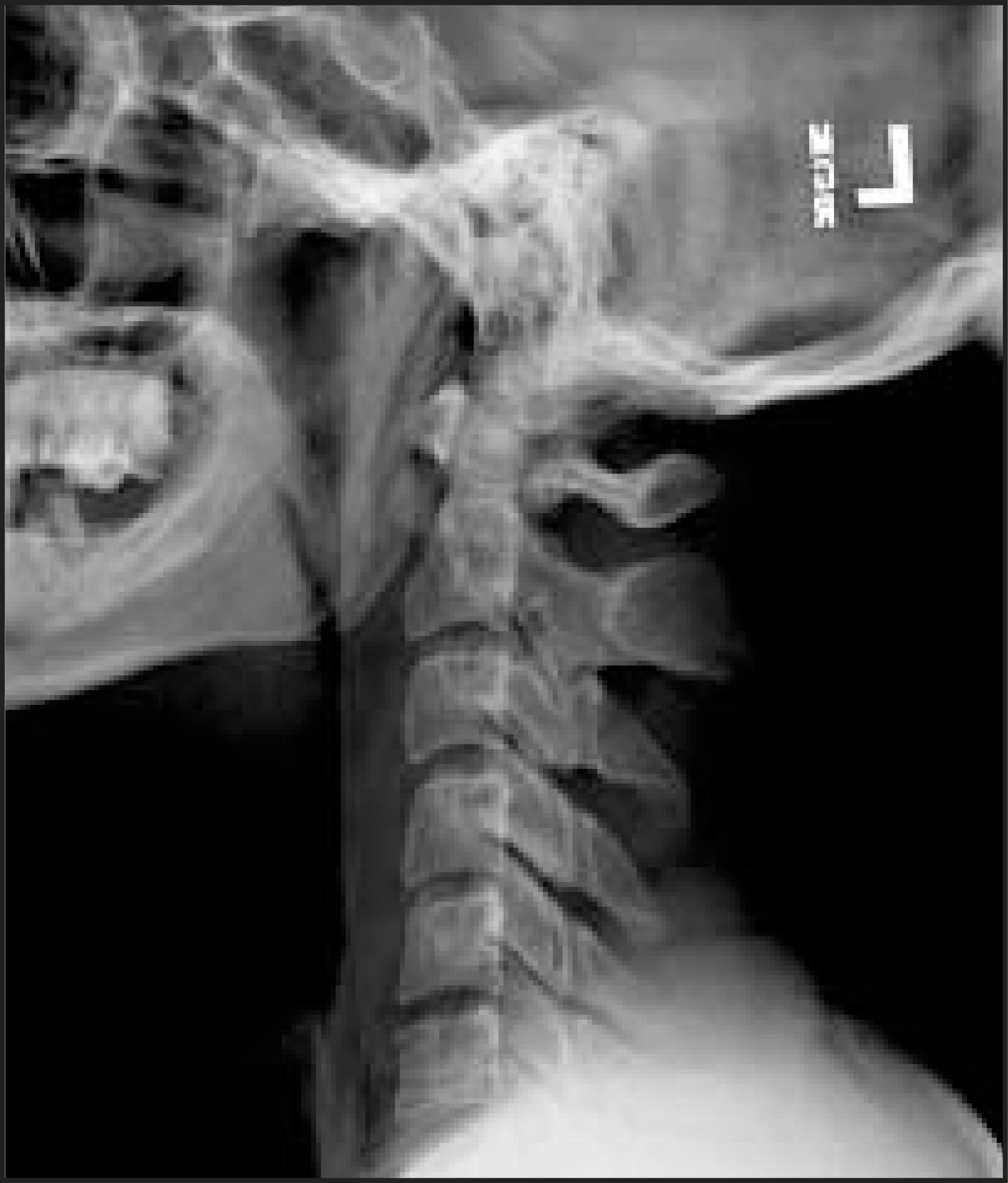
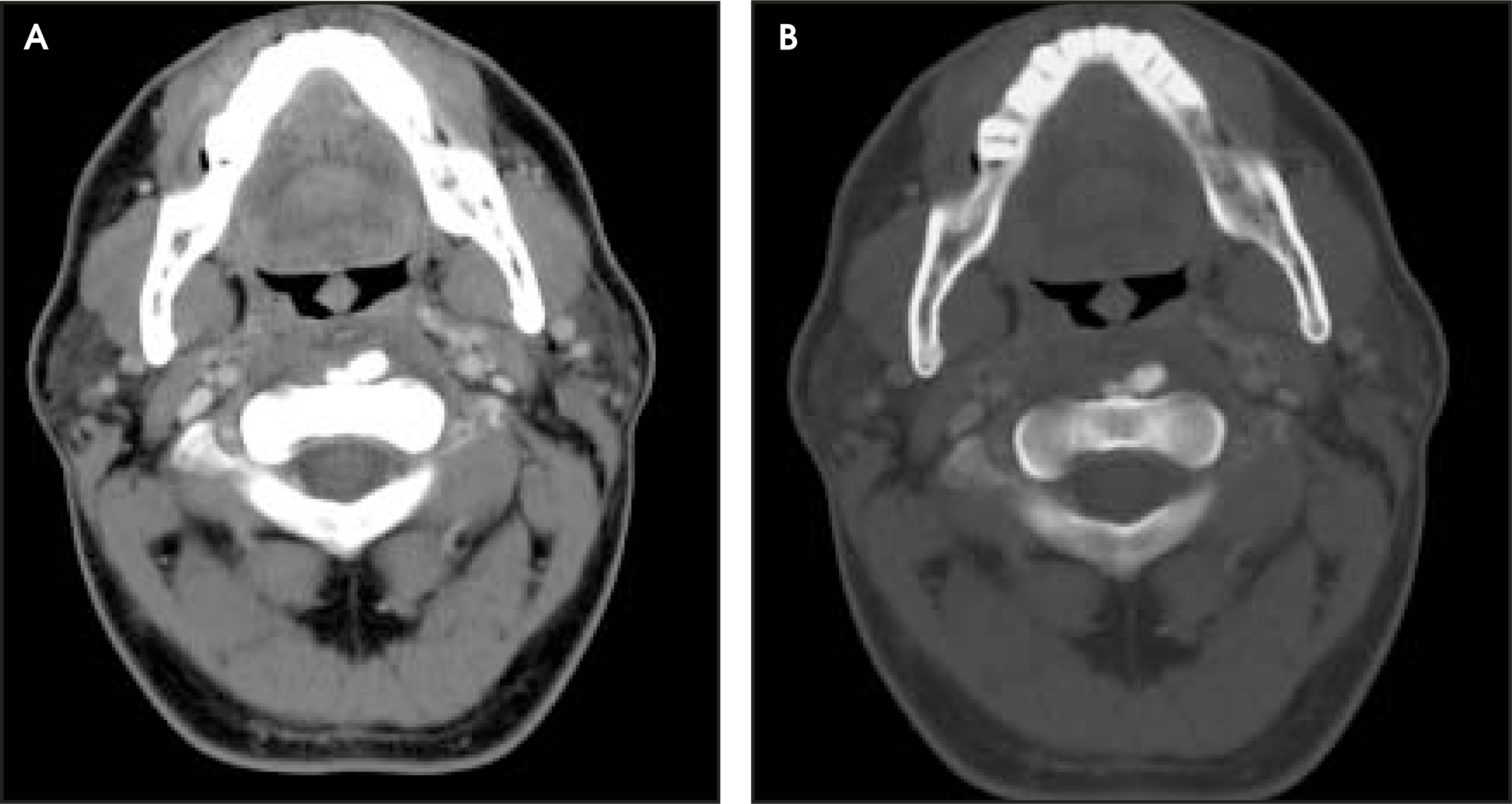
There is loss of lordotic curvature of the cervical spine with prominence of prevertebral soft tissues and a small focal area of calcification seen anterior to the C2 vertebral body (Figure 1). A CT scan revealed a prevertebral calcification and a low attenuation area without ring enhancement (Figure 2). There was neither an associated mass nor lymphadenopathy.
DISCUSSION
The longus colli muscle is located in the prevertebral space, extending from the anterior tubercle of the atlas to its inferior attachments in T3. The muscle is formed by vertical, inferior oblique, and superior oblique portions. The superior oblique portion, which inserts in the transverse processes of C3-C5 and extends superomedially into the atlas, is the one that becomes involved in this condition.1,2
Crystals of hydroxyapatite are formed by fibrocartilage cells through an unknown stimulus in the tendon. There is no ossification. During the deposition phase, the crystals appear in the tendon, and pain in this phase is inconsistent and can last for years. In the acute phase, the deposit undergoes spontaneous resolution with the development of moderate to severe pain for days to a few weeks.3
Inflammation, infection, and neoplasia should be considered in those patients presenting with neck pain and no prior history of trauma. Calcific tendinitis of the longus colli muscle is an uncommon cause of neck pain in this group of patients, although this entity may be significantly underdiagnosed. 4
The first communication of this disease in the literature appeared in 1964. Hartley 6 reported a case of acute onset of neck pain, odynophagia, and limited range of motion associated with prominence of prevertebral soft tissues and presence of calcifications on conventional radiography. In 1967, Fahlgren and collaborators 6 reported 12 cases of this condition, describing its clinical presentation, radiographic findings, and final outcome. They were the first to link the presence of amorphous calcifications anterior to C2 with the superior tendon of the longus colli muscle.
Calcium hydroxyapatite deposition occurs more frequently in relation to large joints, such as the shoulder or hip, and more rarely at the knee or elbow. 7 The exact cause of crystal deposition is unknown. There seems to be a genetic and metabolic predisposition associated with chronic trauma, inflammation, and tendon degeneration. Acute symptoms develop when these contained deposits rupture, provoking an acute inflammatory process that usually lasts 2 to 3 weeks and responds to the administration of nonsteroidal anti-inflammatory medication.8
The diagnosis of this condition is made based on the clinical presentation combined with the findings on imaging studies. Based on the cases presented in the literature, this condition affects middle-aged patients with no gender predilection. It is important to know that there is no history of trauma in relation to the onset of symptoms, although sometimes there is a remote history of trauma or history of minor trauma that does not correlate with the intensity of the symptoms. The most frequent presentation is neck pain, limited range of motion, and odynophagia. In some cases, there may be a low-grade fever. It is not uncommon to see mild leukocytosis and elevation of the C-reactive protein and sedimentation rate.9
The characteristic radiographic findings consist of presence of calcifications at level C1-C2 and prominence of prevertebral soft tissues on a lateral view. The calcifications are not always visible on plain films.10 Similar appearance can be produced by an inferior accessory ossicle of the anterior arch of the atlas, the inferior portion of the arch of this vertebra, and the lateral masses of C2 on a rotated projection. A calcified stylohyoid ligament can be confused with intratendinous calcification also.11 CT is more sensitive for depiction of intratendinous calcifi-cations, showing also the edema of the retropharyngeal space. The appearance of the calcification varies from punctuate to a dense, prominent concretion.2 MR was not performed in this case; in the cases described in the literature, inflammation in the prevertebral space is evident. This imaging modality is not the best imaging study to detect soft-tissue calcifications.2,7 Bone scans are not helpful for the diagnosis of this condition.8
The patient experienced significant relief during the first hours of nonsteroidal anti-inflammatory treatment. After 72 hours of treatment, the patient reported almost complete absence of symptoms. He remained afebrile during hospitalization. Blood cultures were negative. Leukocyte count decreased to normal values, from 15,800 on the day of admission to 7660 on the day of discharge. The patient was discharged with a 14day supply of naproxen. On clinic follow-up 2 weeks later, he reported complete resolution of symptoms with presence of full range of motion.
CONCLUSION
The clinical presentation of neck pain and limited range of motion associated with odynophagia and without history of trauma should raise the suspicion of prevertebral calcific tendinitis of the longus colli muscle. A lateral cervical spine radiograph and CT of the neck can provide the imaging characteristics typical of the disease that would help the referring physician to narrow the differential diagnosis. Early diagnosis is important to avoid invasive diagnostic/therapeutic procedures. Nonsteroidal anti-inflammatory medication provides a fast and effective relief of symptoms with total resolution in 10 to 15 days.
References
- Som P, Curtin H, Som P, Curtin H. Head and Neck Imaging. 1996:738-746.
- Artenian D, Lipman J, Scidmore G. Acute neck pain due to tendonitis of the longus colli: CT and MRI findings. Neuroradiology. 1989;31:166-169.
- Eastwood J, Hudgins P, Malone D. Retropharyngeal effusion in acute calcific tendinitis: Diagnosis with CT and MR imaging. AJNR Am J Neuroradiol. 1998;19:1789-1792.
- Widlus D. Calcific tendonitis of the longus colli muscle: A cause of atraumatic neck pain. Ann Emerg Med. 1985;14:1014-1017.
- Hartley J. Acute cervical pain associated with retropharyngeal calcium deposit. J Bone Joint Surg Am. 1964;46A:1753-1754.
- Fahlgren H, Jana S, Lofstedt S. Retropharyngeal tendonitis. Acta Neurologica Scand. 1967;43(suppl):188.
- De Maeseneer M, Vreugde S, Laureys S. Calcific tendinitis of the longus colli muscle. Head Neck. 1997;19:545-548.
- Ring D, Vaccaro A, Scuderi G. Acute calcific retropharyngeal tendinitis. J Bone Joint Surg Am. 1994;76:1636-1642.
- Haun C. Retropharyngeal tendinitis. AJR Am J Roentgenol. 1978;130:1137-1140.
- Newmark H, Blackford D, Roberts D. Computed tomography of acute cervical spine tendinitis. J Comput Tomog. 1986;10:373-375.
- Keats T. The inferior accessory ossicle of the anterior arch of the atlas. AJR Am J Roentgenol. 1967;101:834-836.
Citation
. RCOM RADIOLOGICAL CASE OF THE MONTH. Applied Radiology. 2004;33(9):38-41. doi:10.37549/AR1280.