RCOM RADIOLOGICAL CASE OF THE MONTH
Applied Radiology — Vol. 33 , Issue 10 , pp. 11 -11
DOI: 10.37549/AR1283
Published: October 1, 2004
Categories
CASE SUMMARY
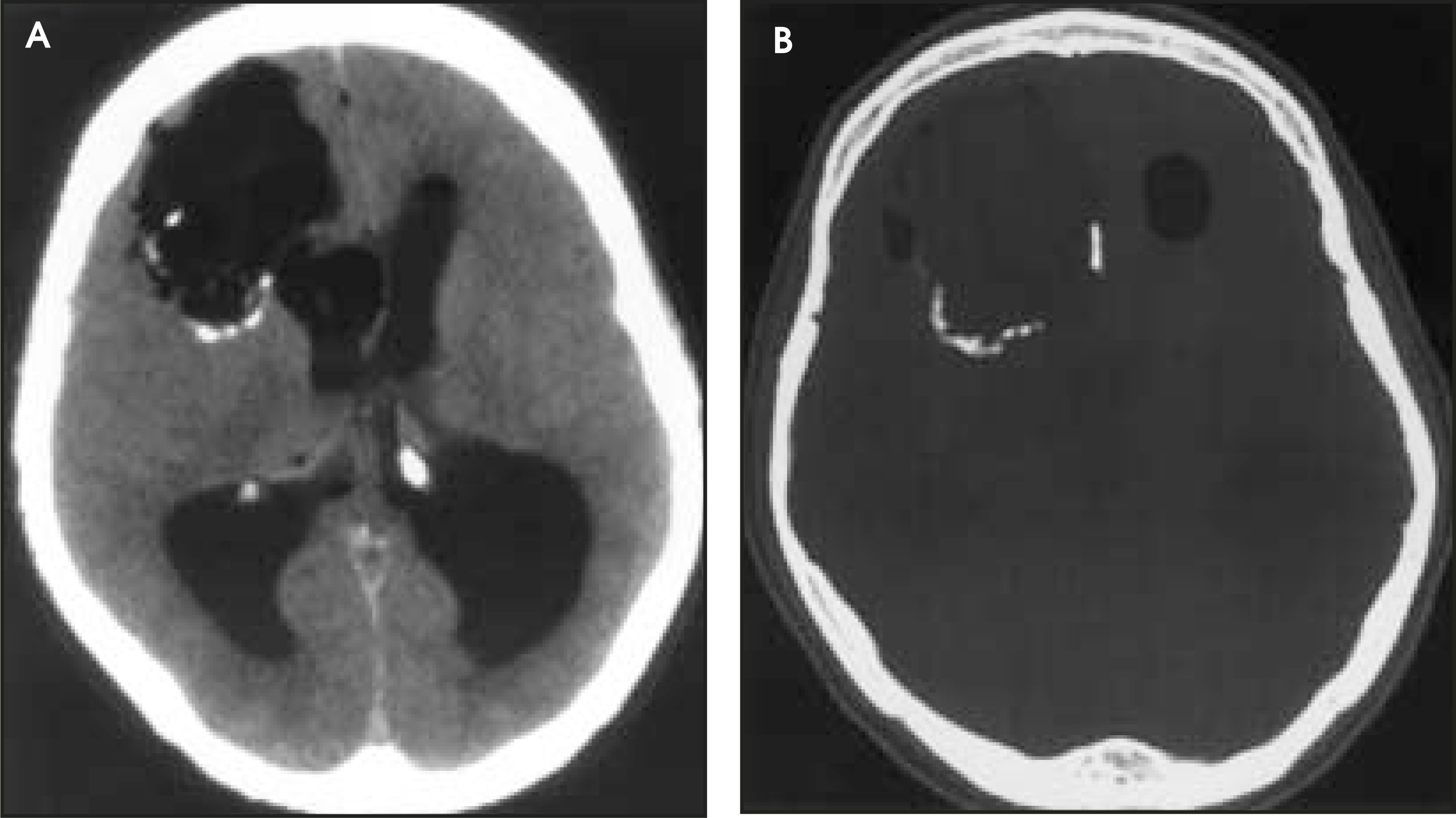
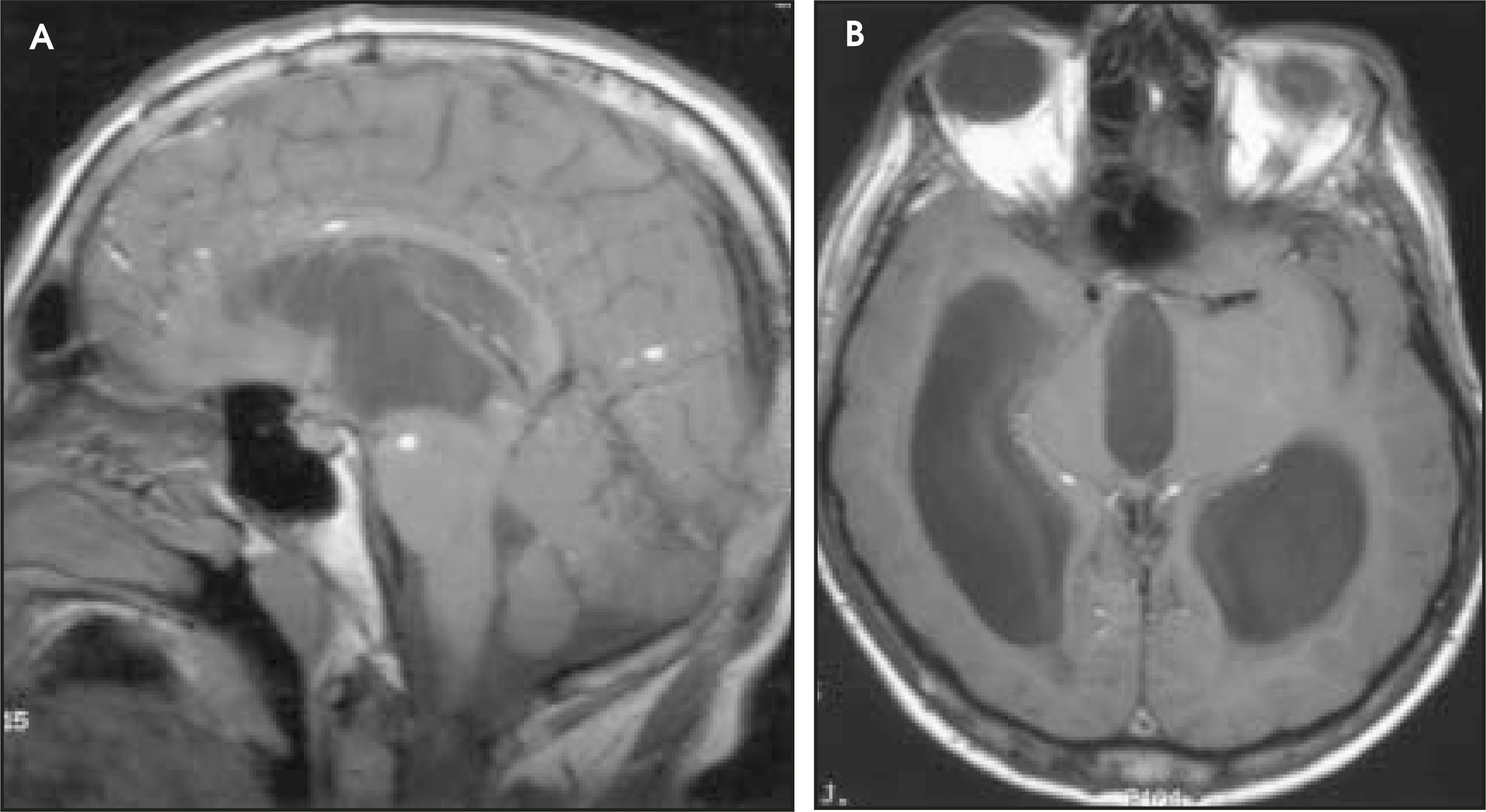
A 42-year-old man with a 19-year history of seizure disorder was transferred to the emergency department at our institution by an outside rural hospital. He reportedly presented to the rural hospital in a comatose state with decorticate posturing. A computed tomography (CT) scan of the brain showed severe hydrocephalus, which was treated by emergency ventriculostomy placement. When he arrived at our institution, he was alert and oriented to person, place, and time, although his answers to questioning were inappropriate at times. Head CT and brain magnetic resonance imaging (MRI) scans were performed (Figures 1 and 2).
DIAGNOSIS
Dermoid cyst with rupture into the right lateral ventricle
IMAGING FINDINGS
Axial CT images of the brain using parenchymal windows revealed a large hypoattenuating mass in the right frontal lobe that was isodense to the ventricular system and appeared to be communicating with the right frontal horn (figure 1A). A subtle ill-defined density within the mass was identified and was later shown by pathology to represent ectodermal material (hair follicles, squamous epithelium, and mesodermal cells). A CT image using bone windows showed a partial rim of calcification at the posterior and medial aspect of the cyst (figure 1B). In addition, a fat-fluid level was identified within the frontal horn of the left lateral ventricle. When using CT windows optimized for brain parenchyma, it may be difficult to differentiate fatty deposits from pneumocephalus; however, using a filming technique designed for bone detail reliably separates lipid from gas, as is well-demonstrated in this case.
On T1-weighted MRI sagittal and axial images, multiple foci of high signal intensity were seen scattered throughout the subarachnoid spaces of the brain parenchyma and brainstem (Figure 2). Hemorrhage may also give a similar appearance but can be eliminated from the differential diagnosis by its lack of chemical shift and uniquely different T2 characteristics. Although it was not seen in this patient, fatty droplets may be seen in remote locations, such as the lumbar spine in a patient with rupture of an intracranial dermoid cyst.
DISCUSSION
It is believed that dermoid cysts result from the inclusion of epithelial cells and skin appendages during the time of neural groove closure between the 3rd and 5th weeks of embryonic life.1 These tumors comprise 0.7% to 1.8% of intracranial tumors, although the most common location is within the spinal canal. Dermoid cysts typically grow very slowly and are often asymptomatic. While patients with rupture of an intracranial dermoid cyst may be asymptomatic, they can also present with headaches, seizures, transient cerebral ischemia with associated arterial spasm, hydrocephalus, or meningitis.2 If a direct sinus tract from the cyst is present, meningitis may be bacterial. However, in the event of a rupture, the clinician should be aware of the possibility of chemical meningitis. This is believed to occur when the cholesterol, a breakdown product of keratin, causes a nonspecific inflammatory response by the meninges.3
CONCLUSION
The findings associated with a ruptured dermoid cyst provide an eloquent example of the importance of a multimodality approach to intracranial tumors. Utilizing various CT window algorithms may prove invaluable in elucidating subtle features such as fat-fluid levels in the ventricular system. Additionally, the finding of multiple punctate foci of increased signal intensity within the subarachnoid spaces virtually clinches the diagnosis of a ruptured dermoid cyst in such a clinical setting.
References
- Russell D, Rubinstein L. Pathology of Tumours of the Nervous System. . 1989:690-695.
- Calabrò F, Capellini C, Jinkins J. Rupture of spinal dermoid tumors with spread of fatty droplets in the cerebrospinal fluid pathways. Neuroradiology. 2000;42:572-579.
- Scearce T, Shaw C, Bronstein A, Swanson P. Intraventricular fat from a ruptured sacral dermoid cyst: Clinical, radiographic, and pathological correlation. J Neurosurg. 1993;78:666-668.
Citation
. RCOM RADIOLOGICAL CASE OF THE MONTH. Applied Radiology. 2004;33(10):11-11. doi:10.37549/AR1283.