RCOM RADIOLOGICAL CASE OF THE MONTH
Applied Radiology — Vol. 33 , Issue 10 , pp. 37 -40
DOI: 10.37549/AR1286
Published: October 1, 2004
Categories
CASE SUMMARY
The patient is a 2-year-old boy with developmental delay due to sequelae from the resorption of large cephalohematomas sustained as a result of vacuum extraction during delivery. He initially had seizures, which resolved approximately 1 year previously but experienced recurrence of seizure activity after he was taken off his antiseizure medications.
DIAGNOSIS
Bilirubin encephalopathy
IMAGING FINDINGS
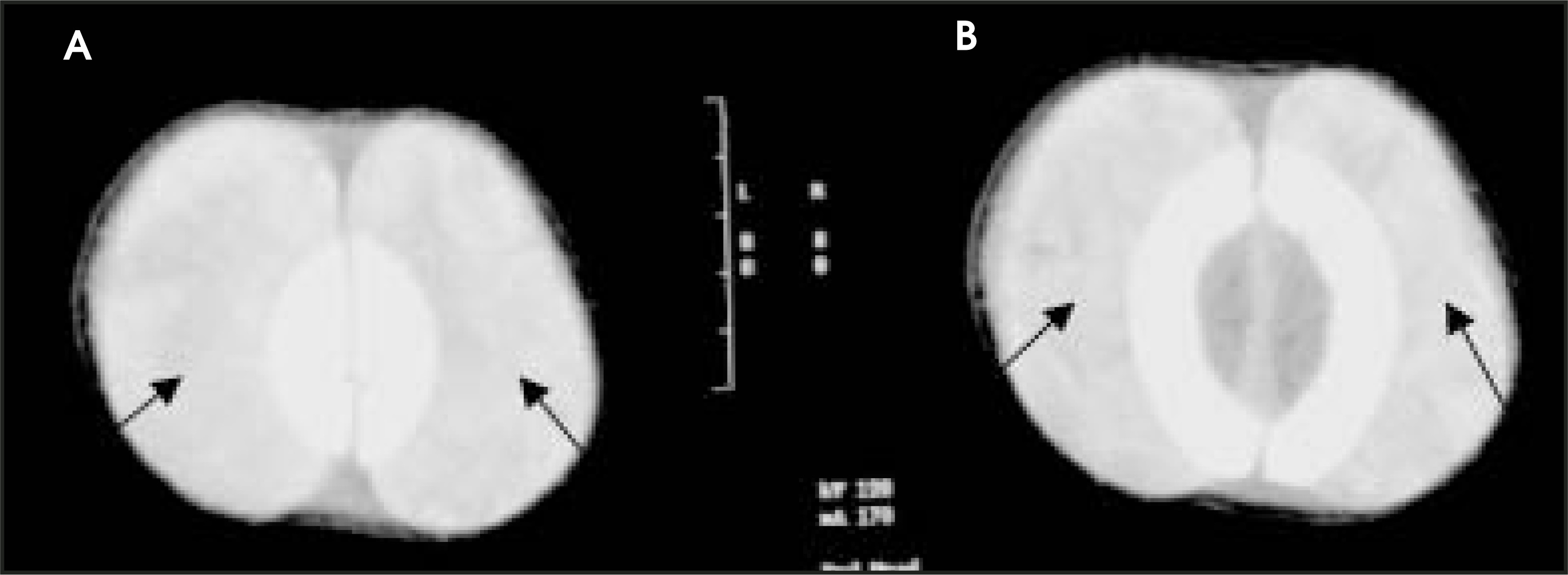
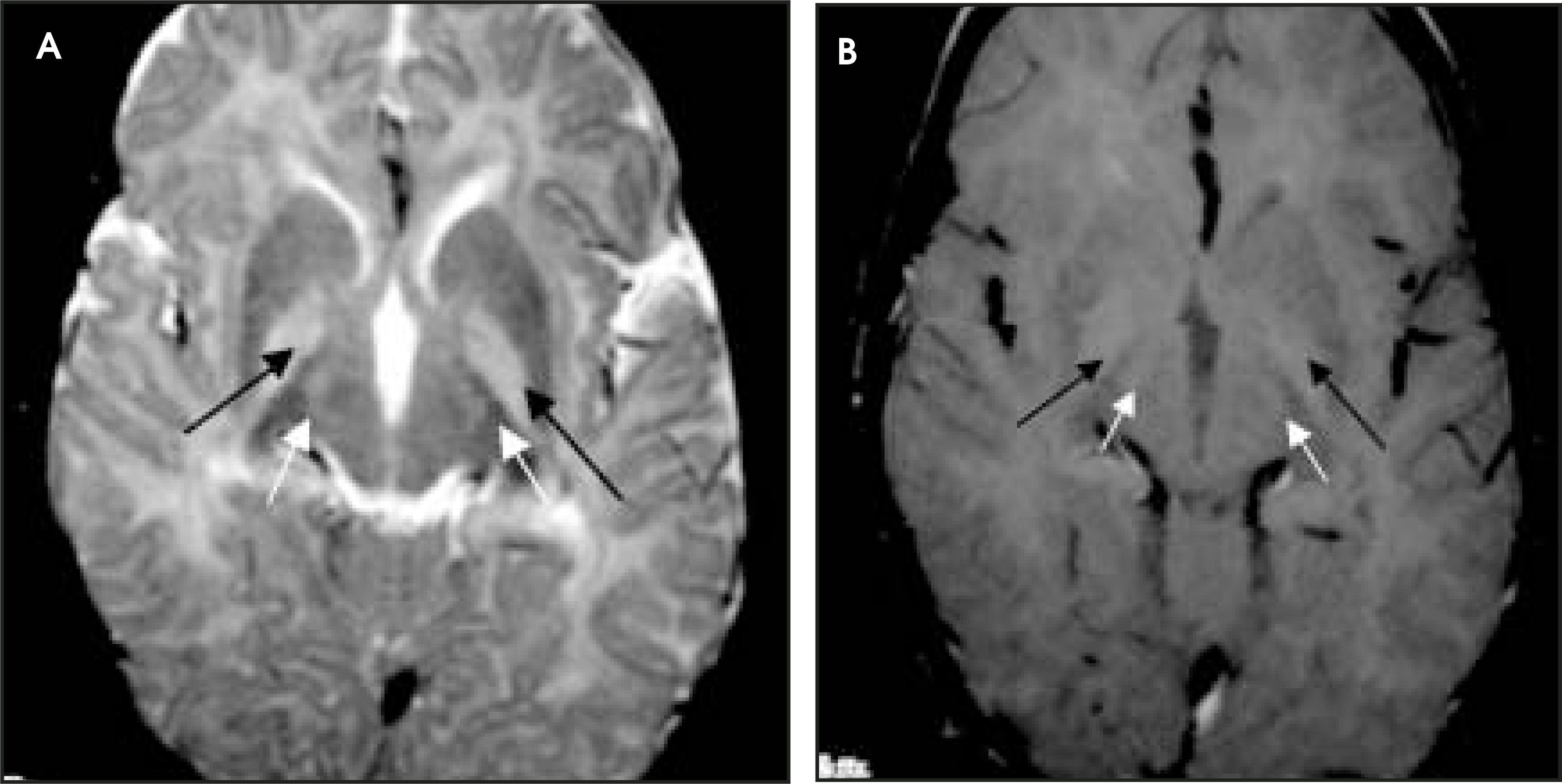
Radiographically, there are no specific computed tomography (CT) or ultrasound findings described for bilirubin encephalopathy. In this case, however, there was the associated finding of large bilateral cephalo-hematomas on noncontrast axial CT (Figure 1). Magnetic resonance imaging (MRI) examination during the acute stages reveals symmetric increased signal intensity on both T1- and T2-weighted images in the typical distribution of kernicterus.1 In the chronic stage, symmetric increased signal intensity on both proton-density and T2-weighted images is seen in the globus pallidus bilaterally and subthalamic nuclei, and hippocampal atrophy may also be found on MR imaging2,3 (Figure 2). At least one study has found that increased signal at the posteromedial border of the globus pallidus in patients with cerebral palsy is strong evidence of brain damage caused by kernicterus.4
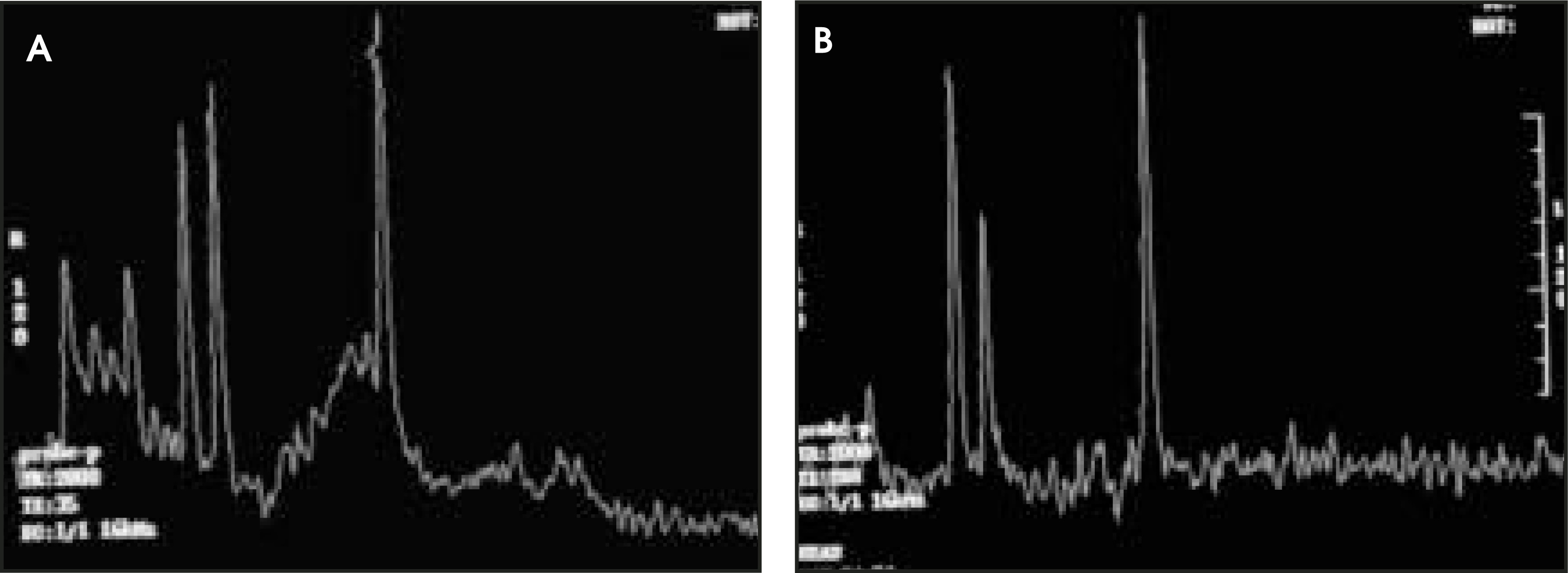
The proton spectroscopy findings of bilirubin encephalopathy are not well defined. According to the authors’ examination of the literature, a typical spectroscopic appearance of hyperbilirubinemia (or the sequelae from it) has not been described in detail. We found a somewhat nonspecific decrease in the N-acetylaspartate (NAA) peak in the region of a 1.68 × 1.68 × 2.0 voxel placed over the left basal ganglia (Figure 3). While this most likely represents a region of decreased NAA concentration in the basal ganglia due to bilirubin deposition, additional spectroscopic data from other patients would be necessary to characterize a typical appearance of kernicterus on proton spectroscopy.
DISCUSSION
In 1903, Dr. Christian Schmorl used the term kernicterus to describe the neuropathologic yellow staining (secondary to unconjugated bilirubin) of the basal ganglia of infants who died with neonatal jaundice. The terms kernicterus and bilirubin encephalopathy are commonly used interchangeably. It is not known, however, if bilirubin may stain brain structures without the accompanying microscopic or clinical evidence of neuronal injury.5 Therefore, bilirubin encephalopathy should be used to describe the clinical neurologic sequelae seen following marked hyperbilirubinemia, presumably caused by kernicterus.
The neurological manifestations of acute bilirubin encephalopathy have two distinct phases. The first phase usually presents in the first few days of life in term infants and as late as the seventh day of life in premature infants. Typically, these infants present with somnolence, hypotonia, and loss of the Moro reflex. This is then followed several days later by an irreversible stage characterized by hypertonia of the extensor muscle groups. This is characterized by retrocollis (backward arching of the neck) and opisthotonus (backward arching of the trunk).6 Additionally, fever and a high-pitched cry are often noted. Patients who survive the neonatal period demonstrate the chronic manifestations of bilirubin encephalopathy. These characteristic findings include choreoathetosis, sensorineural hearing loss, dental dysplasia, gaze abnormalities (particularly vertical gaze palsy) and mild mental retardation.2,7
As mentioned previously, kernicterus is the yellow staining of specific areas of brain tissue in the neonate secondary to accumulation of unconjugated bilirubin. Bilirubin staining is seen most commonly in the basal ganglia (particularly the globus pallidus), subthalamic nuclei, and hippocampus and multiple cranial nerve nuclei (facial, cochlear, oculomotor, and vestibular nuclei).8 Other areas of the brain are also involved, including the reticular formation of the pons, the dentate nucleus, the inferior olives, and anterior horn cells of the spinal cord.8
Bilirubin is normally bound to plasma albumin, which renders it nontoxic. However, in its free, unconjugated form, bilirubin is toxic. Histopathologically, areas of bilirubin deposition demonstrate neuronal loss, gliosis, astrocytosis, and demyelination.9
There are many causes of hyperbilirubinemia, which can lead to kernicterus. The most common cause is any type of hemolytic anemia, usually due to Rh or ABO incompatibility. Other types of hemolysis are also potential causes of hyperbilirubinemia, such as in this case in which the formation of extensive hematomas followed by breakdown of the blood products resulted in hemolytic hyperbilirubinemia (Figure 1). Other causes include congenital or acquired disorders of bilirubin metabolism. Factors implicated in potentiating the risk of developing bilirubin encephalopathy include low birth weight, hypothermia, anoxia, acidosis, sepsis, hypoalbuminemia, and meningitis.10
CONCLUSION
The term bilirubin encephalopathy describes the neuro-logic sequelae following marked hyperbilirubinemia. The most common cause of hyperbilirubinemia is hemo-lytic anemia secondary to Rh or ABO incompati-bility. Other causes include hemolysis following hema-toma, and congenital or acquired disorders of metab-olism. The utilization of MRI offers the most specific findings of bilirubin encephalopathy.
References
- Penn A, Enzmann D, Hahn J. Kernicterus in a full term infant. Pediatrics. 1994;93:1003-1006.
- Martich-Kriss V, Kollias S, Ball W. MR findings in kernicterus. AJNR Am J Neuroradiol. 1995;16:819-821.
- Steinborn M, Seelos K, Hueck A. MR findings in a patient with kernicterus. Eur Radiol. 1999;9:1913-1915.
- Sugama S, Soeda A, Eto Y. Magnetic resonance imaging in three children with kernicterus. Pediatr Neurol. 2001;25:328-331.
- Connolly A, Volpe J. Clinical features of bilirubin encephalopathy. Clin Perinat. 1990;17:371-379.
- VanPraagh R. Diagnosis of kernicterus in the neonatal period. Pediatrics. 1961;28:870-876.
- Maisels M, Baltz R, Bhutani V. Neonatal jaundice and kernicterus. Pediatrics. 2001;108:763-765.
- Ahdab-Barmada M, Moosy J. The neuropathology of kernicterus in the premature neonate: Diagnostic problems. J Neuropathol Exp Neurol. 1984;43:45-56.
- Beckwitt T, Miller C, Guttenberg M. A clinical pathologic reappraisal of kernicterus. Pediatrics. 1982;69:267-272.
- Lucey J. The unsolved problem in kernicterus in the susceptable low birth weight infant. Pediatrics. 1972;49:646-647.
Citation
. RCOM RADIOLOGICAL CASE OF THE MONTH. Applied Radiology. 2004;33(10):37-40. doi:10.37549/AR1286.