RCOM RADIOLOGICAL CASE OF THE MONTH
Applied Radiology — Vol. 33 , Issue 11 , pp. 54 -55
DOI: 10.37549/AR1293
Published: November 1, 2004
Categories
CASE SUMMARY
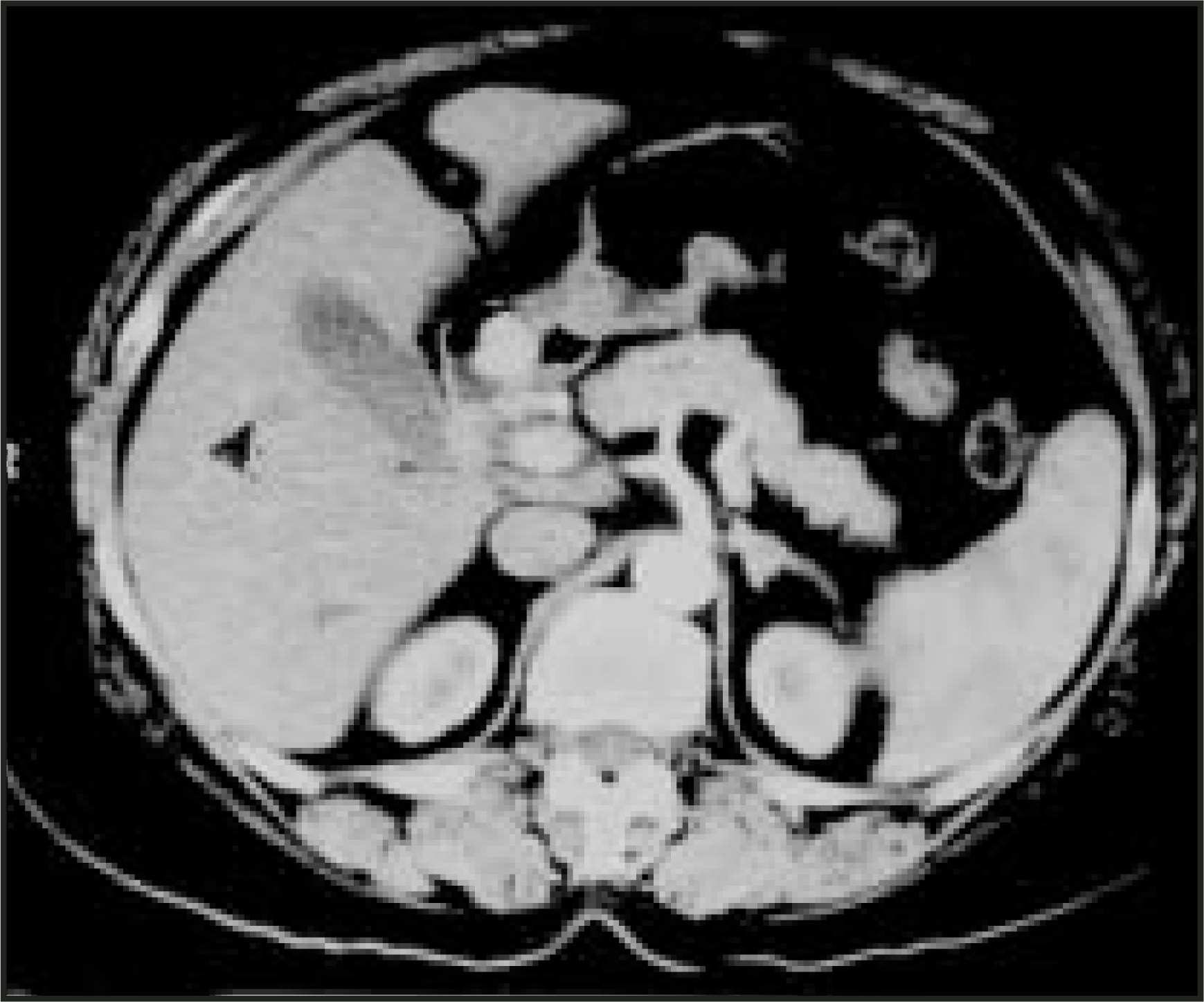
A 50-year-old woman presented with a history of abdominal pain, vomiting, loose bowel movements, and bronchospasm. Physical examination did not reveal any abnormality. Hematologic parameters were normal. A urine 5-HIAA level was found to be raised. An upper gastrointestinal endoscopy (Figure 1) and dual-phase computed tomography (CT) of the abdomen were performed (Figure 2).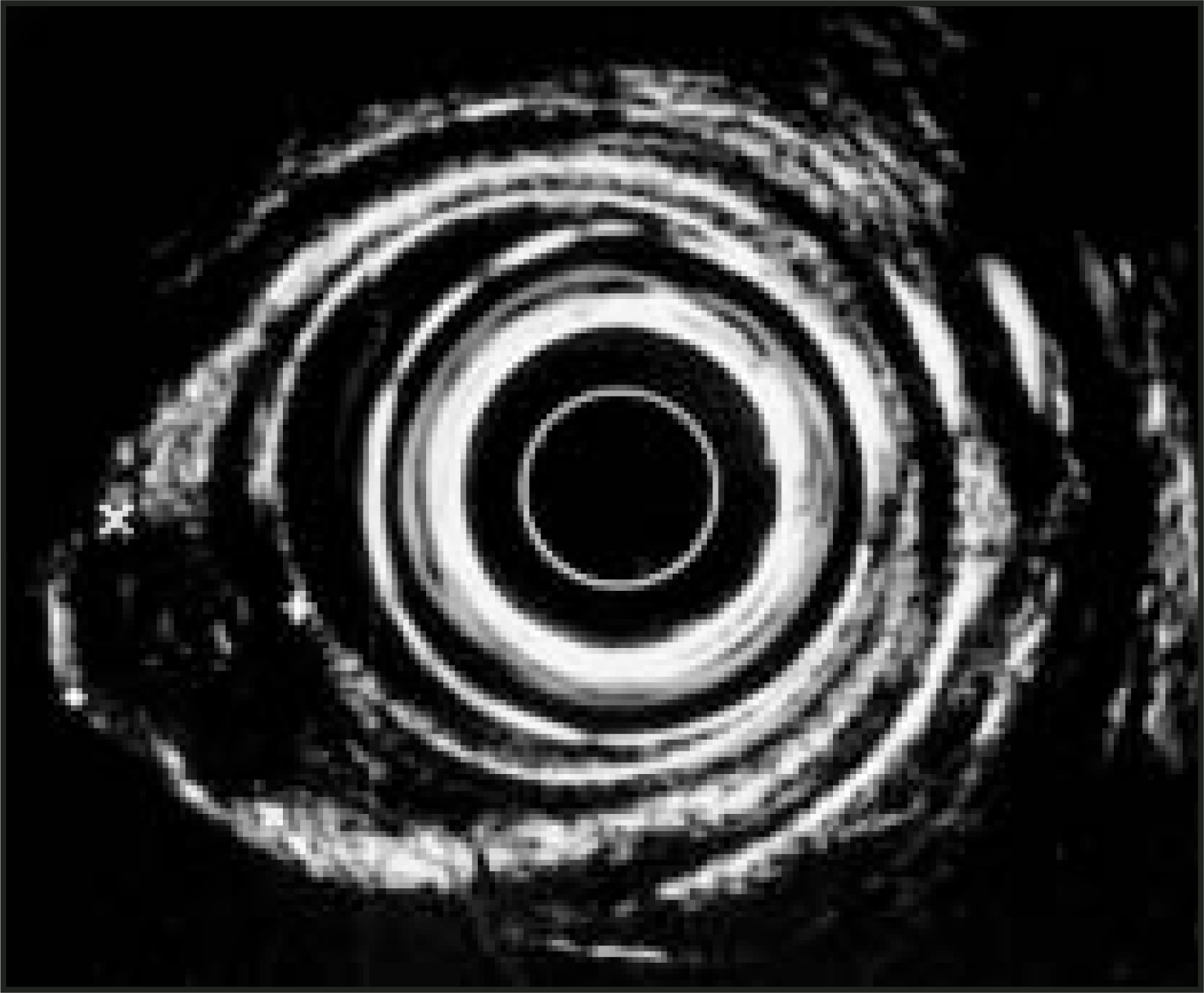
DIAGNOSIS
Duodenal bulb carcinoid tumor
IMAGING FINDINGS
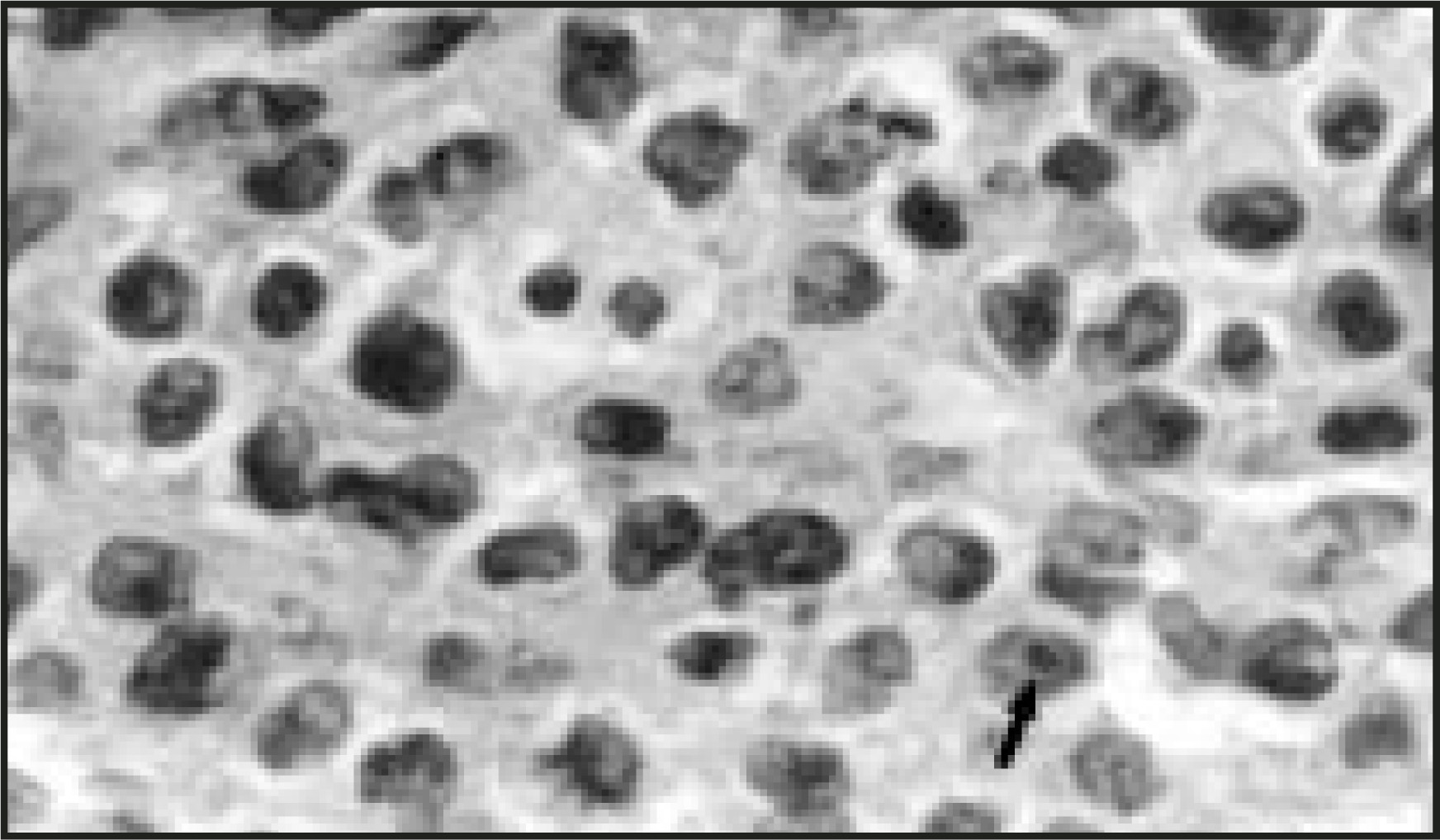
An upper gastrointestinal endoscopy revealed a solitary polypoidal lesion at the base of the duodenal bulb; the overlying mucosa was intact. Endosonography revealed an 11- × 8-mm well-defined hypoechoic tumor confined to submucosal hyperechoic layer; the underlying hyperechoic muscularis propria was intact (Figure 1). Dual-phase CT of the abdomen was performed (slice thickness 5 mm, table increment 7 mm, reconstruction interval 3 mm). In the arterial phase, the tumor was intensely enhancing and protruded into the duodenal bulb lumen. In addition, there was a solitary hypervascular metastasis in segment 5 of the liver (Figure 2). In view of confinement of tumor to submucosa, endoscopic resection of tumor was performed. The specimen revealed features consistent with carcinoid tumor (Figure 3 and Figure 4). For the solitary hepatic metastasis, radiofrequency ablation was planned.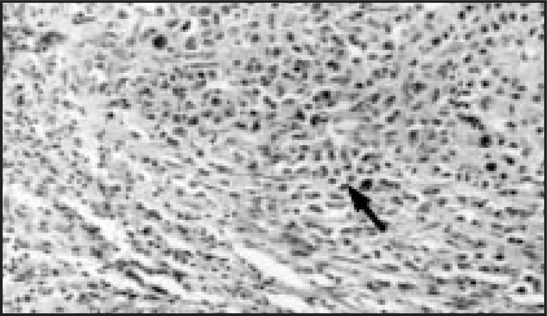
DISCUSSION
Primary duodenal carcinoid tumors are very rare1-3; they account for 2% to 3% of all gastrointestinal carcinoid tumors.4 Apart from the carcinoid tumor, the other primary malignant tumors of duodenum are lymphoma, adenocarcinoma and sarcoma.5 Carcinoid tumor is a unique neoplasm, being neither completely malignant nor completely benign.4 Most gastrointestinal carcinoids are localized in the appendix (30%).6,7 Duodenum is the second least common site (least common being the stomach) for carcinoid, 57% are seen in the duodenal cap, 23% in the second portion, and 3% in the third portion.8
These tumors may remain asymptomatic or may manifest as carcinoid syndrome because of hepatic metastases. The characteristic findings of carcinoid that are associated with increased risk of metastatic disease are involvement of muscularis propria, size greater than 2 cm, and presence of mitotic figures.9 Lesions <2 cm that are localized to the submucosa are treated by local surgical resection. However, lesions >2 cm or those exhibiting muscularis propria invasion require radical surgery.2 Endoscopic resection can be curative for the duodenal carcinoid less than 1 cm and confined to submucosal layer. However, this treatment in the thin-walled duodenum is considered to be dangerous and difficult because of peculiar submucosal growth of the tumor.
Traditionally, most primary duodenal carcinoid tumors were detected in postmortem examination because of very small size.5 However, since the development of endoscopy and endosonography, the detection rate has been increased.4,6 With the development of fiber-optic endoscopy, it is possible to detect these tumors at an early stage.4,5 Very uncommonly they are detected preoperatively, especially by CT.
Endosonography provides invaluable information about tumor size and depth of invasion, whereas dual-phase CT provides information about metastatic disease, preoperatively.10 In this case, both endosonography and dual-phase CT provided us with the most useful information about the depth of invasion and about the presence of metastatic disease.
CONCLUSION
Dual-phase CT helps in detection of small hypervascular metastasis and very rarely can detect duodenal carcinoid tumors. We report a case of duodenal bulb carcinoid that has been detected by both endosonography and dual-phase CT, which was resected endoscopically.
References
- Charles R, Kelley M, Campeti F. Primary duodenal tumors: A study of 31 cases. Arch Intern Med. 1963;111:23-33.
- Morgan J, Marks C, Heam D. Carcinoid tumors of the gastrointestinal tract. Ann Surg. 1974;180:720-727.
- Gutman H, Deutsch A, Leiser A. Pimary duodenal carcinoid with malignant carcinoid syndrome: A case report. Am J Gastroenterol. 1986;81:112-114.
- Yoshikane H, Suzuki T, Yoshioka N. Duodenal carcinoid tumor: Endosonographic imaging and endoscopic resection. Am J Gastroenterol. 1995;90:642-645.
- Wengrower D, Fich A. Primary duodenal carcinoid. Am J Gastroenterol. 1987;82:1069-1070.
- Merrell D, Mausbach C, Garbutt J. Carcinoid tumors of the duodenum: Endoscopic diagnosis of two cases. Gastrointest Endosc. 1985;31:269-271.
- Sanders R, Axtell H. Carcinoids of the gastrointestinal tract. Surg Gynecol Obstet. 1964;119:369-380.
- Warren K, McDonald W, Humelogan C. Periampullary and duodenal carcinoid tumors. Gut. 1964;5:448-453.
- Burke A, Sobin L, Federspiel B. Carcinoid tumors of the duodenum: A clinicoathologic study of 99 cases. Arch Pathol Lab Med. 1990;114:700-704.
- Yoshikane H, Tsukamoto Y, Niwa Y. Carcinoid tumors of the gastrointestinal tract: Evaluation with endoscopic ultrasonography. Gastrointest Endosc. 1193;39:375-383.
Citation
. RCOM RADIOLOGICAL CASE OF THE MONTH. Applied Radiology. 2004;33(11):54-55. doi:10.37549/AR1293.