RCOM RADIOLOGICAL CASE OF THE MONTH
Applied Radiology — Vol. 33 , Issue 12 , pp. 30 -33
DOI: 10.37549/AR1303
Published: December 1, 2004
Categories
CASE SUMMARY
A 29-year-old woman was admitted with a 1-day history of sudden onset of headache, vomiting, difficulty in swallowing, and saliva drooling from one side of the mouth. The patient had neck pain following a neck massage given by her partner 2 weeks prior to admission. The patient also observed that she had double vision, vertigo, and numbness in her right leg, and was swaying to the left. On examination she had left 7, 9, 10, and 12 cranial nerve palsies, nystagmus, and left Horner’s syndrome. She had some sensory disturbances on the right side of the body and hyperreflexia in the right upper/lower limbs. There was reduced sensation for pain in right arm. The patient was being treated conservatively.
DIAGNOSIS
Left hemimedullary syndrome secondary to vertebral artery dissection
IMAGING FINDINGS
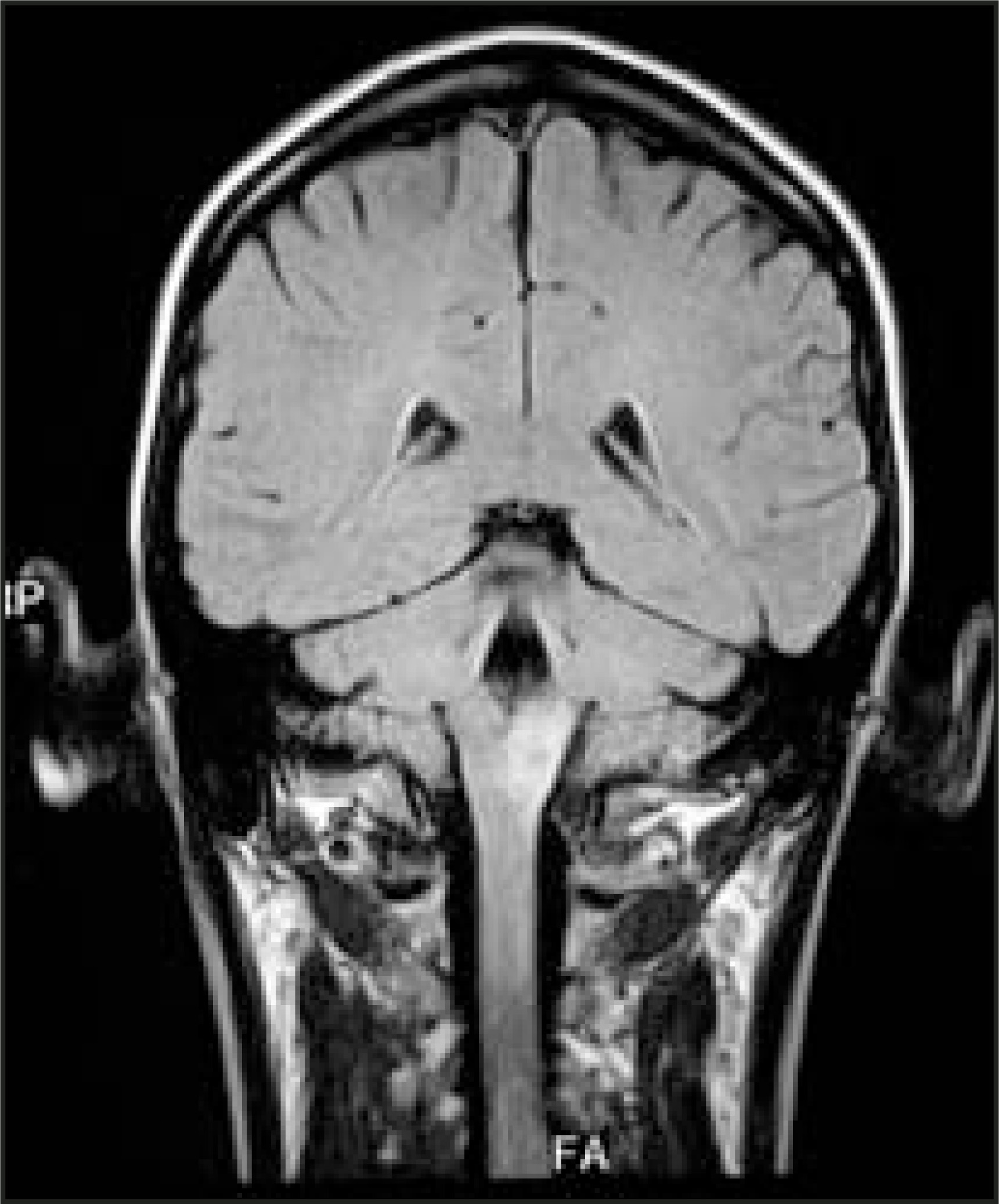
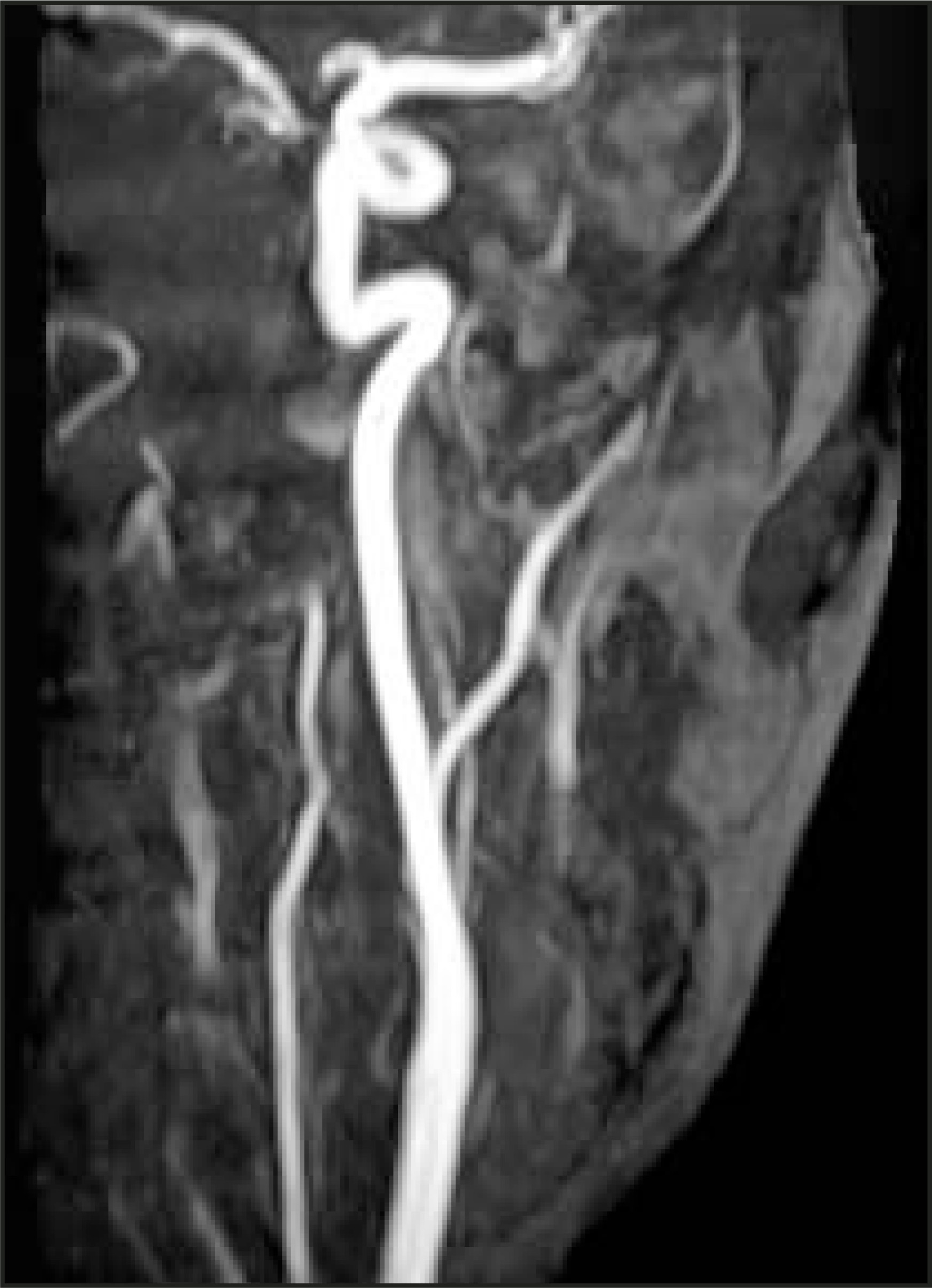
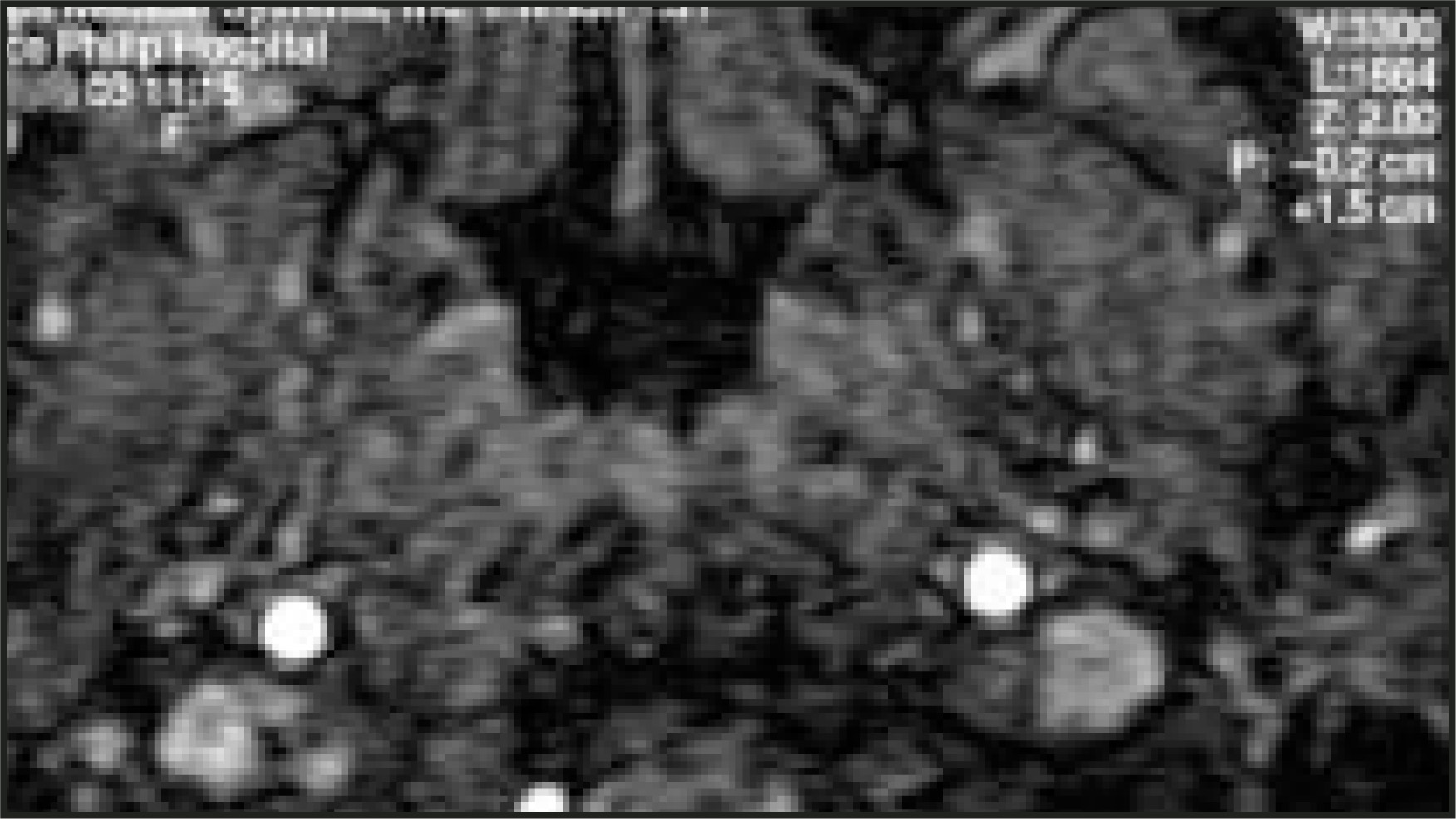
Computed tomography (CT) scan of the brain was normal. Magnetic resonance imaging (MRI) of the brain and a 2-dimensional (2D) MR angiography (MRA) of the head and neck vessels were performed. The T2-weighted axial (Figure 1) and coronal (Figure 2) MRI scans showed abnormal signal of hyperintensity within the left medulla including both dorsolateral and medial areas just below the left middle cerebellar peduncle. There was no abnormal enhancement of this area following contrast administration, and there was no evidence of a significant underlying mass lesion. The MRA showed abrupt termination of the nondominant left vertebral artery at the level of C1 vertebra (Figure 3). The right vertebral artery, though patent, showed an area of focal narrowing at its junction with the basilar artery probably spasm. The MRA source axial scans (radiofrequency-spoiled Fourier-acquired steady state [RF-FAST], echo time = 6.7 msec, repetition time 30 msec) showed that the left vertebral artery was occluded due to a dissection (Figure 4).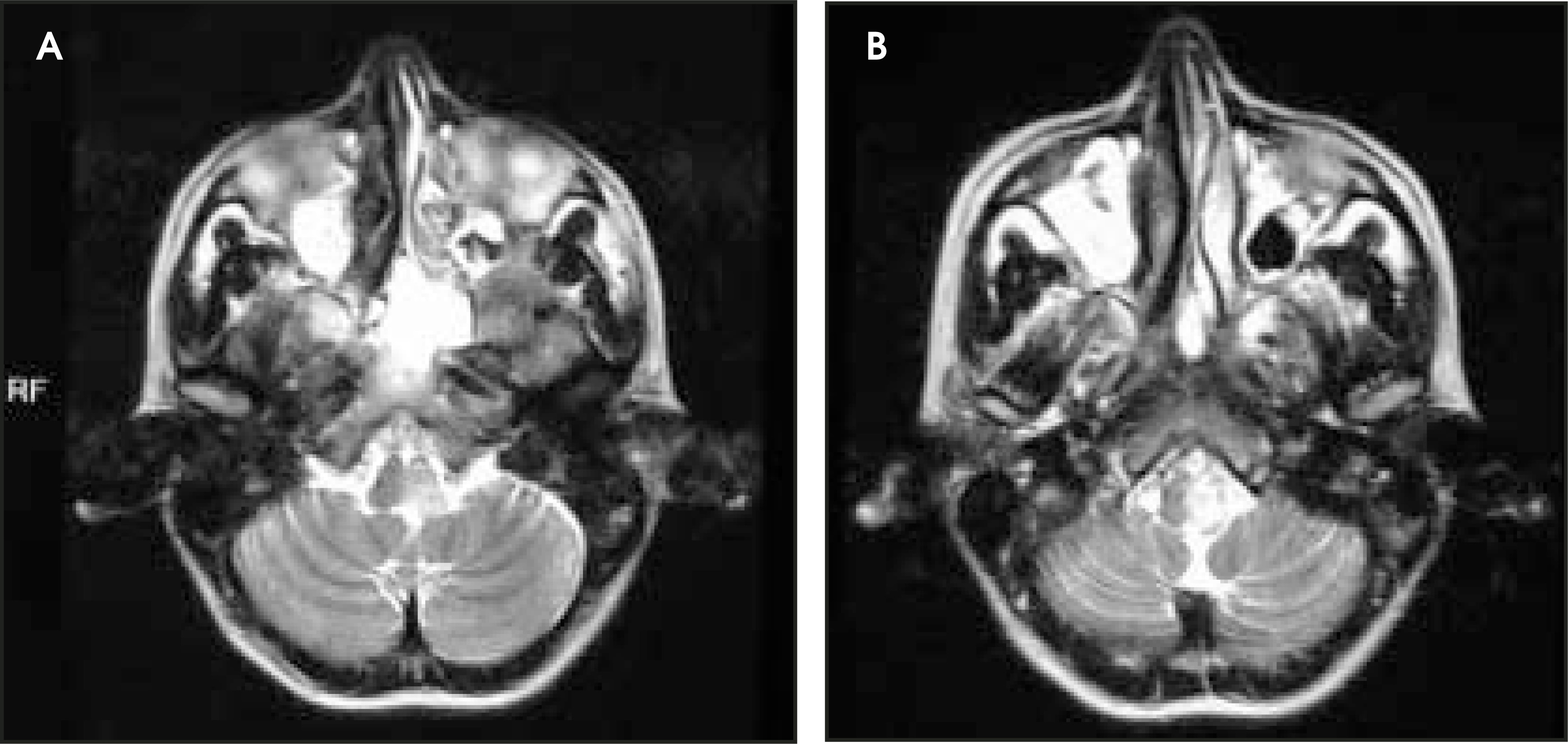
DISCUSSION
Hemimedullary syndrome, which was first described in 1902 and known as Babinski-Nageotte Syndrome,1 is the ischemic infarction of the medulla due to vascular events in the territory of the vertebral arteries. It is the combination of the pathology and clinical features of the medial and lateral medullary syndromes. Medial medullary syndrome is rare, and, while lateral medullary syndrome is not as rare, the combination of these two is extremely rare. In a report by Mossuto-Agatiello et al2 there were 6 cases of hemimedullary syndrome reported in the literature that were confirmed pathologically, and 3 cases were reported with anatomical confirmation.
We report a case of hemimedullary syndrome developed following a neck massage in a young patient. CT scan of the brain was normal, and MRI revealed left hemimedullary infarction. A 2D MRA of the head and neck vessels demonstrated abrupt termination of the left vertebral artery.
Hemimedullary syndrome is infarction of the hemimedulla and includes both medial and lateral medullary syndromes. Vertebral artery dissection (VAD) is the most frequent cause of lateral medullary syndrome (LMS) and is seen in up to 50% of patients.3 The other causes of LMS are stenosis or occlusion of the artery due to atherosclerosis. The most common cause of medial medullary syndrome is thrombosis of vertebral artery; it is less frequently due to thrombosis of anterior spinal artery. Occlusive disease of the vertebral artery has been the cause of hemimedullary syndrome in all of the cases that have been confirmed pathologically so far. Other reported causes of hemimedullary syndrome include hypoplastic left vertebral artery (VA), probably occluded by a thrombus or dissection,3 and bilateral stenosis of VA.4
Arterial dissections may occur spontaneously or may be associated with trauma. The magnitude of the trauma may be quite trivial, such as turning the head or looking upwards; an association between routine types of neck movement in sports and the evolution of VAD is well recognized in children.5 In the case we report here, since the signs and symptoms developed following a neck massage, we believe that the neck massage was the triggering event. In spontaneous VAD, pain develops in the back of the neck in half of patients and the median interval between the onset of neck pain and the appearance of other symptoms is 2 weeks, whereas other symptoms occur only 15 hours after the onset of headache. This patient had neck pain for 2 weeks followed by headache for 24 hours before other symptoms developed. Dissection of the artery is associated with the development of intramural hematoma. Subintimal hematoma most likely causes lumen stenosis or occlusion; thrombus may develop as a result of stenosis and may cause embolic complications.
In medial medullary syndrome, contralateral hemiparesis and hemisensory symptoms, usually in areas other than the face, are by far the most common mode of presentation.7 Lingual paresis due to involvement of the hypoglossal nerve is also commonly seen. Presenting signs and symptoms of lateral medullary syndrome is characterised by contralateral sensory deficits, ipsilateral5,9,10 cranial nerve palsies and sensory and motor deficits affecting the face, Horner’s syndrome, nystagmus, vertigo, and dysphagia. The patient reported here had a combination of signs and symptoms of these two syndromes and, due to the risk of aspiration as a consequence of the lingual paresis, was fed through a nasogastric tube.
The lesions in hemimedullary syndrome are seen mostly in the upper medulla, and, less frequently, in the middle and lower medulla.1 T2-weight-ed scans show high-intensity signal in the dorsolateral medulla in lateral medullary syndrome and in the medial part of the medulla in medial medullary syndrome. In this patient, the high signal intensity spread across the entire hemimedulla.
Most vertebral dissections occur extradurally above the C2 level.8 A small number arise or extend intradurally and can be associated with subarachnoid hemorrhage. Vertebral dissection in this patient was at the level of C1, and there was no subarachnoid hemorrhage. Extracranial color-flow duplex sonography detects the flow abnormalities in patients with extracranial VAD, but due to lack of specific findings, MR imaging and angiography are more suitable for initial diagnosis. However, ultrasonographic examinations have shown to be reliable and sensitive in follow-up evaluation.9 The angiographic findings of arterial dissection are a double lumen (the presence of a false lumen or an intimal flap) and the pearl and string sign; these are considered to be reliable findings. Three-dimensional (3D) CT angiography can also be used to make accurate diagnosis of spontaneous VAD.10 MR techniques are replacing conventional angiography as the gold standard in the diagnosis of dissections of carotid and vertebral arteries, since the resolution of MRA approaches that of conventional angiography and MRI is able to show intramural hematoma. Classic description of dissections of carotid and vertebral arteries with incomplete occlusion on T1-weighted images is narrowed eccentric signal void surrounded by a semilunar signal hyperintensity. This hyperintense signal remains unchanged on different sequences and echoes, as does the MR signal of subacute hemorrhage. The narrowed signal void corresponds to the residual lumen surrounded by the mural hematoma.11,12 The abnormal vessel stands out in the low signal cerebrospinal fluid in intracranial dissections, whereas it is more difficult to detect in extracranial dissections due to the intermediate-to-high signal of the normal perivascular structures and slow flow proximal and distal to the dissection.12 A circular region of increased signal intensity surrounding the vessel, thrombus within the lumen without luminal dilatation, and nonvisualization of the vessel are considered less specific for the diagnosis.13 Fat suppression techniques are important to differentiate small intramural hematomas from the surrounding soft tissues. When 2D time-of-flight MRA shows abrupt termination of signal in the occluded vertebral artery, with axial scans showing intramural hematoma, a diagnosis of VAD can be made satisfactorily.
CONCLUSION
This patient presented with signs and symptoms of a brain-stem lesion, and the CT scan was normal. MR imaging revealed infarction in the left medulla. Though we were unable to appreciate the classic description of arterial dissection on T1-weighted images, a simple 2D MRA of the head and neck vessels performed at the same time as the conventional MRI revealed abrupt termination of the left vertebral artery, and the MRA source axial scan showed an occluded vertebral artery with dissection. Hence, the performance of a 2D MRA, in conjunction with MRI, in such patients is recommended. To our knowledge, this is the first case of hemimedullary syndrome reported following a neck massage. Though the condition is extremely rare, and had not been reported previously following a neck massage, it bears consideration.
References
- Babinski J, Nageotte J. Hemiasynergine, lateropulsion et myosis bulbaires avec hemianaesthesie et hemiplegie croisees. Rev Neurol (Paris). 1902;10:358-365.
- Mossuto-Agatiello L, Kniahynicki C. The hemimedullary syndrome: Case report and review of the literature. J Neurol. 1990;237:208-212.
- Hosoya T, Nagahata M, Yamaguchi K. Prevalence of vertebral artery dissection in wallenberg syndrome: Neuroradiological analysis of 93 patients in the Tohoku district, Japan. Radiat Med. 1996;14:241-246.
- Nakane H, Okada Y, Sadoshima S, Fujishima M. Babinski-Nageotte syndrome on magnetic resonance imaging. Stroke. 1991;22:272-275.
- Hasan I, Wapnick S, Tenner M, Couldwell W. Vertebral artery dissection in children: A comprehensive review. Pediatr Neurosurg. 2002;37:168-177.
- Silbert P, Mokri B, Schievink W. Headache and neck pain in spontaneous internal and carotid and vertebral artery dissections. Neurology. 1995;45:1517-1522.
- Kim J, Kim H, Chung C. Medial medullary syndrome report of 18 new patients and review of the literature. Stroke. 1995;26:1548-1552.
- Friedman D, Flanders A. Unusual dissection of the proximal vertebral artery: Description of three cases. AJNR Am J Neuroradiol. 1992;13:283-286.
- Lu C, Sun Y, Jeng J. Imaging in the diagnosis and follow-up evaluation of vertebral artery dissection. J Ultrasound Med. 2000;19:263-270.
- Kurokawa Y, Yonemasu Y, Kano H. The usefulness of 3D-CT angiography for the diagnosis of vertebral artery dissection-report of two cases. Comput Med Imaging Graph. 2000;24:115-119.
- Gelbert F, Assouline E, Hodes J. MRI in spontaneous dissection of vertebral and carotid arteries. Neuroradiology. 1991;33:111-113.
- Mascalchi M, Bianci M, Mangiafico S. MRI and MR angiography of vertebral artery dissection. Neuroradiology. 1997;39:329-340.
- Zuber M, Meary E, Meder J. Magnetic resonance imaging and dynamic CT scan in cervical artery dissections. Stroke. 1994;25:576-581.
Citation
. RCOM RADIOLOGICAL CASE OF THE MONTH. Applied Radiology. 2004;33(12):30-33. doi:10.37549/AR1303.