RCOM RADIOLOGICAL CASE OF THE MONTH
Applied Radiology — Vol. 33 , Issue 12 , pp. 41 -43
DOI: 10.37549/AR1304
Published: December 1, 2004
Categories
CASE SUMMARY
A 20-year-old Brazilian man with a history of a right-knee sports injury 2 years prior presented with 2 weeks of right-knee pain and swelling. He denied any history of fever, chills, or any other constitutional symptoms. Physical examination revealed slightly erythematous swelling of the right knee without point of tenderness or limitation of motion.
DIAGNOSIS
Diffuse pigmented villonodular synovitis
IMAGING FINDINGS
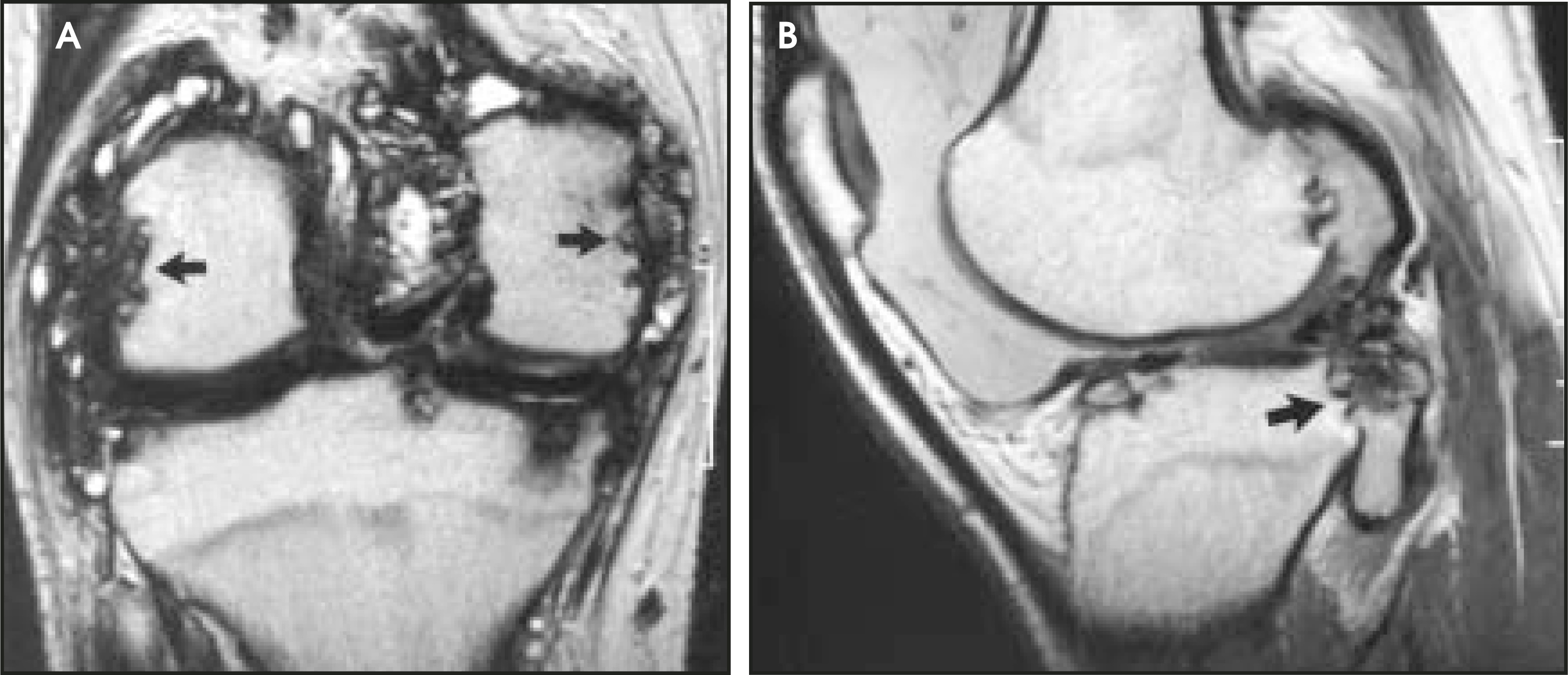
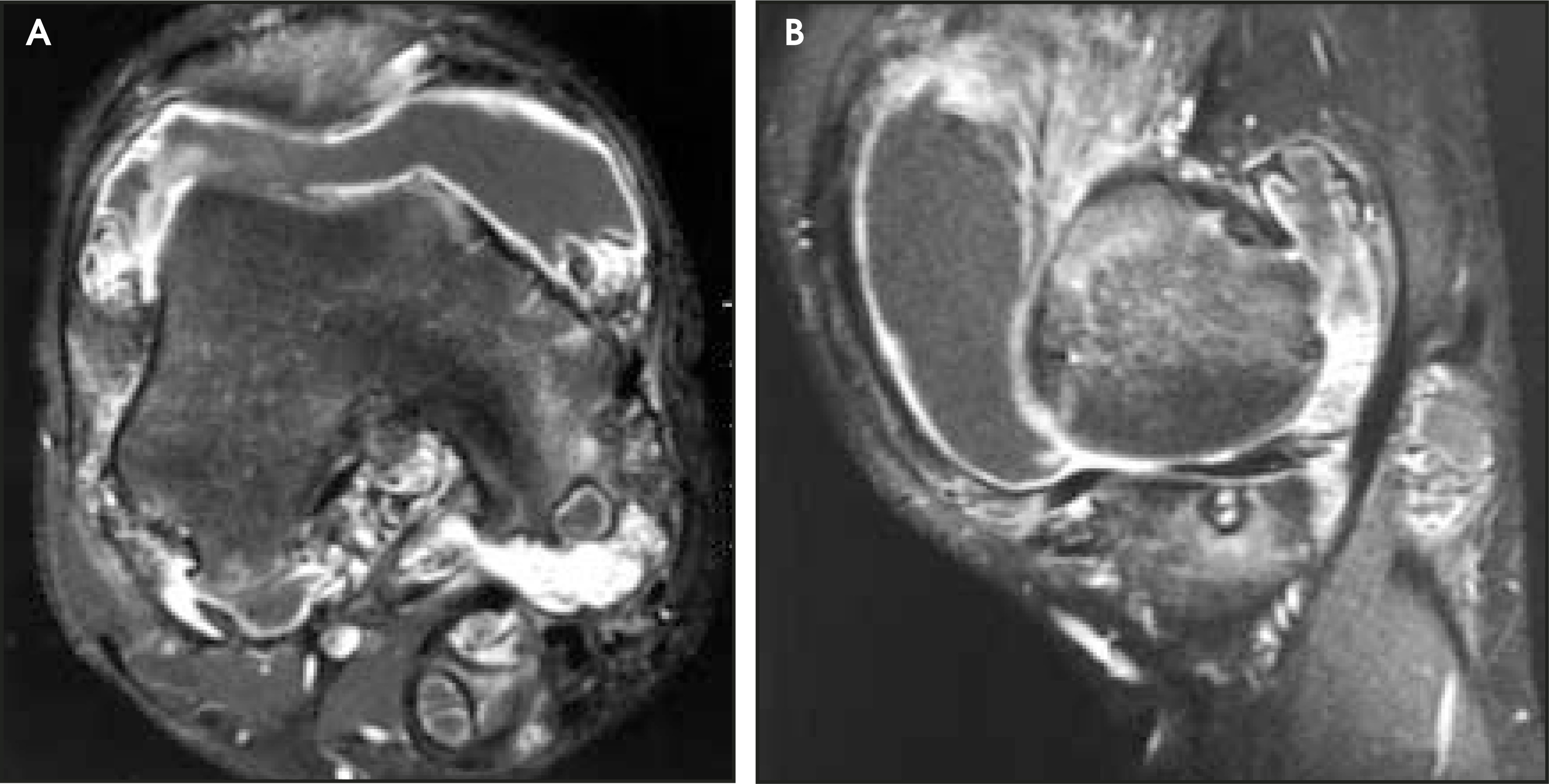
Radiographs were taken and magnetic resonance (MR) imaging studies were performed (Figures 1 through 3). Radiographs of the right knee reveal widening of the femoral intercondylar fossa, joint effusion, and peripheral erosive changes (Figure 1). MR imaging shows nodular soft-tissue masses of low signal intensity along with pressure erosions and a large amount of joint effusion (Figure 2). Postgadolinium images show extensive enhancement of the thickened synovium (Figure 3).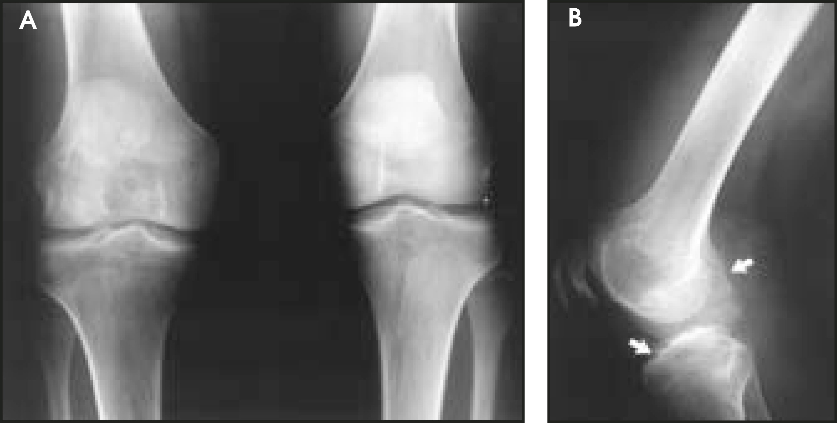
DISCUSSION
Pigmented villonodular synovitis (PVNS) was defined by Jaffe et al1 in 1941. It is a benign, proliferative disorder of synovium resulting in villous and/or nodular changes in synovium-lined joints, tendon sheaths, and bursae. The lesion occurs in both localized and, more commonly, diffuse forms.2 Diffuse PVNS occurs most frequently in young adults and usually affects the large joints of the lower limb, most frequently the knee, followed by the hip.3 The lesion is slightly more common in men; the majority are unilateral and monoarticular.4 The most common presenting complaint is mild but progressive pain that is insidious at onset. Mechanical derangement with locking and palpable masses may also be present.
Grossly, diffuse PVNS consists of exuberant, frondlike, thickened synovium with a characteristic tan or reddish color caused by deposition of hemosiderin. Microscopically, it is characterized by fibrous stroma containing multinucleated giant cells, lymphocytes, xanthoma cells, and pigmentation due to both intra- and extracelluar hemosiderin, and lipid deposition. The hyperplastic synovium can invade the subchondral bone and produce cysts.5 The cause is unknown; however, several theories are accepted, including a chronic inflammatory process, repetitive trauma, benign neoplasia, and disorder of lipid metabolism.1,6,7 Malignant transformation has been reported, although it is extremely rare and controversial.8
Radiographs may be normal or may reveal periarticular soft-tissue swelling with an absence of calcifications. Normal bony mineralization, and preservation of the joint space are characteristic.9 Bony changes begin with erosion of the articular cartilage, often near the chondro-osseous junction, with subsequent extension of the process through the cartilage and the underlying cortical bone into the cancellous bone. This gives rise to the juxta-articular cysts evident radiographically.10 A variety of imaging methods have been used to diagnose PVNS, each having varying degrees of diagnostic value. Arthrography, used more commonly in the pre-MRI period, may show filling defects due to intra-articular nodules, but is not widely used.11 Computed tomography (CT) is especially useful in delineating bony erosions and in revealing high-attenuation tisue due to iron deposit in the synovium. However, CT is limited compared with MRI in its delineation of soft-tissue lesions and its sensitivity to inflammatory tissue.12
With its high resolution and excellent soft-tissue contrast, MRI has become the imaging method of choice. On MRI, mass-like synovial proliferations have lobulated margins and the paramagnetic effect of hemosiderin results in the reduction of signal intensity in all pulse sequences, which is enhanced by high-resolution field-strength and gradient-echo sequences.13 However, the appearance varies widely, depending on the relative proportions of lipid, hemosiderin, fibrous stroma, septation, pannus formation, fluid, and articular erosions.14 Pigmented villonodular synovitis characteristically shows intense gadolinium enhancement, reflecting the high vascularity of the synovial proliferation.15
Definitive diagnosis relies on synovial biopsy. Treatment of choice is synovectomy.16 Recurrence is presumably due to incomplete excision. Recurrence rates are lower in open procedures than in arthroscopic synovectomies. On the other hand, postoperative morbidity is lower in arthroscopically treated patients than in open surgeries.17 Nonsurgical therapies, such as local injection of steroids, Y-90 radiosynoviorthesis, or external beam radiation, are controversial.18-20
CONCLUSION
Diffuse PVNS is a benign proliferative disorder of the synovium of unknown etiology that is prevalent in lower limbs in young adults. MRI has proved to be the most helpful tool in the investigation, and, in uncertain cases, an arthroscopic biopsy is easily obtainable. Treatment mainly consists of surgical excision of affected tissue.
References
- Jaffe H, Lichtenstein L, Sutro C. Pigmented villonodular synovitis, bursitis, and tenosynovitis. Arch Pathol. 1941;31:731-765.
- Pantazopoulos T, Savrou Z, Stamos C. Bone lesions in pigmented villonodular synovitis. Acta Orthop Scand. 1975;46:579-592.
- Byers P, Cotton R, Deacon O. The diagnosis and treatment of pigmented villonodular synovitis. J Bone Joint Surg Br. 1968;50:290-305.
- Durr H, Stabler A, Maier M. Pigmented villonodular synovitis. Review of 20 cases. J Rheumatol. 2001;28:1620-1630.
- Mirra J. Bone Tumors: Clinical, Radiologic, and Pathologic Correlations. 1989;Vol 1:1766-1775.
- Smith J, Pugh D. Roentgenographic aspects of articular pigmented villonodular synovitis. AJR Am J Roentgenol. 1962;87:1146-1156.
- Rao A, Vigorita V. Pigmented villonodular synovitis (giantcell tumor of the tendon sheath and synovial membrane). A review of eighty-one cases. J Bone Joint Surg Am. 1984;66-A:76-94.
- Kalil R, Unni K. Malignancy in pigmented villonodular synovitis. Skeletal Radiol. 1998;27:392-395.
- Bravo S, Winalski C, Weissman B. Pigmented villonodular synovitis. Radiol Clin North Am. 1996;34:311-326.
- Dorwart R, Genant H, Johnston W. Pigmented villonodular synovitis of synovial joints: Clinical, pathologic, and radiologic features. AJR Am J Roentgenol. 1984;143:877-885.
- Lowenstein M, Smith J, Cole S. Infrapatellar pigmented villonodular synovitis: Arthrographic detection. AJR Am J Roentgenol. 1980;135:279-282.
- Butt W, Hardy G, Ostlere S. Pigmented villonodular synovitis of the knee: Computed tomographic appearances. Skeletal Radiol. 1990;19:191-196.
- Llauger J, Palmer J, Roson N. Nonseptic monoarthritis: Imaging features with clinical and histopathologic correlations. RadioGraphics. 2000;20:S263-278.
- Hughes T, Sartoris D, Schweitzer M. Pigmented villonodular synovitis: MRI characteristics. Skeletal Radiol. 1995;24:7-12.
- Narvaez J, Narvaez J, Ortega R. Hypointense synovial lesions on T2-weighted images: Differential diagnosis with pathologic correlation. AJR Am J Roentgenol. 2003;181:761-769.
- Flandry F, Hughston J, Jacobson K. Surgical treatment of diffuse pigmented villonodular synovitis of the knee. Clin Orthop. 1994;300:183-192.
- Zvijac J, Lau A, Hechtman K. Arthroscopic treatment of pigmented villonodular synovitis of the knee. Arthroscopy. 1999;15:613-617.
- Rydholm U. Pigmented villonodular synovitis. Acta Orthop Scand. 1998;69:203-210.
- Gumpel J, Shawe D. Diffuse pigmented villonodular synovitis: Non-surgical management. Ann Rheum Dis. 1991;50:531-533.
- O’Sullivan B, Cummings B, Catton C. Outcome following radiation treatment for high-risk pigmented villonodular synovitis. Int J Radiat Oncol Biol Phys. 1995;32:777-786.
Citation
. RCOM RADIOLOGICAL CASE OF THE MONTH. Applied Radiology. 2004;33(12):41-43. doi:10.37549/AR1304.