RCOM RADIOLOGICAL CASE OF THE MONTH
Applied Radiology — Vol. 33 , Issue 12 , pp. 34 -37
DOI: 10.37549/AR1305
Published: December 1, 2004
Categories
CASE SUMMARY
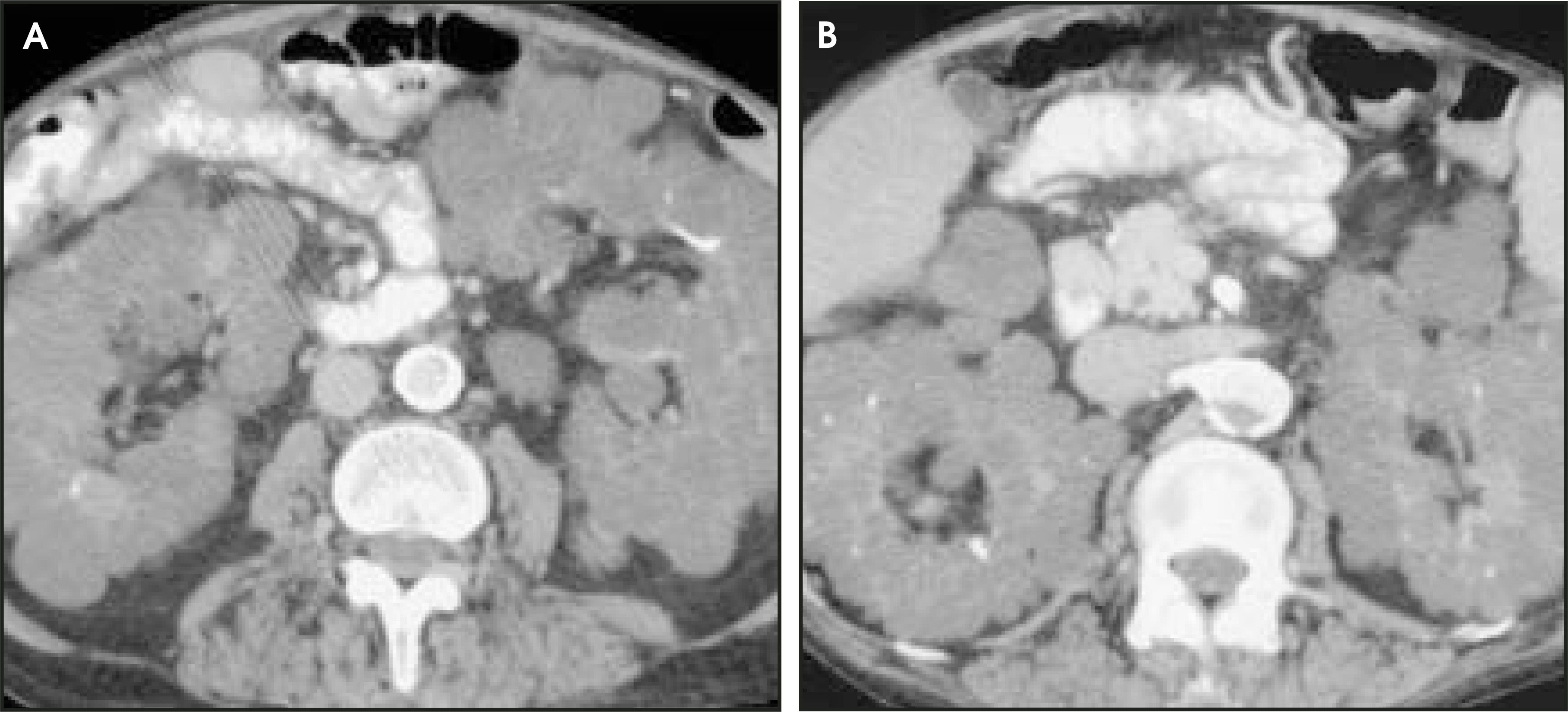
A 71-year-old man with renal failure secondary to autosomal dominant polycystic kidney disease had been on hemodialysis for 22 years. Bilaterial hand pain and swelling prompted radiographic evaluation (Figures 1 and 2).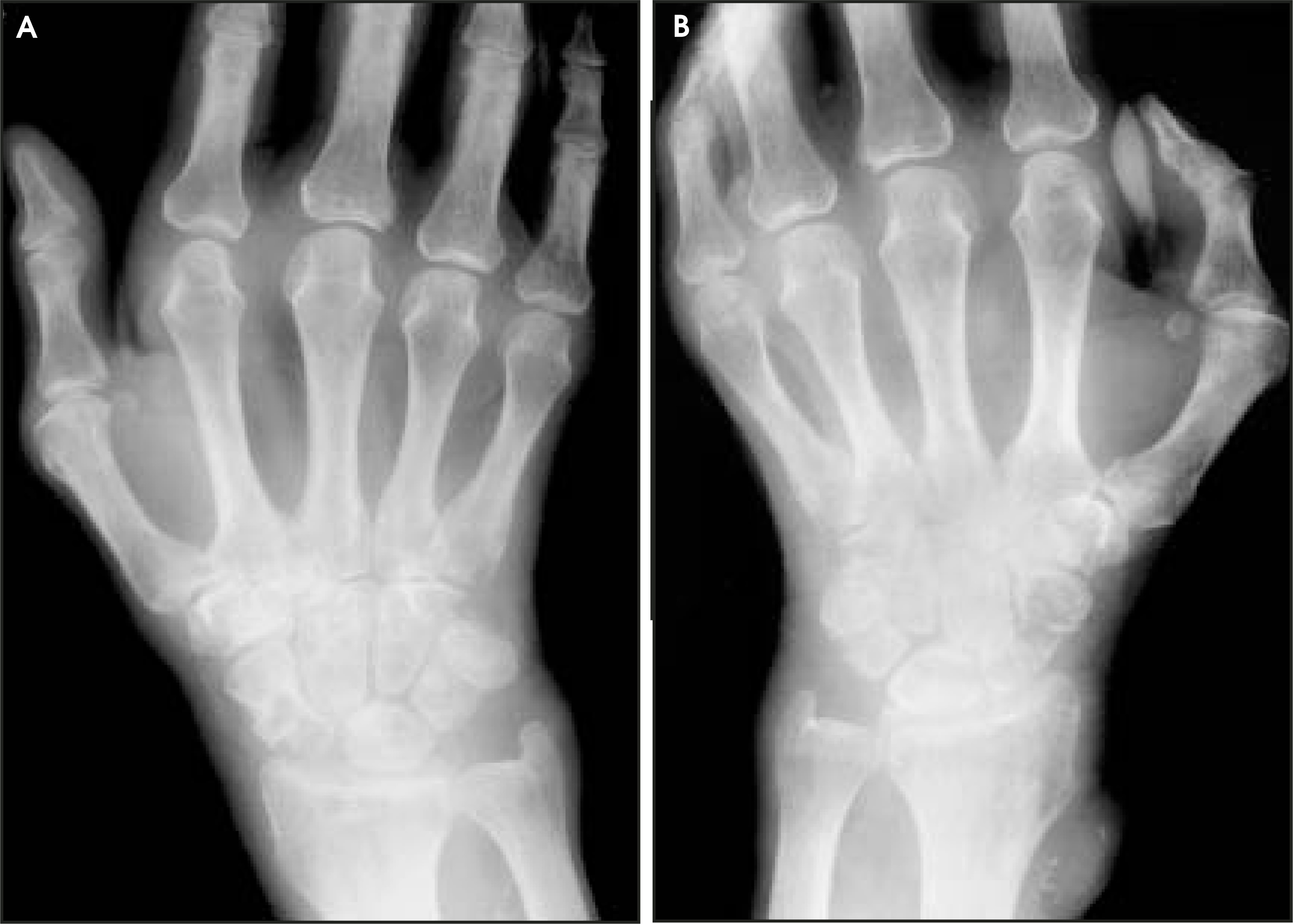
DIAGNOSIS
Dialysis-related amyloid arthropathy
IMAGING FINDINGS
Radiography of both wrists showed bilateral soft-tissue swelling and numerous osseous erosions bilaterally (Figure 1). The patient had evidence of polycystic kidney disease on CT (Figure 2). Differential considerations: pigmented villonodular synovitis (PVNS), noncalcified synovial chondromatosis, tuberculosis, and silicone synovitis. Pigmented villonodular synovitis and noncalcified synovial chondromatosis are almost invariably unilateral as opposed to amyloid arthropathy, which tends to be bilateral. Pigmented villonodular synovitis and synovial chondromatosis have predominantly lower extremity predilection, unlike the upper extremity prevalence of amyloid arthropathy. Both PVNS and synovial chondromatosis are noted for absent juxta-articular osteoporosis. Tuberculosis, unlike amyloid arthropathy, is usually unilateral; however, their radiographic appearances are similar. The diagnosis of silicone synovitis is predicated on the presence of a worn prosthesis.
DISCUSSION
Amyloidosis is a heterogeneous group of disorders characterized by the extracellular deposition of protein fibrils in organs or tissues.1 The precursors of the amyloid fibrils are circulating plasma proteins, and a wide variety of proteins can form from these fibrils.1 Historically, amyloidosis was classified as primary or secondary. This disorder is now categorized according to the type of protein contained within the chains that form the abnormal deposits. Table 1 shows an abbreviated summary of the current classification nomenclature of amyloidosis2 as established by an international symposium held in Norway in 1990.2,3 The capital letter A (for amyloid) is always the first letter, followed (without a space) by an abbreviation for the first letter(s) of the amyloid protein in the tissue deposits.
Beta 2-microglobulin (ß2M) was first described by Gejyo, et al4 in 1985. It is a unique form of amyloid that is deposited in the bones and joints of patients on chronic hemodialysis. Recently, it has also been found to affect patients on continuous peritoneal dialysis and patients with chronic renal insufficiency without a history of dialysis.5 Risk factors for developing ß2M-amyloid arthropathy include older patient age at the time of initiation of dialysis, longer duration of chronic renal failure, loss of residual renal function, and cumulative years on dialysis. The principal component in this dialysis-associated arthropathy is ß2M, a small component of a major cell surface histocompatibility complex.6 In dialysis patients, serum levels of this substance have been found to be 20 to 60 times higher than normal. This substance is normally filtered by the glomerulus and catabolized by the proximal tubules. Serum levels of ß2M rise because of the tubular dysfunction and failure of the conventional dialysis membranes to filter the substance. It is not known, however, why the amyloid forms fibrils and is deposited within tissues, particularly in the musculoskeletal system, where it has a strong predilection for synovial membranes.6
Amyloid deposition usually involves the bones and joints, commonly presenting as carpel tunnel syndrome, chronic arthropathy, destructive spondyloarthropathy, and bone cysts with or without pathologic fractures. Clinically, the articular changes tend to be bilateral and most frequently affect the shoulders, hips, wrists, and knees.7 Pain and stiffness are usually present, and severe soft-tissue swelling often occurs. The fibrillar amyloid is deposited along the flexor tendons in the wrists and the transverse carpal ligament. The amyloid deposits press upon the median nerve, resulting in carpal tunnel syndrome.8 External pressure erosions on intracapsular cortices occur secondary to infiltration of the periarticular soft tissues. Subchondral bone cysts are frequent and are filled with amyloid. These cysts are a pathognomonic finding of ß2M amyloid arthropathy.9,10 Histologically, the synovium is hyperplastic, while the joint capsule and adjacent tendons and ligaments are diffusely replaced by eosinophilic amorphous material. Similar deposits can be found in the walls of small blood vessels in the visceral form of ß2M amyloidosis. These luminal, subendothelial, amyloid nodules can then result in ischemia supplied by those vessels.
Plain-film findings of dialysis-associated arthropathy include nodular soft-tissue swelling and juxta-articular osteoporosis. The prominence and nodularity of the periarticular, soft-tissue swelling is partially due to thickening of adjacent tendons and amorphous deposits in the periarticular tissues. Prominent subchondral cysts often occur in the absence of significant cartilage space narrowing. The cysts of amyloid arthropathy usually are multiple and well-defined, can be multiloculated, and may or may not have sclerotic edges.2 On magnetic resonance (MR) images, synovial articular amyloid and amyloid within cysts demonstrate long T1 and short T2 relaxation times. On all sequences, the signal intensity is heterogeneous and between that of muscle and fibrocartilage.2
Efforts to correct this problem generally focus on the use of more permeable dialysis membranes and absorbent columns that are better able to filter Aß2M.3 Renal transplantation is generally not a solution. One series estimated that 26% of renal allografts have deposits of either Aß2M (due to the failure of the transplant) or the AA type (due to recurrent infection).11
CONCLUSION
Amyloid deposition is an unusual cause of pain and arthropathy in patients with chronic renal failure or in patients on dialysis. The arthropathy results from extracellular deposition of protein fibrils in organs or tissues. The radiographic presentation is usually manifested by signs of nodular soft-tissue swelling and juxta-articular osteoporosis. The appearance may be similar to other synovial processes such as PVNS, synovial osteochondromatosis, or tuberculosis. Treatment consists of finding dialysis filters to increase the absorption of the Aß2M and the symptomatic treatment of this disorder. The prompt identification of this entity should assist in determining what follow-up is necessary and should contribute to the formation of an appropriate clinical treatment plan.
DISCLAIMER
The opinions and assertions herein are the private views of the authors and are not to be construed as official or as reflecting the view of the Department of the U.S. Air Force, U.S. Department of Defense, or the U.S. government.
References
- Husby G. Amyloidosis. Arthritis Rheum. 1992;22:67-71.
- Goldman A, Bansal M. Amyloidosis and silicone synovitis. Radiol Clin N Am. 1996;34:375-394.
- Husby G. Nomenclature a classification of amyloid and amyloidosis. J Intern Med. 1992;232:511-512.
- Gejyo G, Yamada T, Odani S. A new form of amyloid protein associated with chronic hemodialysis was identified as B2-microglobulin. Biochem Biophys Res Commun. 1985;129:701-706.
- Zingraff J, Noel L, Bardin T. B2-microglobulin amyloidosis in chronic renal failure. N Engl J Med. 1990;323:1070-1071.
- Arakawa M, Gezyo F. B2-microglobulin-associated amyloidoses. J Intern Med. 1992;232:531-532.
- Gielen J, van Holsbeeck M, Hauglustaine D. Growing bone cysts in long-term hemodialysis. Skeletal Radiol. 1990;19:43-49.
- McClure J, Bartley C, Ackrill P. Carpel tunnel syndrome caused by amyloid containing B2-microglobulin. A new amyloid and a complication of long-term hemodialysis. Annals Rheum Dis. 1986;45:1007-1011.
- Isaacs M, Bansal M, Flombaum C. Case 772. Skeletal Radiol. 1933;22:129-133.
- Danesh F, Ho L. Dialysis-related amyloidosis: History and clinical manifestations. Semin Dialysis. 2001;14:80-85.
- Harrison K, Alpers C, Davis C. De novo amyloidosis in a renal allograft: A case report and review of the literature. Am J Kid Dis. 1993;22:468-473.
Citation
. RCOM RADIOLOGICAL CASE OF THE MONTH. Applied Radiology. 2004;33(12):34-37. doi:10.37549/AR1305.