RCOM RADIOLOGICAL CASE OF THE MONTH
Applied Radiology — Vol. 33 , Issue 12 , pp. 38 -40
DOI: 10.37549/AR1306
Published: December 1, 2004
Categories
CASE SUMMARY
A 22-year-old white woman presented with a 2-month history of ataxia and personality changes, consisting mainly of abulia, headache, and neck pain. Her medical history included significant hypertension and long-standing mental retardation. The physical examination was significant for severely diminished visual acuity in her right eye as well as bilateral papilledema.
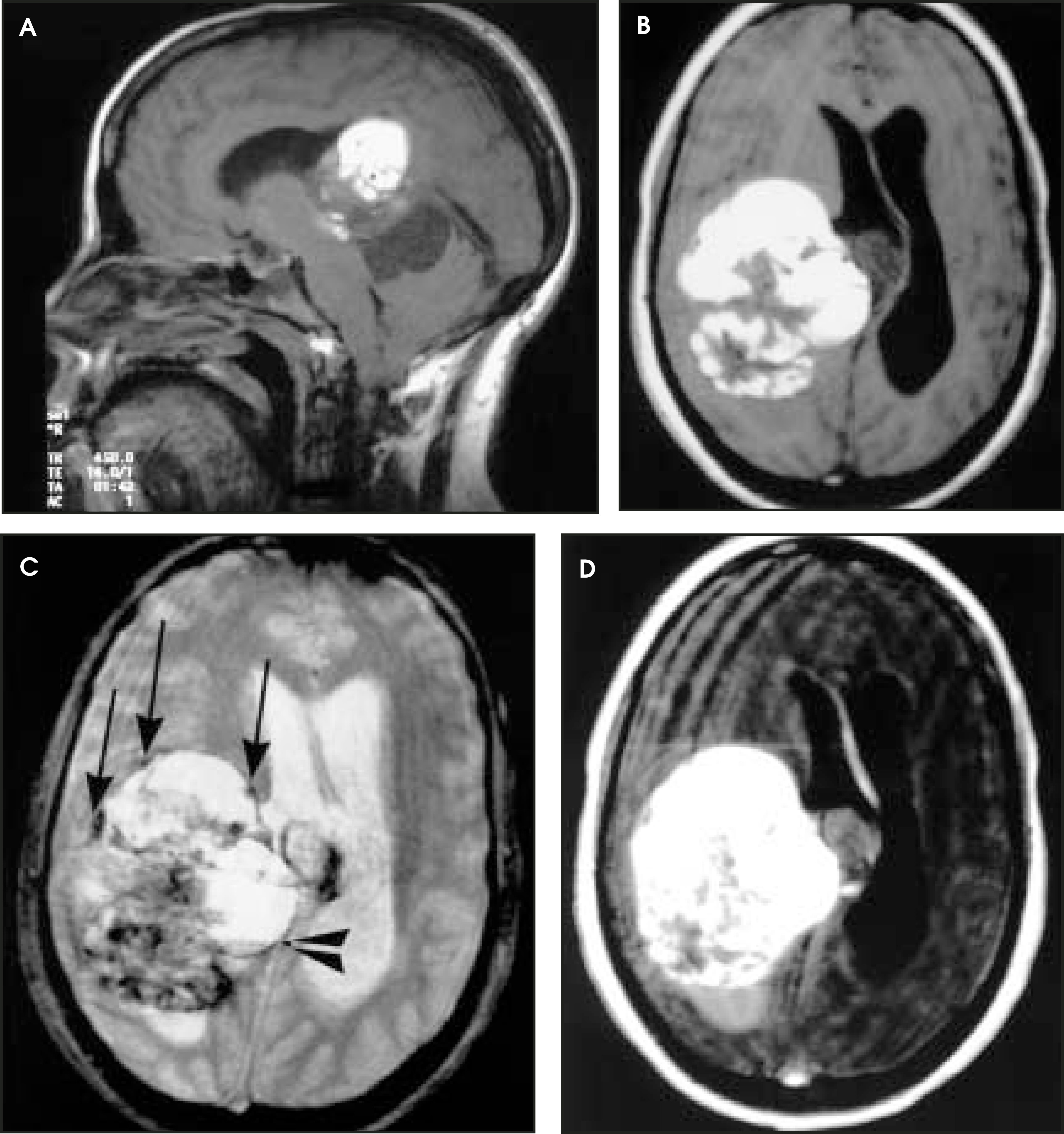
Contrast-enhanced computed tomography (CT) revealed a complex solid/cystic mass (Figure 1). Magnetic resonance imaging was performed to obtain further information (Figure 2).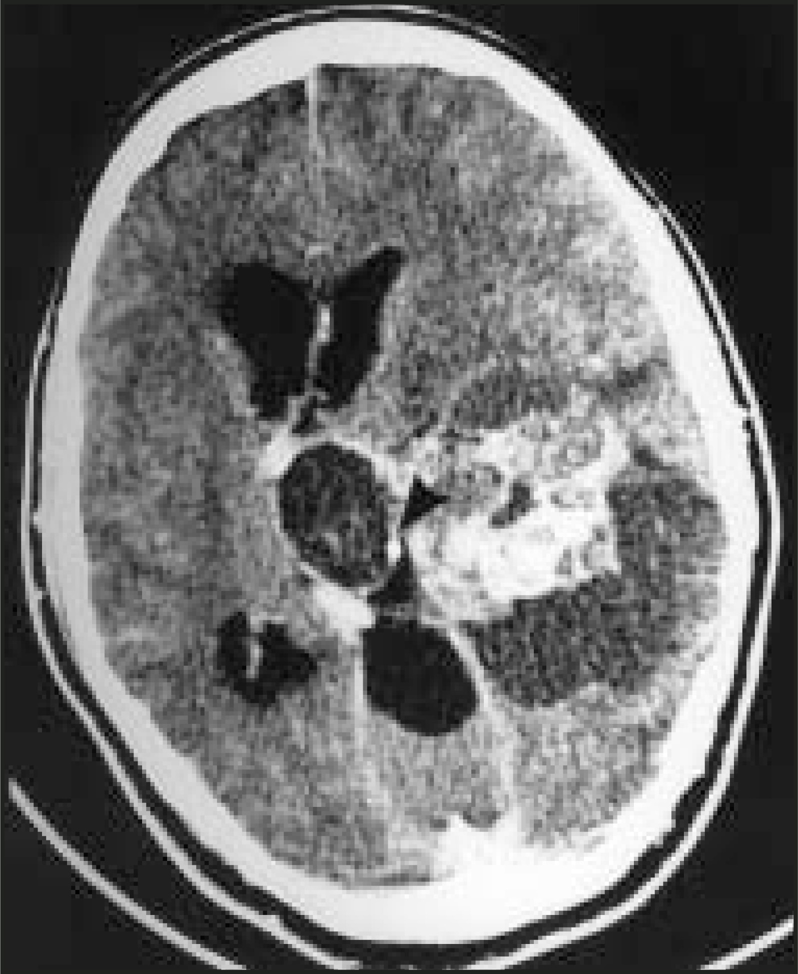
DIAGNOSIS
Intraventricular cavernous angioma (IVCA)
IMAGING FINDINGS
The patient’s preoperative contrast-enhanced CT image indicated a heterogenous cystic and solid mass. The mass involved the temporal and parietal parenchyma as well as the lateral and third ventricles. Small foci of cyst wall calcifications were present. The solid components enhanced, and the ventricles were enlarged (Figure 1).
In addition, the patient underwent preoperative MRI (Figure 2). The nonenhanced T1-weighted image revealed high signal intensity within the lesion. Transtentorial cyst extension was seen with associated transtentorial herniation. Afluid-fluid level was present, suggesting acute intralesion hemorrhage. Peripheral hemosiderin was present. The cystic components mildly enhanced (Figure 2).
CASE FOLLOW-UP
The patient underwent a right posterior temporal craniotomy. Grossly, the tumor exhibited a fibrous surface very reminiscent of dura, and the contents appeared to be organized blood of varying ages. Gross specimen, removed in toto, was 7.5 × 4 × 3.5 cm. Pathologic tissue diagnosis was cavernous angioma with literature referenced.1 Postoperatively, the patient did well and was without new deficits. She was discharged in good condition on postoperative day 4. She was seen 1 month later, at which time her visual acuity had normalized and she had no new complaints or deficits.
DISCUSSION
Intraventricular cavernomas are rare lesions.2 Occasionally, they will emerge in the subdural, subarachnoid, epidural, or intraventricular spaces.3 Congenital in origin, cavernomas do not disseminate or metastasize; therefore, their ability to grow in a previously unoccupied cavity leads to the more frequent presentation from mass effect than from onset of acute hemorrhage or seizure.4 After a literature review, we believe this case represents the largest IVCA fully resected without residual postoperative compromise of function. In fact, the lesion’s size forced the existence of an extraventricular, or intraparenchymal, component. The case is especially unique when considered with the patient’s remarkable postoperative course.
The radiologic differential diagnosis for lesions that originate in the third ventricle is broad, and preoperative imaging of this case was useful when the findings from the CT and MR examinations were combined. In accordance with the pathophysiology, and as seen with this lesion, the vascular nature of the IVCA may result in contrast enhancement, and prior multiple small hemorrhages with metabolic breakdown may result in radio-opaque calcifications.5 Therefore, the hyperdense CT image seen with IVCA may be from agitated vascular blood pooling, recent hemorrhage, or calcifications.6 Cystic components may also be present, appearing as numerous hypodense areas.7
MR imaging often shows a characteristic mixed signal of central hyperdensity that stems from the presence of methemoglobin as well as areas of hypodensity from the constructed fibrosis and calcifications.2 Gadolinium infusion will usually enhance due to the lesion’s vascularity and is usually coupled with a hypodense rim of hemosiderin. The cystic nature, calcifications, and short T1 content may lead to confusion with craniopharyngioma or teratoma. There may be a curious lack of mass effect that would be expected based on lesion size, which aids in the lesion’s often late presentation. Of course, diagnosis in this case was dependent upon the histologic diagnosis, but preoperative imaging aided in the decision to proceed to surgery, especially when discussion of possible aggressive neoplasm and nonoperative intervention arose. CT and MR imaging will continue to be integral utilities in the preoperative investigation of these unusual lesions.
CONCLUSION
Intraventicular cavernomas are rare lesions. In this case, CT and MR analysis was very helpful in determining a preoperative differential diagnosis and aiding in the eradiacation of this patient’s lesion.
References
- Reyns N, Assaker R, Louis E, Lejeune J. Intraventricular cavernomas: Three cases and review of the literature. Neurosurgery. 1999;44:648-655.
- Tannouri F, Divano L, Caucheteur V. Cavernous haemangioma in the cavernous sinus: Case report and review of the literature. Neuroradiology. 2001;43:317-320.
- Zabramski J, Wascher T, Spetzler R. The natural history of familial cavernous malformations: Results of an ongoing study. J Neurosurg. 1994;80:422-432.
- Murakami S, Sotsu M, Morooka S, Suzuki T. Chronic encapsulated intracerebral hematoma associated with cavernous angioma: A case report. J Neurosurg. 1990;26:700-702.
- Chadduck W, Binet E, Farrell F. Intraventricular cavernous hemangioma: Report of three cases and review of the literature. Neurosurgery. 1985;16:189-197.
- Sinson G, Zager E, Grossman R. Cavernous malformations of the third ventricle. Neurosurgery. 1995;37:37-42.
- Ogawa A, Katakura R, Yoshimoto T. Third ventricle cavernous angioma: Report of two cases. Surg Neurol. 1990;34:414-420.
Citation
. RCOM RADIOLOGICAL CASE OF THE MONTH. Applied Radiology. 2004;33(12):38-40. doi:10.37549/AR1306.