RCOM RADIOLOGICAL CASE OF THE MONTH
Applied Radiology — Vol. 34 , Issue 3 , pp. 45 -47
DOI: 10.37549/AR1325
Published: March 1, 2005
Categories
CASE SUMMARY
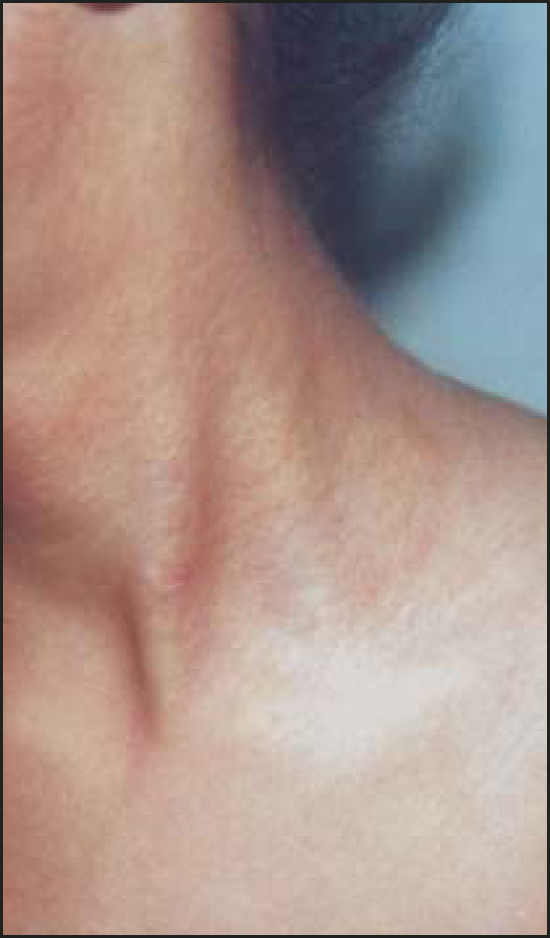
A 12-year-old boy born out of nonconsanguineous marriage presented to the radiology department to undergo a routine evaluation for the purpose of medical insurance. Ultrasound examination of the abdomen was performed (Figure 1). A cutaneous rash was noted, and the patient was referred to the dermatology department (Figure 2). Systemic examination was normal.
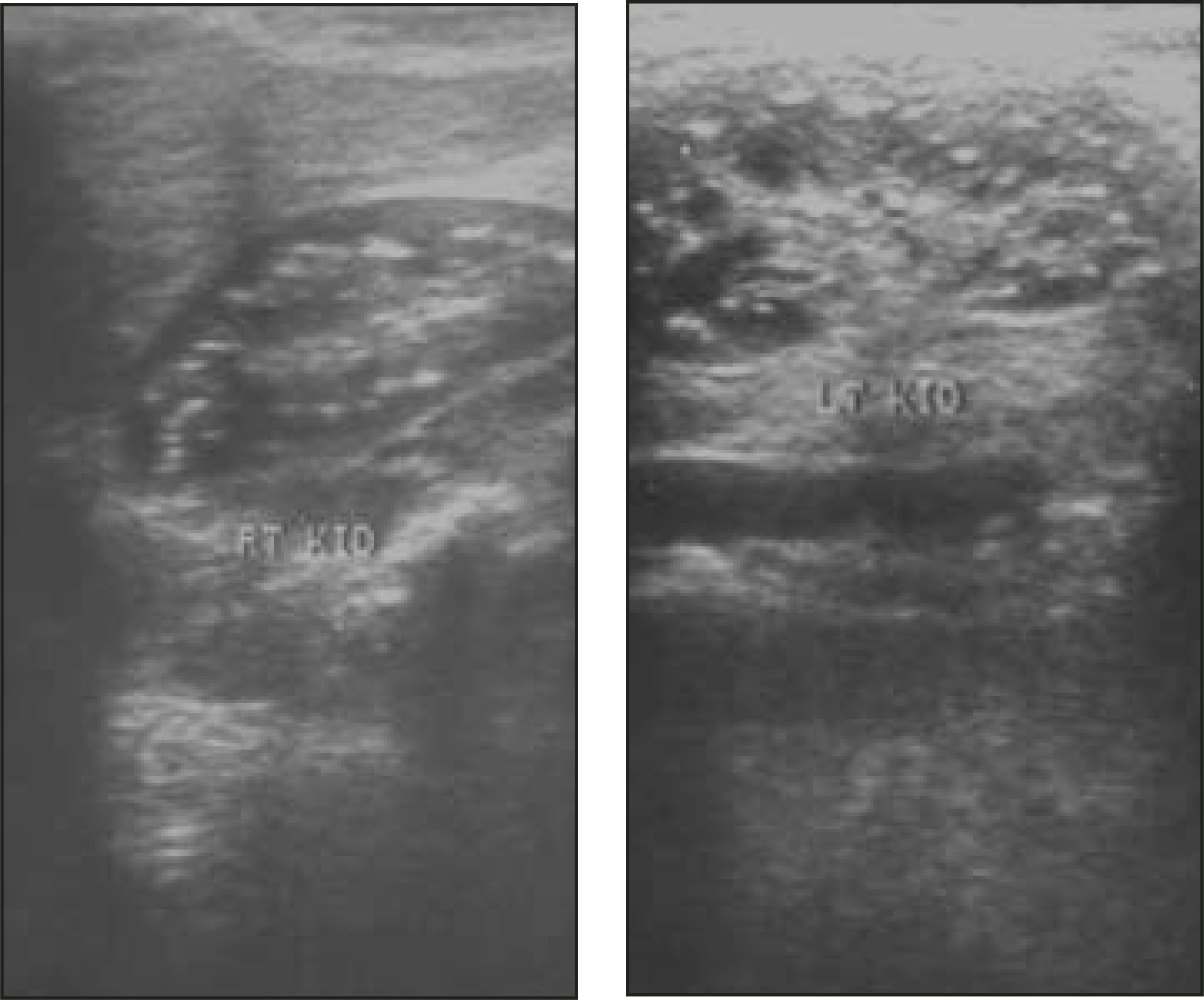
In view of the nephrocalcinosis, a history was taken, which was notable for premature birth (35 weeks gestation), the use of frusemide in infancy, vitamin D overdose, and a family history of urinary calculi. Opthalmologic examination showed bilateral colloid bodies and peripapillary atrophy with angioid streaks.
DIAGNOSIS
Pseudoxanthoma elasticum (PXE; Grönblad-Strandberg syndrome) with nephrocalcinosis
IMAGING FINDINGS
Ultrasound examination of the abdomen showed multiple small echogenic foci with distal shadowing that was seen bilaterally in the renal parenchyma at the corticomedullary junction. This finding was suggestive of calcification. No other significant abnormality was detected on ultrasonography (Figure 1).
The patient had asymptomatic, skin-colored, flat-topped papules involving the neck and periumblical areas for the last 6 months. Examination revealed 1- to 3-mm yellowish-white papules coalescing to form plaques distributed in the flexures, neck, and the periumbilical area with characteristic “chicken-skin” appearance on the neck (Figure 2). Oral mucosa, scalp, palm, and sole were not involved.
LABORATORY AND PATHOLOGICAL RESULTS
Biochemical investigations revealed a normal hemogram; serum calcium, potassium, uric acid, phosphorus, albumin, urea, and creatinine levels were also normal. Urine examination revealed a 24-hour protein level of 500 mg and normal pH, and there were no blood cells on microscopic examination. Urinary excretion of calcium was estimated (as a calcium:creatinine ratio) and was normal. Stool was negative for occult blood.
Electrocardiogram and 2-dimensional echocardiography of the heart were normal (not shown). Blood pressure was within normal limits. Skin biopsy was performed from the neck, and the specimen was stained with Verhoeff-van Gieson stain, which revealed clumped-up, darkly staining elastic fibers in the mid dermis. Von-Kossa stain revealed basophilic deposits of calcium.
CASE FOLLOW-UP
The patient was asked to follow up regularly. Subsequently, a detailed examination of the family and first-degree relatives was undertaken, which revealed biopsy-proven PXE, without any other complication, in the sister of the patient reported here. The patient was advised to avoid any form of head trauma to prevent retinal hemorrhage. Follow-up after 1 year did not reveal any further complication.
DISCUSSION
Pseudoxanthoma elasticum (Grönblad-Strandberg syndrome) is a heritable multisystemic disorder characterized by yellow, linear, reticular “cobblestone” or “chicken-skin” appearing involvement of the neck and the flexures with associated ocular and cardiovascular involvement. The disease varies in its degree of expression and is underdiagnosed due to physicians’ lack of familiarity with the condition.1 Visceral calcification in the form of pulmonary, renal, pancreatic, and splenic calcification has been rarely reported in PXE.2 Hyperechoic spots suggestive of calcification in the renal parenchyma have been reported in both adult and pediatric patients with PXE. The majority of these patients had a normal serum calcium profile.3,4
In children, the disease is usually diagnosed late, with an average age of onset of 13 years.1 Our patient presented at an earlier age and could be diagnosed based on the classic cutaneous features of PXE. Bilateral nephrocalcinosis with proteinuria was another unique finding in our patient. Renovascular hypertension is seen in 25% of adult patients, but is uncommon in children. This is because of increased rennin activity as a consequence of the obstruction of the renal arteries on account of nephrocalcinosis.5,6 Renovascular hypertension was not seen on our patient.
The cause of calcification in PXE is unknown, but it has been proposed that the knocking out of the matrix calcification gene that generally prevents calcification might be responsible, with altered elastic fibers in the basement membrane of the arteries providing the nidus for cal-cification.7 In our case, an attempt was made to exclude other causes of nephrocalcinosis, but all investigations (including the ultrasonography, ocular evaluation, and skin biopsy) pointed toward a diagnosis of PXE. As serum calcium was normal, it can be presumed that the nephrocalcinosis might be an extension of the secondary dystrophic calcification of the damaged tissues. The prognostic value of nephrocalcinosis is as yet undetermined, but this form of renal calcification may help in the early diagnosis of this rare disorder, especially in children who manifest the classic dermatological signs at a later age.4-6
CONCLUSION
Pseudoxanthoma elasticum, a heritable multisystemic disorder, varies in its degree of expression and is underdiagnosed due to the lack of knowledge of the condition among physicians. Though nephrocalcinosis was a chance finding in our case, this could enable an early diagnosis of PXE, with timely modification of potential risk factors and close monitoring of renal function.
References
- Sherer D, Sapadin A, Lebwohl M. Pseudoxanthoma elasticum: An update. Dermatology. 1999;199:3-7.
- Suarez M, Garcia J, Orense M. Sonographic aspects of pseudoxanthoma elasticum. Pediatr Radiol. 1991;21:538-539.
- Gresser U, Stuatner B, Zoller W. Kidney involvement in pseudoxanthoma elasticum—Sonography shows early calcinosis of the kidney parenchyma [In German]. Bildgebung. 1987-89;56(5):179-180.
- Crespi G, Derchi L, Saffioti S. Sonographic detection of renal changes in pseudoxanthoma elasticum. Urol Radiol. 1992;13:223-225.
- Gubler M, Antignac C, Broye R. Diffuse arterial calcified elastopathy—A new cause of renovascular hypertension in children. Ind J Pediatr Nephrol. 1985;6:47-54.
- Dynock R. Pseudoxanthoma elasticum: Report of a case with renovascular hypertension. Aust J Dermatol. 1979;20:82-84.
- Uitto J, Boyd C, Lebwohl M. International centennial meeting on pseudoxanthoma elasticum. J Invest Dermatol. 1998;110:840-842.
Citation
. RCOM RADIOLOGICAL CASE OF THE MONTH. Applied Radiology. 2005;34(3):45-47. doi:10.37549/AR1325.