RCOM RADIOLOGICAL CASE OF THE MONTH
Applied Radiology — Vol. 34 , Issue 3 , pp. 42 -44
DOI: 10.37549/AR1326
Published: March 1, 2005
Categories
CASE SUMMARY
A 9-year-old boy presented to the emergency room with right knee pain after a fall.
DIAGNOSIS
Blount’s disease
IMAGING FINDINGS
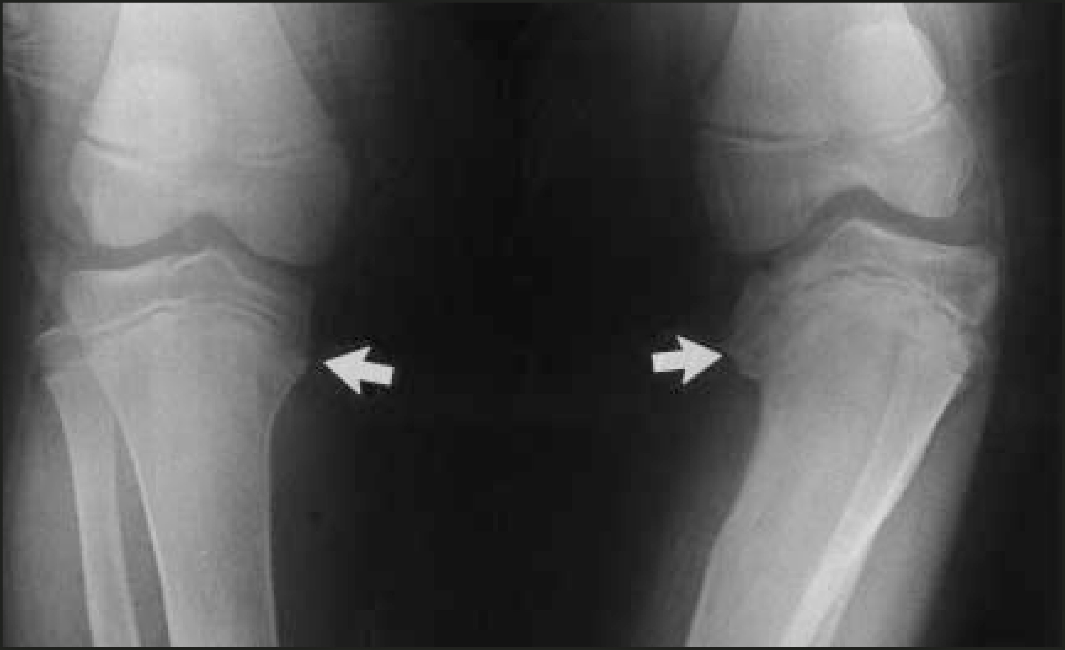
The anteroposterior radiograph of both knees shows an irregularity of the medial epiphysis of the tibias bilaterally (Figure 1). The epiphysis on the left is more medially sloped than on the right. There is mild varus angulation of both metaphyses, which is more pronounced on the left. A bony protuberance or “beak” is identified at the medial aspect of the left tibial metaphysis. Both tibial growth plates are widened and irregular. There is no evidence of acute fracture or dislocation.
DISCUSSION
Blount’s disease, or tibia vara, is a growth disorder of the medial aspect of the proximal tibial physis with abrupt medial angulation of the proximal tibia distal to the epiphysis, which leads to varus angulation of the proximal tibia.1 Several types of tibia vara have been recognized and include infantile, juvenile, adolescent, and a rare type caused by focal fibrocartilaginous dysplasia. The infantile type, or early-onset tibia vara, has an age of onset between 1 and 3 years and is usually bilateral and symmetric. The juvenile and adolescent types are both considered late-onset tibia vara and have an age of onset between 4 and 10 years and >11 years, respectively. The later onset types are much less common than the infantile form and are more often unilateral. Clinically, children with infantile tibia vara present with painless bowing and length discrepancy of the lower extremities. A bony nontender protuberance may be palpable on the medial aspect of the proximal tibia. In late-onset tibia vara, leg shortening may be associated with pain and tenderness over the medial prominence of the proximal tibia.2
The finding that obesity and early walking predispose to tibia vara suggests that abnormal pressure on the medial aspect of the proximal tibial growth plate may play a role in the pathogenesis of infantile tibia vara. It has been suggested that infantile tibia vara may have a familial occurrence. One study reports a family history of the disease in 14 of 32 patients.3 The infantile type is also more commonly seen in females and children of African-American descent. The juvenile and adolescent types develop from unknown causes; however, trauma or infection are believed to play a role.1,2
The diagnosis of Blount’s disease is based on several radiographic changes in the proximal tibia best visualized with standing anteroposterior images of both legs. Typically, radiographs reveal sharp varus angulation in the metaphysis, a widened and irregular physeal line medially, a medially sloped and irregularly ossified epiphysis, and “beaking” of the medial cortical wall of the metaphysis with lucent cartilage islands within the “beak.”4 Measurement of the metaphyseal-diaphyseal angle has been used to differentiate Blount’s disease from developmental bowing and to make a more accurate diagnosis. The metaphyseal-diaphyseal angle is described as the angle between a line drawn parallel to the top of the proximal tibial metaphysis and a line drawn perpendicular to the long axis of the tibial shaft. Studies suggest that a metaphyseal-diaphyseal angle >11° indicates Blount’s disease, whereas an angle <11° suggests physiologic bowing.5 Typical radiographic findings in late-onset tibia vara include narrowing of the medial aspect of the tibial epiphysis with an irregularly thickened physis. The overall appearance is that of a prematurely closing medial physis.1
In 1952, Langenskiold1 found that the radiographic appearance of infantile tibia vara undergoes changes with maturation of the skeleton. He classified these changes into 6 stages, which demonstrate progressive beaking, depression, and fragmentation of the medial tibial metaphysis. Stages I through IV do not provide prognostic information. However, stages V and VI indicate progression of the disease and are typically followed by increasing varus deformity.1
Histologic changes of the growth plate in patients with Blount’s disease suggest a delay in cartilage ossification in the medial metaphysis and epiphysis, which causes varus deformity if growth in the lateral aspect of the growth plate continues. Characteristic histologic features include islands of densely packed, hypertrophied cartilage cells, acellular cartilage, and abnormally large groups of blood vessels.1
Nonsurgical and surgical treatments are available for Blount’s disease, depending on the age of the patient and severity of the disease. In mild cases (stages I and II) in children <3 years old, a knee-ankle-foot-prosthetic brace can be used. Older patients, patients who show no improvement after 1 year of brace use, and those with more severe disease usually require an osteotomy procedure. The goal of these procedures is to restore a more normal configuration of the articular surface of the proximal tibia and to improve the mechanical axis of the leg. Although surgical intervention is usually successful in treating Blount’s disease, weight control is important in preventing recurrence.1
CONCLUSION
Blount’s disease is an abnormality of the growth of the proximal inner tibia, which is best diagnosed radiographically. It is important to distinguish Blount’s disease from other causes of lower extremity bowing in children because, if left untreated, it can result in progressive varus deformity and early arthritis.
References
- Langenskiold A. Tibia vara. A critical review. Clin Orthop. 1989;246:195-207.
- Thompson G, Carter J. Late-onset tibia vara (Blount’s disease). Current concepts. Clin Orthop. 1990;255:24-35.
- Schoenecker P, Meade W, Pierron R. A retrospective review and recommendations for treatment. J Pediatr Orthop. 1985;5:181-186.
- Johnston C. Infantile tibia vara. Clin Orthop. 1990;255:13-23.
- Cheema J, Grissom L, Harcke H. Radiographic characteristics of lower-extremity bowing in children. RadioGraphics. 2003;23:871-880.
Citation
. RCOM RADIOLOGICAL CASE OF THE MONTH. Applied Radiology. 2005;34(3):42-44. doi:10.37549/AR1326.