RCOM RADIOLOGICAL CASE OF THE MONTH
Applied Radiology — Vol. 34 , Issue 4 , pp. 38 -40
DOI: 10.37549/AR1327
Published: April 1, 2005
Categories
CASE SUMMARY
A 23-year-old woman with a 4-week history of headaches presented to the hospital for a computed tomographic (CT) scan of the brain. She was 19 weeks pregnant upon presentation. Her obstetrician had treated the headaches with antimigraine medication and treated a sinus infection with Macrobid (P&G Pharmaceuticals, Cincinnati, OH) without relief. A hypodensity was seen adjacent to the left frontal lobe on the CT scan (Figure 1). The following day, the patient returned for a noncontrast magnetic resonance imaging (MRI) scan of the brain, which confirmed a left frontal epidural mass (Figure 2).
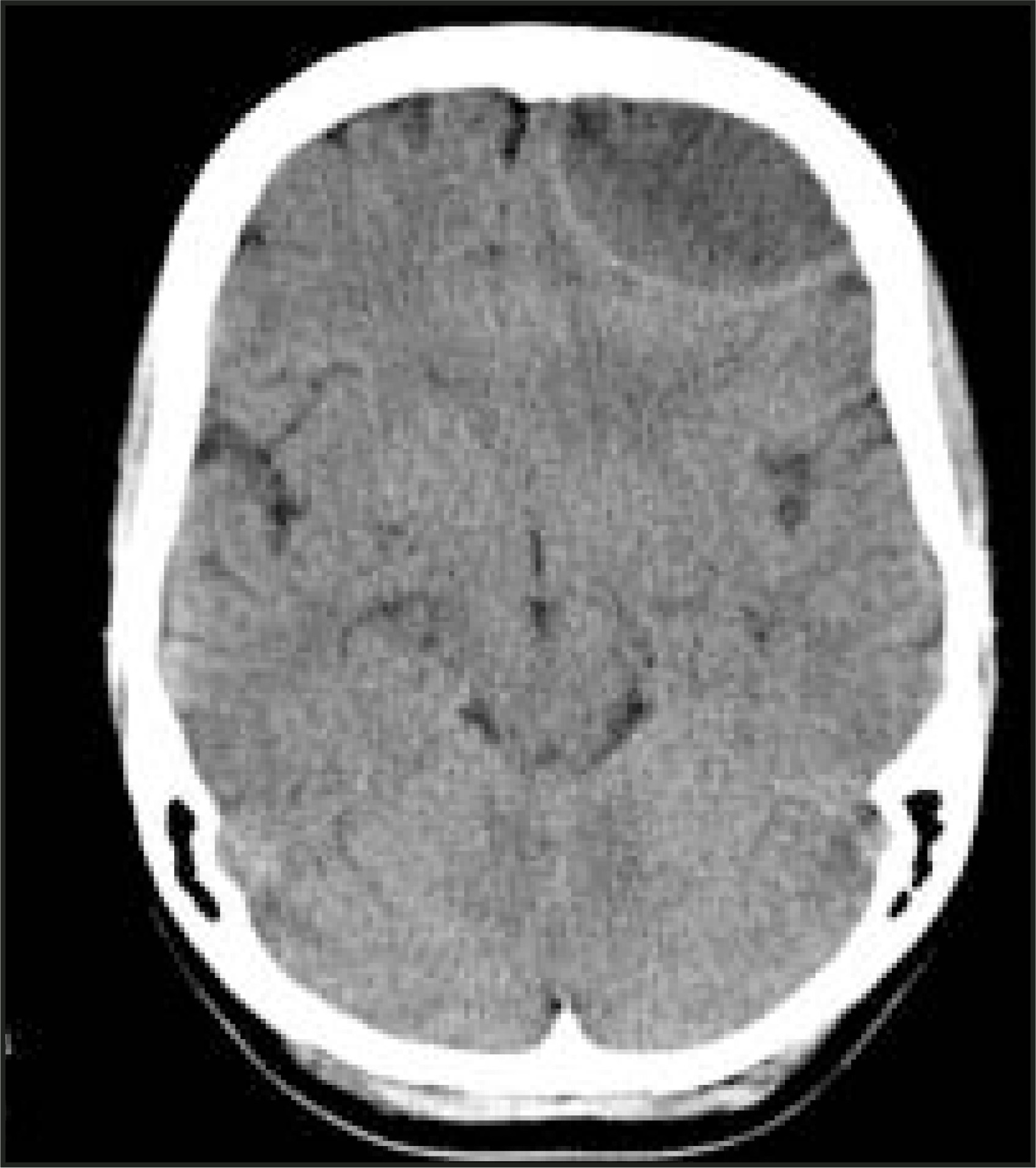
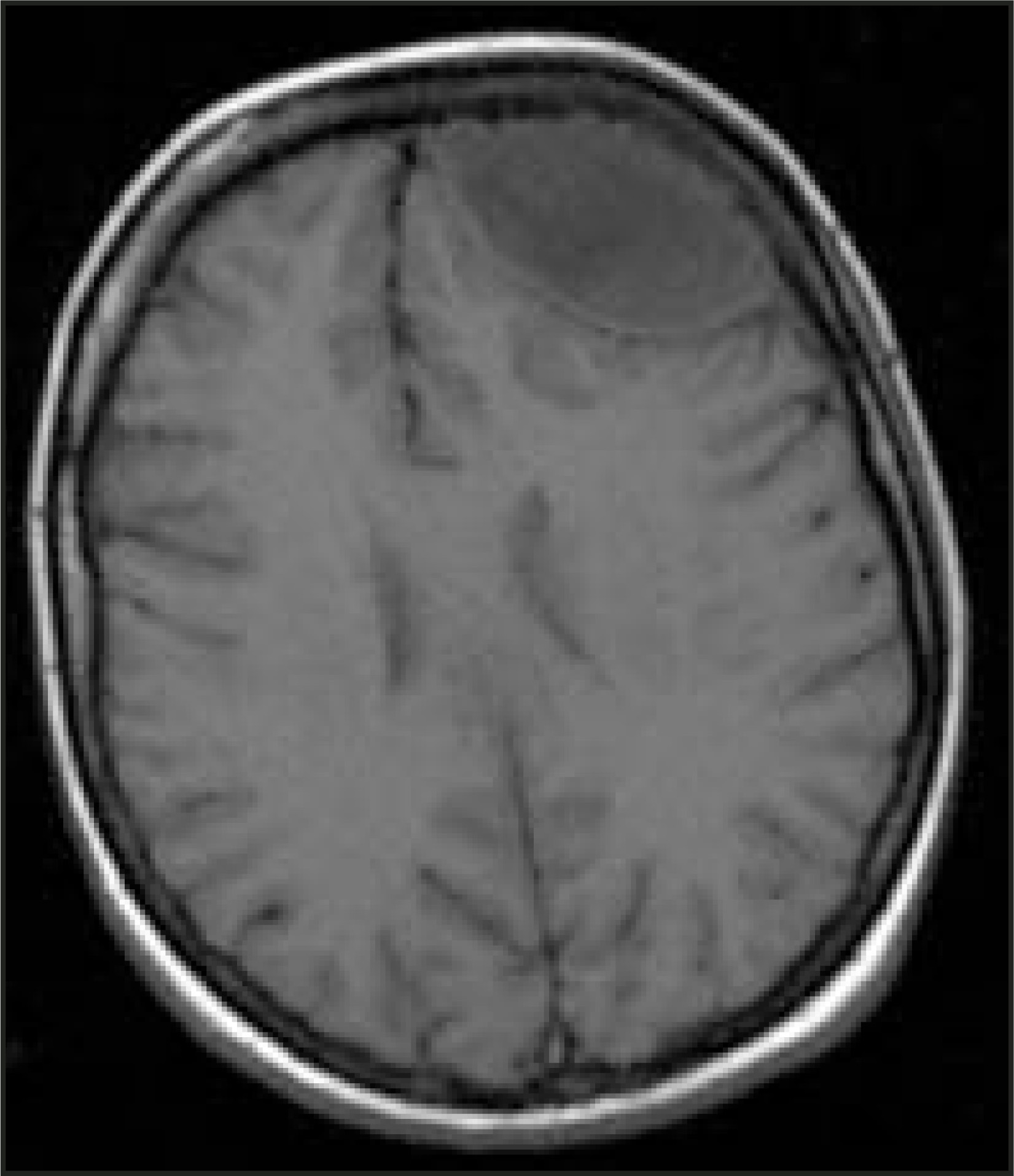
The patient was admitted for work-up of this mass, and for treatment planning because of her pregnancy. On admission, she was alert and in no acute distress. She did not have focal neurological findings on physical examination. Following consultation with otolaryngology, obstetrics, infectious disease, and neurosurgery, it was decided that the best treatment option for this pregnant patient was surgical evacuation of the abscess. Her operative cultures revealed streptococcus and mixed anaerobes and aerobes. Following treatment, her pregnancy proceeded without apparent subsequent complication.
DIAGNOSIS
Intracranial epidural abscess secondary to sinusitis
IMAGING FINDINGS
The noncontrast CT scan revealed an extra-axial elliptical hypodensity that caused mass effect on the left frontal lobe and frontal horns of the lateral ventricles (Figure 1). There was minimal midline shift (left to right). Maxillary, ethmoid, and frontal sinusitis was present. An epidural abscess was suspected.
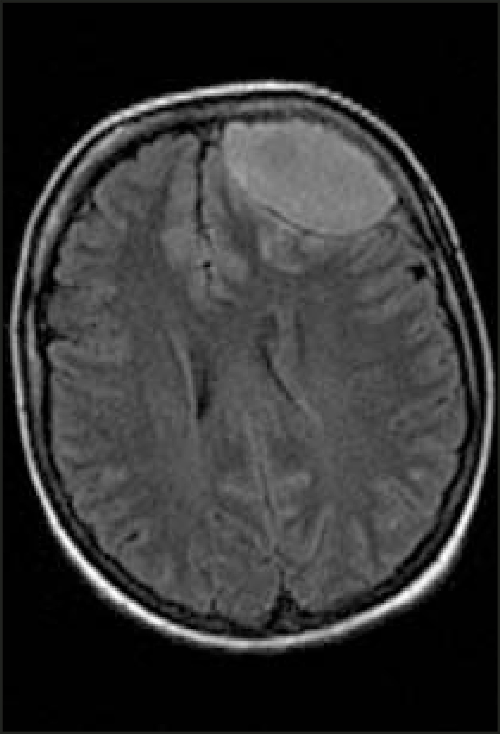
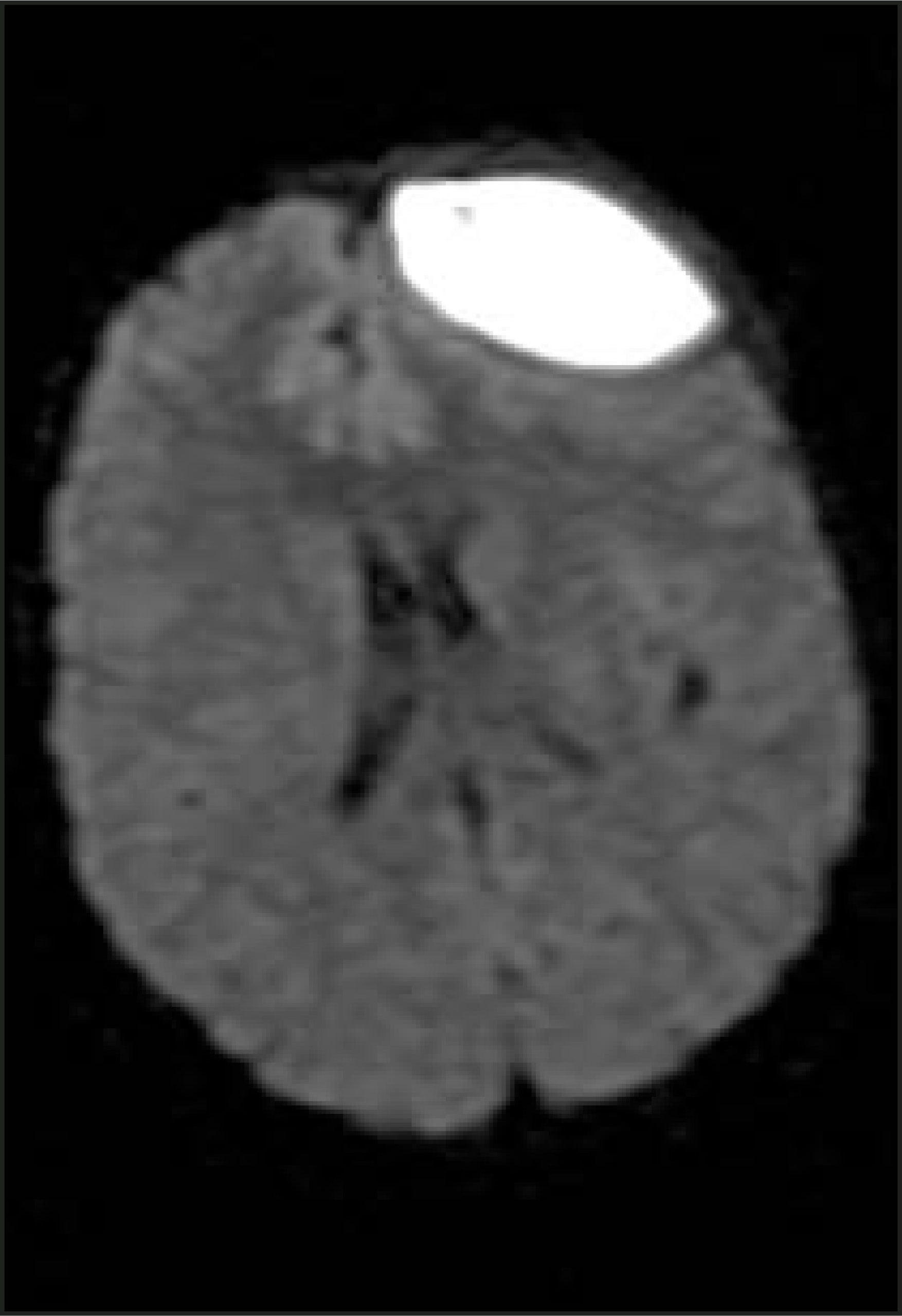
Magnetic resonance (MR) images confirmed a well-circumscribed 5 × 3 cm mass involving the left frontal epidural region. On T1-weighted images, the mass was hypointense (Figure 2), and on the T2 and fluid-attenuated inversion recovery (FLAIR) images (Figure 3), it was hyperintense. Diffusion-weighted imaging (DWI) of the brain was hyperintense in the epidural region (Figure 4) and correspondingly hypointense on apparent diffusion coefficient (ADC) mapping. The mass produced mid-line shift and mass effect on the left lateral ventricle. The mass was confirmed to be a left frontal epidural abscess on the basis of DWI.
DISCUSSION
An epidural abscess is an infection in the epidural space between the dura mater and overlying bone of the skull or the spine. Intracranial epidural abscess is a rare, but potentially fatal, illness. Only 10% of epidural abscesses occur intracranially.1 It is usually caused by direct extension of a pre-existing infection, such as sinusitis, mastoiditis, otitis media, or prior craniotomy.2
Due to the seriousness of this illness, early recognition is imperative. Onset is often insidious with nonspecific symptoms of headache and/or fever. Patients rarely present with focal neurological deficits, making the diagnosis more difficult to establish. Imaging techniques are important in the rapid detection of intracranial epidural abscesses. While CT is the standard modality for diagnosing sinusitis, MRI is more accurate in detecting intracranial abscesses.3 An intracranial abscess is usually hypointense on T1-weighted images and hyperintense on T2-weighted images.3 Diffusion-weighted MRI has been found to be useful in differentiating brain abscesses from noninfectious processes that appear similar on other imaging modalities, such as cystic or necrotic tumors.4 Brain abscesses appear hyperintense on DWI.5
The treatment for intracranial epidural abscess is both medical and surgical. Empirical antibiotic therapy against streptococci, staphylococci, and anaerobes should be started. Intracranial epidural abscesses must be surgically drained with collection of the fluid for culture.
CONCLUSION
Intracranial epidural abscess secondary to sinusitis is a potentially fatal infection that can be reliably recognized through the utilization of multiple MRI sequences combined with CT findings of sinusitis. Diffusion-weighted imaging of the brain plays a role in imaging interpretation beyond the traditional detection of acute stroke. In the setting of infection, MRI with diffusion-weighted imaging can confirm the diagnosis of abscess and allow expedited surgical evacuation combined with immediate intravenous antibiotics.
References
- Fauci A, Braunwald E, Isselbacher K. Harrison’s Principles of Internal Medicine. 1998.
- Handel S, Klein W, Kim Y. Intracranial epidural abscess. Radiology. 1974;111:117-20.
- Younis R, Vinod K, Davidson B. The role of computed tomography and magnetic resonance imaging in patients with sinusitis with complications. Laryngoscope. 2002;112:224-229.
- Chang S, Lai P, Chen W. Diffusion-weighted MRI features of brain abscess and cystic or necrotic brain tumors: Comparison with conventional MRI. J Clin Imaging. 2002;26:227-236.
- Tsai Y, Chang W, Shen C. Intracranial suppuration: A clinical comparison of subdural empyemas and epidural abscesses. Surg Neurol. 2003;59:191-196.
Citation
. RCOM RADIOLOGICAL CASE OF THE MONTH. Applied Radiology. 2005;34(4):38-40. doi:10.37549/AR1327.