RCOM RADIOLOGICAL CASE OF THE MONTH
Applied Radiology — Vol. 34 , Issue 4 , pp. 38 -40
DOI: 10.37549/AR1329
Published: April 1, 2005
Categories
CASE SUMMARY
A 53-year-old man presented complaining of chronic, nonspecific abdominal pain. An abdominal ultrasound was performed to exclude gallstones.
DIAGNOSIS
Mature cystic teratoma (dermoid cyst) of the mesentery
IMAGING FINDINGS
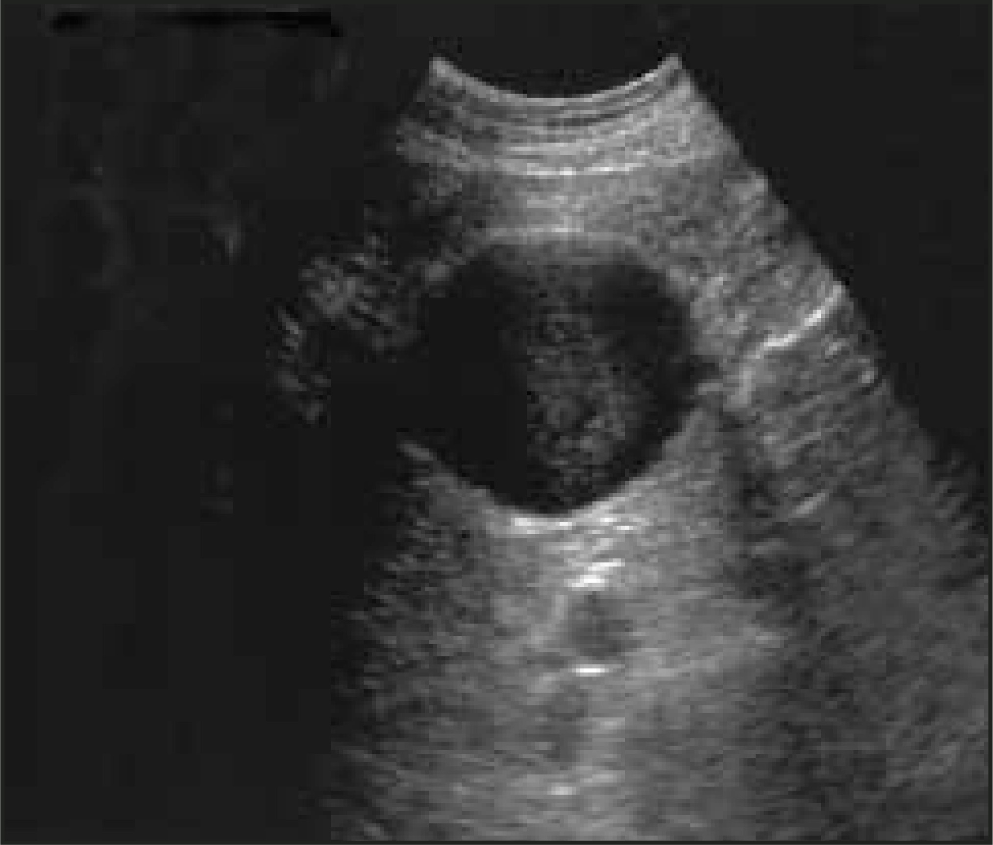
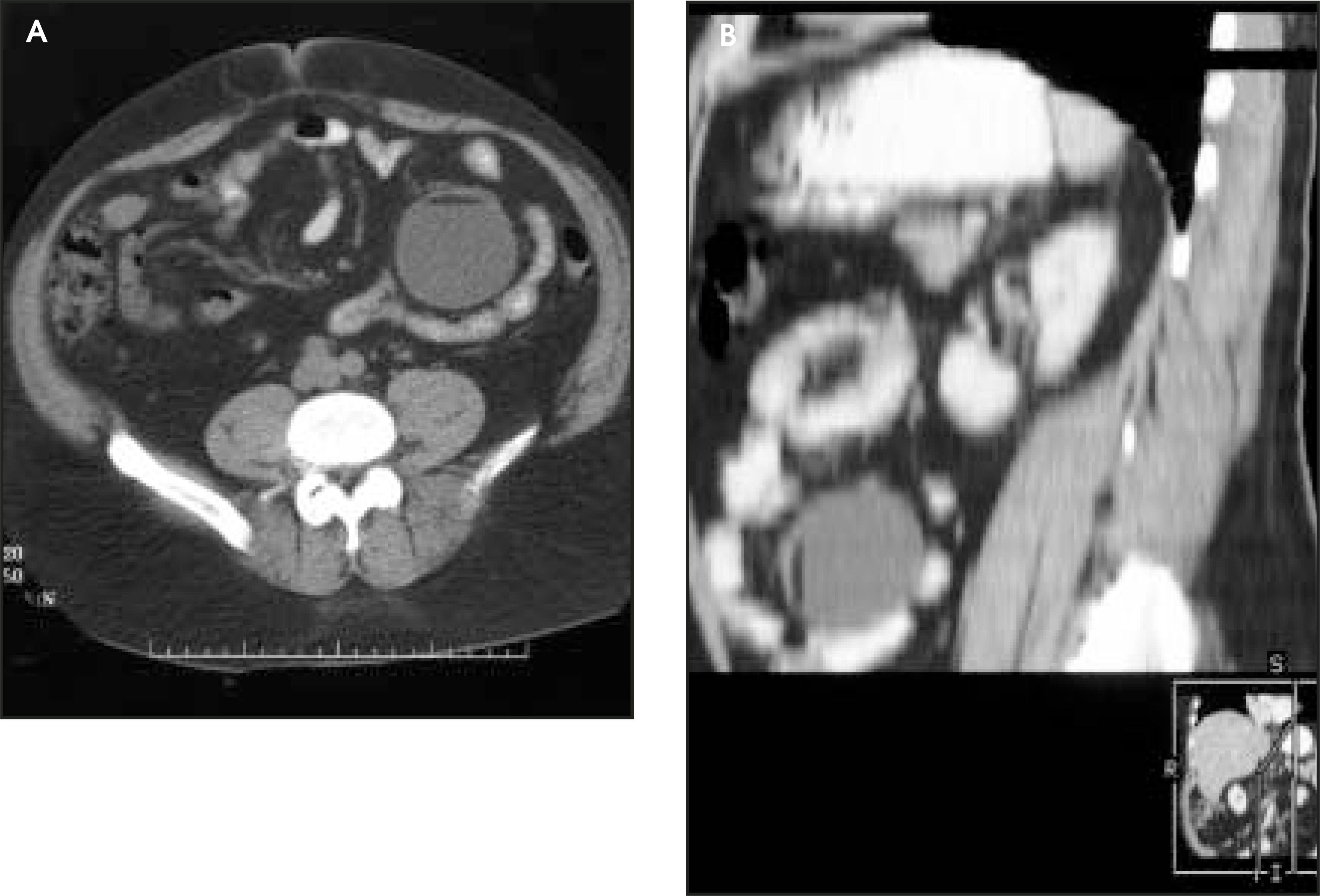
A well-defined, thin-walled complex mass was identified in the left mid abdomen on ultrasonography (Figure 1). Computed tomography (CT) revealed a round, well-circumscribed cystic mass centered in the mesentery in the left abdomen at the level of the iliac crest (Figure 2). It was separate from the bowel and retroperitoneal structures, and contained an internal fat-fluid level. There were no associated calcifications, lymphadenopathy, or free fluid. The patient declined surgery or further diagnostic workup. A diagnosis of dermoid cyst was made on the basis of the specific imaging findings.
DISCUSSION
Dermoid cysts (also referred to as mature cystic teratomas) are benign neoplasms that can occur in many locations, but are most commonly seen in the gonads, head and neck, anterior mediastinum, sacrococcygeal area, retroperitoneum, and central nervous system (CNS). Dermoids have been reported throughout the gastrointestinal (GI) tract and associated organs, including the cecum,1 rectum,2 and pancreas.3 Generally, however, dermoids of the GI tract are rare. Only 2 previous reports of mesenteric dermoids could be located in the English language literature.4,5
Dermoids have been described using all imaging modalities, but the specificity for diagnosis of fat and calcifications makes CT the modality of choice. One study found CT evidence of fat in 93% of cases, teeth or other calcifications in 56%, and tufts of hair in 65%.6 A Rokitansky protuberance, or “dermoid nipple,” was seen in 81%. A fat-fluid level was found in 12% of ovarian dermoids, and is considered diagnostic.
The differential diagnosis of a cystic mesenteric mass includes duplication cysts, cystic mesothelioma, cystic spindle-cell tumor, and liquefying mesenteric hematoma. Liposarcoma and mesenteric lipodystrophy can present as a single fat-containing mesenteric mass. However, mesenteric lipodystrophy would not be cystic, and liposarcoma does not contain fat-fluid levels. The only other cystic mass that might contain a fat-fluid level is a hydatid cyst, but, in the abdomen, this would occur in the liver.
Dermoids are usually asymptomatic, with most symptoms due to local compression, rupture, or infection. Rupture in ovarian dermoids can lead to peritonitis; while in CNS dermoids, it can lead to the spread of fatty, potentially infected material through the subarachnoid, ventricular, or subdural compartments. Rarely, production of hormones in functional tissue components can occur within a dermoid, leading to complications such as Cushing’s disease from overproduction of adrenocorticotropic hormone. Malignant transformation of ovarian dermoid cysts is rare, with the incidence at approximately 1% to 2% of all ovarian neoplasms. Squamous cell carcinoma is most frequent, but adenocarcinoma, undifferentiated carcinoma, and fibrosarcoma also occur.7We are aware of no reported cases of malignant transformation of dermoids in the GI tract. Surgical excision is recommended in cases in which the diagnosis is uncertain, or where the mass is symptomatic.
CONCLUSION
Dermoid cysts have a varied appearance on imaging studies. By being attuned to certain characteristic features, however, a radiologist may be able to make a specific diagnosis with confidence, even when the location of the mass is extremely unusual, as seen in this case.
References
- Schuetz M, Elsheikh T. Dermoid cyst (mature cystic teratoma) of the cecum. Histologic and cytologic features with review of the literature. Arch Pathol Lab Med. 2002;126:97-99.
- Sakurai Y, Uraguchi T, Imazu H. Submucosal dermoid cyst of the rectum: Report of a case. Surg Today. 2000;30:195-198.
- Vermeulen B, Widgren S, Gur V. Dermoid cyst of the pancreas. Case report and review of the literature. Gastroent Clin Biol. 1990;14:1023-1025.
- Aderjou E, Adekunle O, Madubuko G. Mesenteric dermoid cyst simulating hepatocellular carcinoma in a Nigerian male: A case report. East Afr Med J. 1980;57:508-511.
- Torreggiani W, Brenner C, Micallef M, O’Laoide R. Case report: Caecal volvulus in association with a mesenteric dermoid. Clin Radiol. 2001;56:430-432.
- Buy J, Ghossain M, Moss A. Cystic teratoma of the ovary: CT detection. Radiology. 1989;171:697-701.
- Stamp G, McConell E. Malignancy arising in cystic ovarian teratomas. A report of 24 cases. Brit J Obst Gynaecol. 1983;90:671-675.
Citation
. RCOM RADIOLOGICAL CASE OF THE MONTH. Applied Radiology. 2005;34(4):38-40. doi:10.37549/AR1329.