RCOM RADIOLOGICAL CASE OF THE MONTH
Applied Radiology — Vol. 34 , Issue 5 , pp. 50 -54
DOI: 10.37549/AR1337
Published: May 1, 2005
Categories
CASE SUMMARY
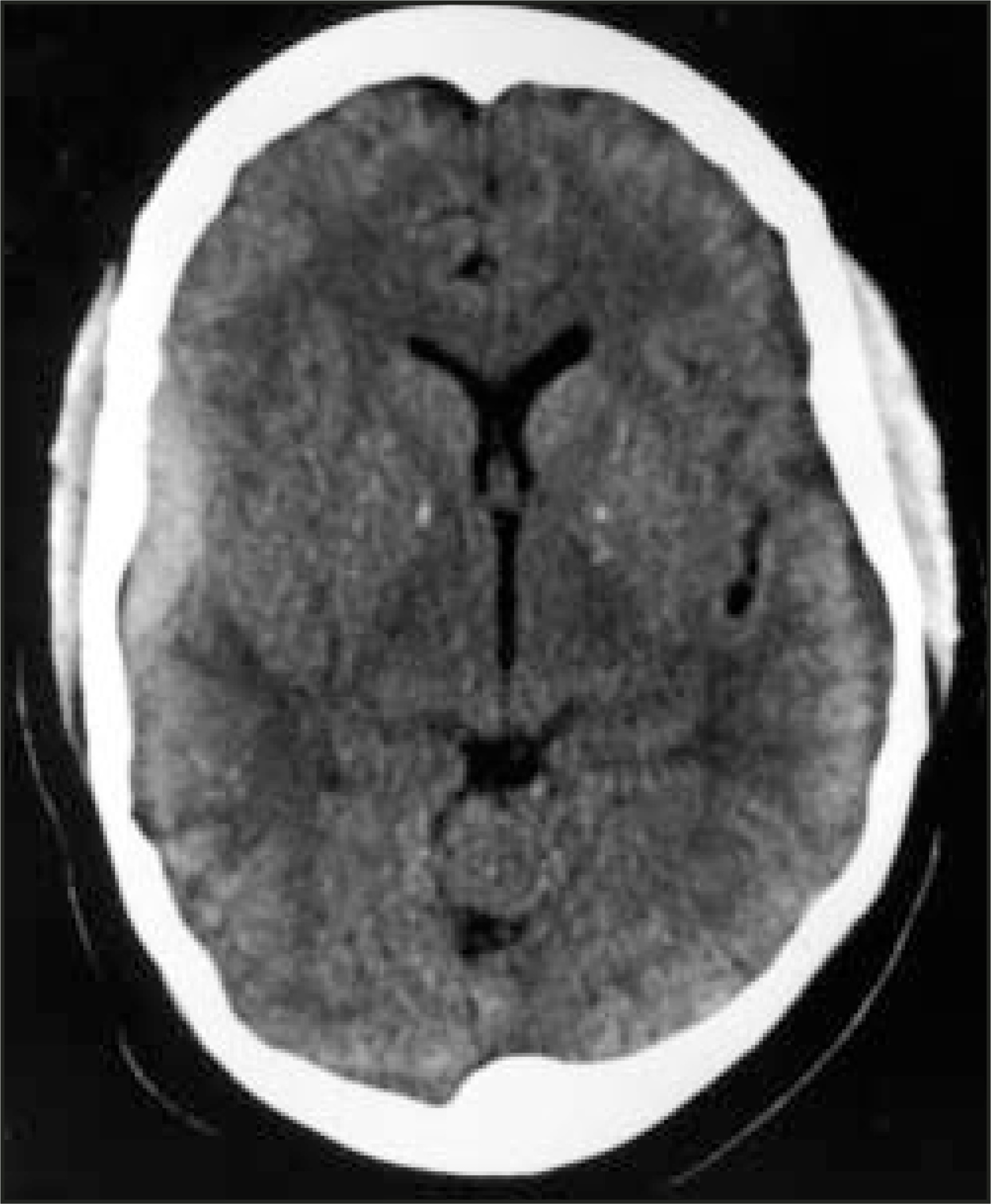
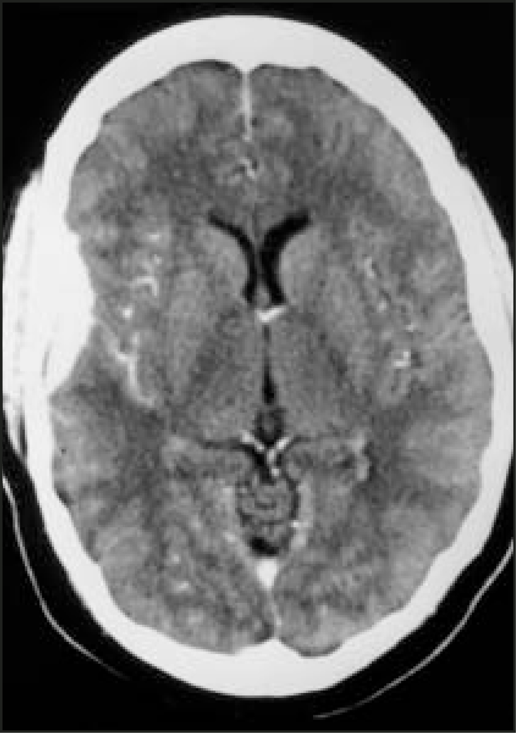
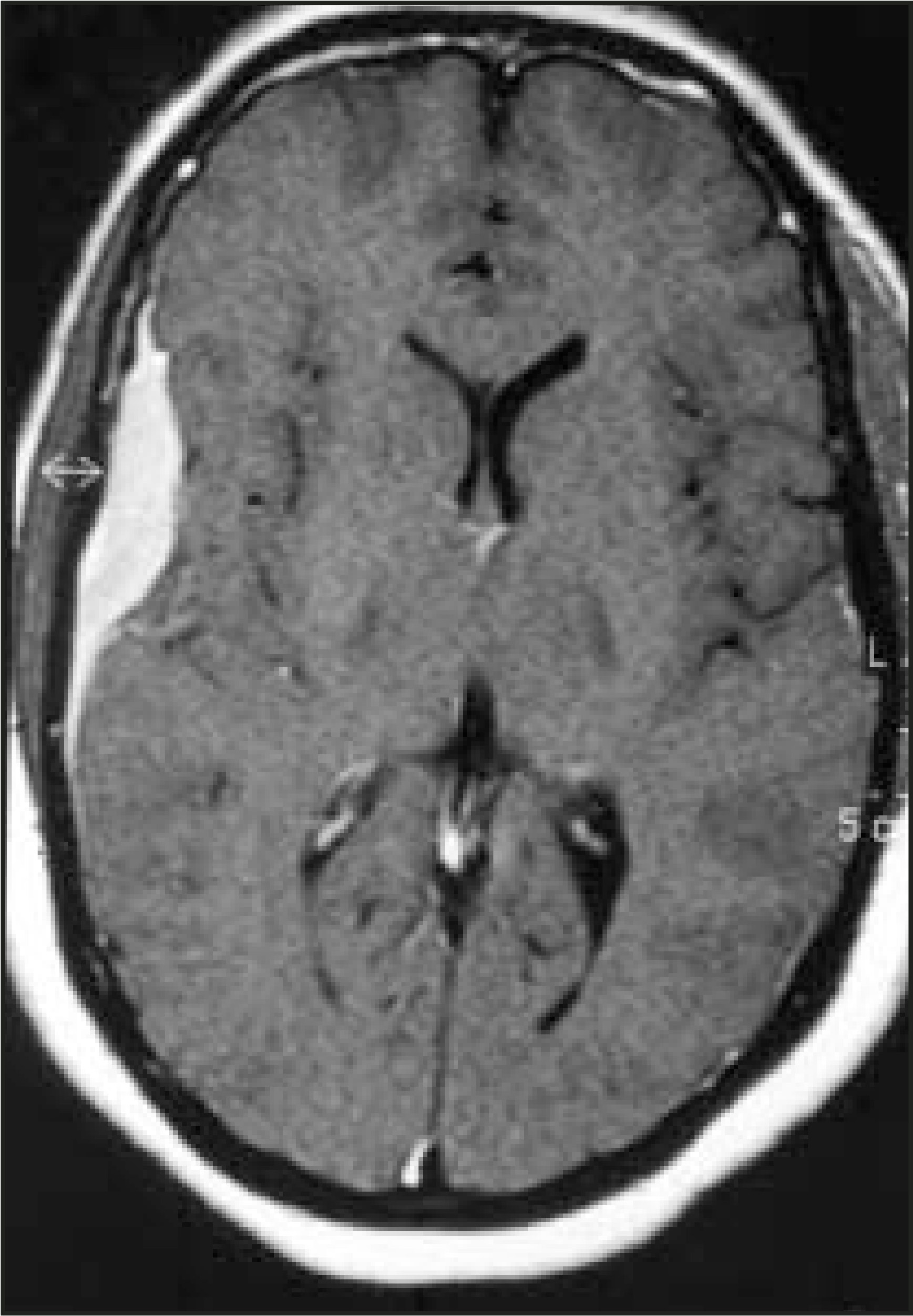
A 48-year-old woman presented to the emergency department for a single episode of seizures. She had no significant medical history. Physical and detailed neurologic examinations were normal without any focal signs. Computed tomography (CT) scan of the brain was performed (Figures 1 and 2) followed by magnetic resonance imaging (MRI; Figure 3).
DIAGNOSIS
B-cell lymphoma of mucosa-associated lymphoid tissue (MALT lymphoma)
IMAGING FINDINGS
Unenhanced CT of the brain revealed an extra-axial elliptical hyperdense mass along the right temporal convexity (Figure 1). Contrast-enhanced CT showed diffuse enhancement of this mass (Figure 2). Contrast-enhanced axial T1-weighted MRI revealed a homogeneously enhancing right temporal extra-axial mass with a dural tail (Figure 3). The patient subsequently underwent craniotomy and tumor resection.
DISCUSSION
Meningeal involvement by lymphoma is most often a manifestation of disseminated disease and occurs with lymphomas of aggressive type. Primary central nervous lymphomas represent only approximately 2% of intracranial neoplasms and 2% of all lymphomas. Primary leptomeningeal lymphomas are even more rare and are distinct clinicopathologic entities, separate from intraparenchymal lymphomas. Although infiltration into adjacent meninges can sometimes occur with intracerebral lymphoma, localization exclusively to the meninges is exceedingly rare. According to the cases reported in the literature, primary leptomeningeal lymphomas can occur in two ways. One is an infiltrative type, the majority of which are of high grade. Lachance et al1 described 9 such cases and estimated that such lesions represent 7.6% of all primary central nervous system lymphomas. They called this infiltrative entity “primary leptomeningeal lymphoma.”
The second variety of primary lymphoma is a localized type that involves the meninges, the majority of which are low grade. As these tumors are dural based, clinically and radiologically, they closely resemble meningioma or subdural hematoma. Only 12 such cases have been described in the literature to date. Ten of these cases were low-grade B-cell MALT lymphomas.2-6 Kumar et al2 postulated that these tumors arise from meningoepithelial cells, which are present throughout the arachnoid membrane with more concentration at the arachnoid villi within the dural venous sinuses.2 Two of the cases of localized variety were of low-grade follicular lymphomas.7,8
CONCLUSION
Primary localized leptomeningeal lymphoma is a rare entity. These tumors are low grade, display indolent behavior, have a favorable clinical course, and should be considered in a differential diagnosis of the extracerebral extra-axial tumor.
References
- Lachance D, O’Neill B, Macdonald D. Primary leptomeningeal lymphoma: Report of 9 cases, diagnosis with immunocytochemical analysis, and review of the literature. Neurology. 1991;41:95-100.
- Kumar S, Kumar D, Kaldjian E. Primary low-grade B-cell lymphoma of the dura: A mucosa associated lymphoid tissue-type lymphoma. Am J Surg Pathol. 1997;21:81-87.
- Freudenstein D, Bornemann A, Ernemann U. Intracranial malignant B-cell lymphoma of the dura. Clin Neuropathol. 2000;19:34-37.
- Goetz P, Lafuente J, Revesz T. Primary low-grade B-cell lymphoma of mucosa-associated lymphoid tissue of the dura mimicking the presentation of an acute subdural hematoma. J Neurosurg. 2002;96:611-614.
- Altundag M, Ozisik Y, Yalcin S. Primary low grade B-cell lymphoma of the dura in an immunocompetent patient. J Exp Clin Cancer Res. 2000;19:249-251.
- Kambham N, Chang Y, Matsushima A. Primary low-grade B-cell lymphoma of mucosa-associated lymphoid tissue (MALT) arising in dura. Clin Neuropathol. 1998;17:311-317.
- Hodgson D, David K, Powell M. Intracranial extracerebral follicular lymphoma mimicking a sphenoid wing meningioma. J Neurol Neurosurg Psychiatry. 1999;67:251-252.
- Beriwal S, Hou J, Miyamoto C, Garcia-Young J. Primary dural low grade BCL-2 negative follicular lymphoma: A case report. J Neurooncol. 2003;61:23-25.
Citation
. RCOM RADIOLOGICAL CASE OF THE MONTH. Applied Radiology. 2005;34(5):50-54. doi:10.37549/AR1337.