RCOM RADIOLOGICAL CASE OF THE MONTH
Applied Radiology — Vol. 34 , Issue 6 , pp. 36 -39
DOI: 10.37549/AR1342
Published: June 1, 2005
Categories
CASE SUMMARY
A 45-year-old woman presented with decreasing oral intake secondary to abdominal pressure, easy fatigability, and increasing abdominal girth over the last year. She also complained of “crampy,” diffuse abdominal pain. She denied any recent abdominal trauma, fever, chills, nausea, or vomiting. Her medical history was significant for a diagnosis of mucinous cystadenocarcinoma of the appendix with metastasis to the omentum and extreme intraperitoneal carcinomatosis in 1998, with subsequent appendectomy, colon resection, omentectomy, and chemotherapy. Physical examination revealed an extremely distended and tense abdomen, severe caput medusae, a readily reducible umbilical hernia, and anicteric sclera.
DIAGNOSIS
Pseudomyxoma peritonei (PMP)
IMAGING FINDINGS
Computed tomography (CT) is the modality of choice because it allows relatively accurate localization and quantification of PMP.1 CT characteristics of PMP include multiple complex cystic masses of fat density in the peritoneum, which may have rims of calcifications, characteristic scalloping of the liver (and occasionally spleen) margins (Figure 1), omental thickening, and compression of varying degrees of the visceral organs and structures2 (Figure 2). Ultrasound often reveals gross, nonmobile ascites with septations and echogenicity (Figure 3). When correlated clinically, these radiologic features are highly specific for PMP.2 In patients with incidental findings of PMP, plain films of the abdomen may have revealed abdominal calcific plaques, ascites and poorly defined soft-tissue masses. These plain-film findings must be followed by CT studies.3 CT can also be used to follow and re-evaluate patients.
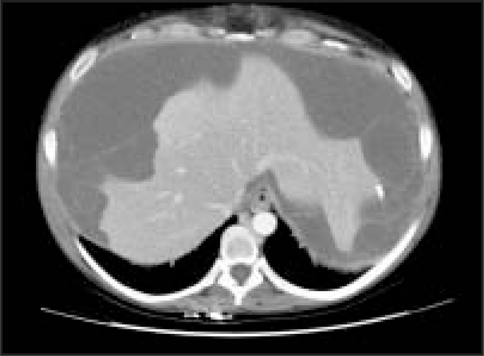
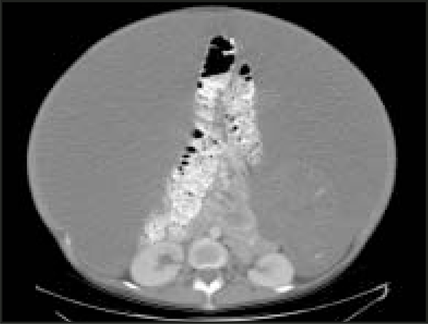
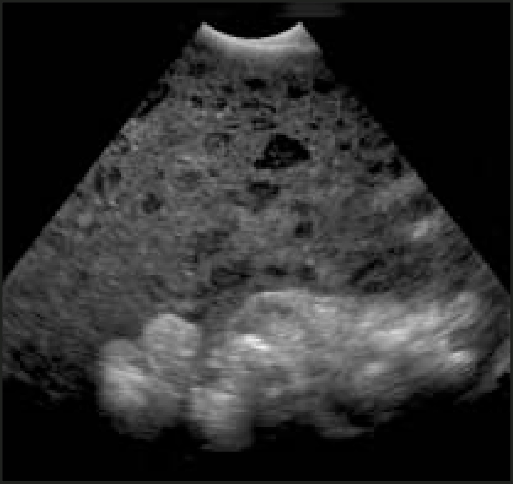
Therapeutic paracentesis is not possible because of the nature of the mucin, but enough material can be removed by ultrasound guidance for a diagnostic paracentesis, if the diagnosis is not certain. This is rarely done because the history, examination, and imaging studies often lead to the diagnosis.
DISCUSSION
Pseudomyxoma peritonei (PMP) is a rare complication of mucinous tumors of appendiceal or ovarian origin, which result in peritoneal and omental implants. While the origin of PMP is controversial, clinical morbidity and mortality results from the fact that copious amounts of extracellular and peritoneal mucin results in distortion and loss of function of visceral organs.2 Subsequent unrelieved compression can lead to adhesions and further morbidity, including small-bowel obstruction, renal or caval obstruction, and death. Because of the viscous, gelatinous nature of the mucus, it cannot be drained by paracentesis. Impending bowel obstruction, renal compromise, and discomfort are relieved by repeated laparotomy for debridement of the mucin and subsequent decompression of the viscera. Recent research indicates that the PMP clinical symptoms may be caused by an overwhelming increase in MUC2-secreting cells as well as the fact that the excessively produced mucin has no place to drain. This raises the possibility of MUC2-targeted therapy.4
Currently, cytoreductive surgery combined with perioperative intraperitoneal chemotherapy is the standard treatment for patients with peritoneal spread of primary appendiceal tumors.5 Aggressive surgical debulking is recommended to reduce the tumor load and to prevent complications such as fistulas, hernias, and visceral compression. Pseudomyxoma peritonei of ovarian origin is treated surgically with hysterectomy, bilateral salpingo-oophorectomy, appendectomy, and general debulking of the mucin.2 Removal of the peritoneal implants is done aggressively, as it has been shown to reduce the frequency of repeat debulking. However, controversy still exists regarding how aggressively high-grade lesions are managed, because aggressive therapy has been associated with higher morbidity without a significant survival benefit.2 In general, therefore, patients with high-grade lesions should be treated symptomatically. Total parenteral nutrition is highly recommended, as it may prolong survival. Intraperitoneal chemotherapy is also recommended. Current chemotherapy protocols include combinations of cisplatin, 5-fluorouracil (5FU) and cyclophosphamide. Intraperitoneal hyperthermic chemotherapy with mitomycin C and 5FU, at 10°F above body temperature, has also shown promise.2 While resection and suppression of disease is the desired outcome, in more advanced cases, palliative debulking and morbidity management become the treatment goals.
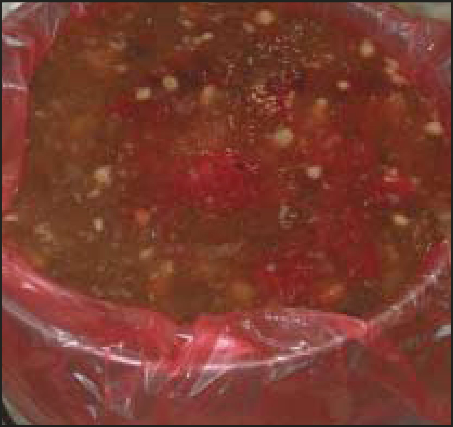
Our patient underwent debulking surgery with intraperitoneal chemotherapy. At surgery, more than 5 “buckets” (approximately 15 liters) of mucin were removed (Figure 4). The patient underwent intraperitoneal chemotherapy perioperatively, mytomycin C once, and 4 days of 5FU. Upon discharge, she still had residual ascites and had plans for physical therapy and rehabilitation.
CONCLUSION
Pseudomyxoma peritonei is a rare complication of a mucinous tumor of appendiceal or ovarian origin. The diagnosis is usually straightforward and can be made with ultrasound or CT. Marked compression of intra-abdominal structures leads to morbidity and, ultimately, mortality. Palliative debulking, as in this case, may temporarily reduce morbidity and ameliorate chemotherapy.
References
- Pickhardt P, Levy A, Rohrmann C, Kende A. Primary neoplasms of the appendix: Radiologic spectrum of disease with pathologic correlation. RadioGraphics. 2003;23:645-662.
- Harshen R, Jyothirmayi R, Mithal N. Pseudomyxoma peritonei. Clin Oncol (R Coll Radiol). 2003;15:73-77.
- Lee H, Agha F, Weatherbee L, Boland C. Pseudomyxoma peritonei. Radiologic features. J Clin Gastroenterol. 1986;8:312-316.
- O’Connell J, Tomlinson J, Roberts A, McGonigle K, Barsky S. Pseudomyxoma peritonei is a disease of MUC2-expressing goblet cells. Am J Pathol. 2002;161:551-564.
- Sugarbaker P. Cytoreductive surgery and peri-operative intraperitoneal chemotherapy as a curative approach to pseudomyxoma peritonei syndrome. Eur J Surg Oncol. 2001;27:239-243.
Citation
. RCOM RADIOLOGICAL CASE OF THE MONTH. Applied Radiology. 2005;34(6):36-39. doi:10.37549/AR1342.