RCOM RADIOLOGICAL CASE OF THE MONTH
Applied Radiology — Vol. 34 , Issue 6 , pp. 40 -43
DOI: 10.37549/AR1347
Published: June 1, 2005
Categories
CASE SUMMARY
A 59-year-old Hispanic man presented with a slowly growing mass in the left temporal region. The area was mildly pruritic but was not associated with fevers, erythema, numbness, or drainage. His medical history was significant for AIDS. He had started salvage antiretroviral therapy for a low CD4 count, which was 350 cells/mm3 at the time of presentation. He also had a history of a left nasal septal tumor that had been treated 2 years prior with surgical excision followed by radiation therapy. Physical examination revealed a firm mass (2.5 × 3.5 cm) in the left temporalis area that was poorly mobile, nonpulsatile, and nontender to palpation. He had neither a lymphadenopathy nor evidence of a recurrent mass within the nasal cavity.
DIAGNOSIS
Extramedullary plasmacytoma
IMAGING FINDINGS
A computed tomographic (CT) scan revealed a 2.7- × 1.1-cm soft-tissue mass superficial to the temporalis muscle without lateral orbital wall or zygomatic bony erosion (not shown). The mass showed homogeneous intermediate signal intensity on both T1- and T2-weighted magnetic resonance (MR) images and slight enhancement after intravenous contrast administration. The lesion obliterated the suprazygomatic buccal space fat, superficial to the temporalis muscle and deep to the temporalis fascia, and extended down to the infrazygomatic buccal space along the surface of the temporalis muscle (Figures 1 and 2).
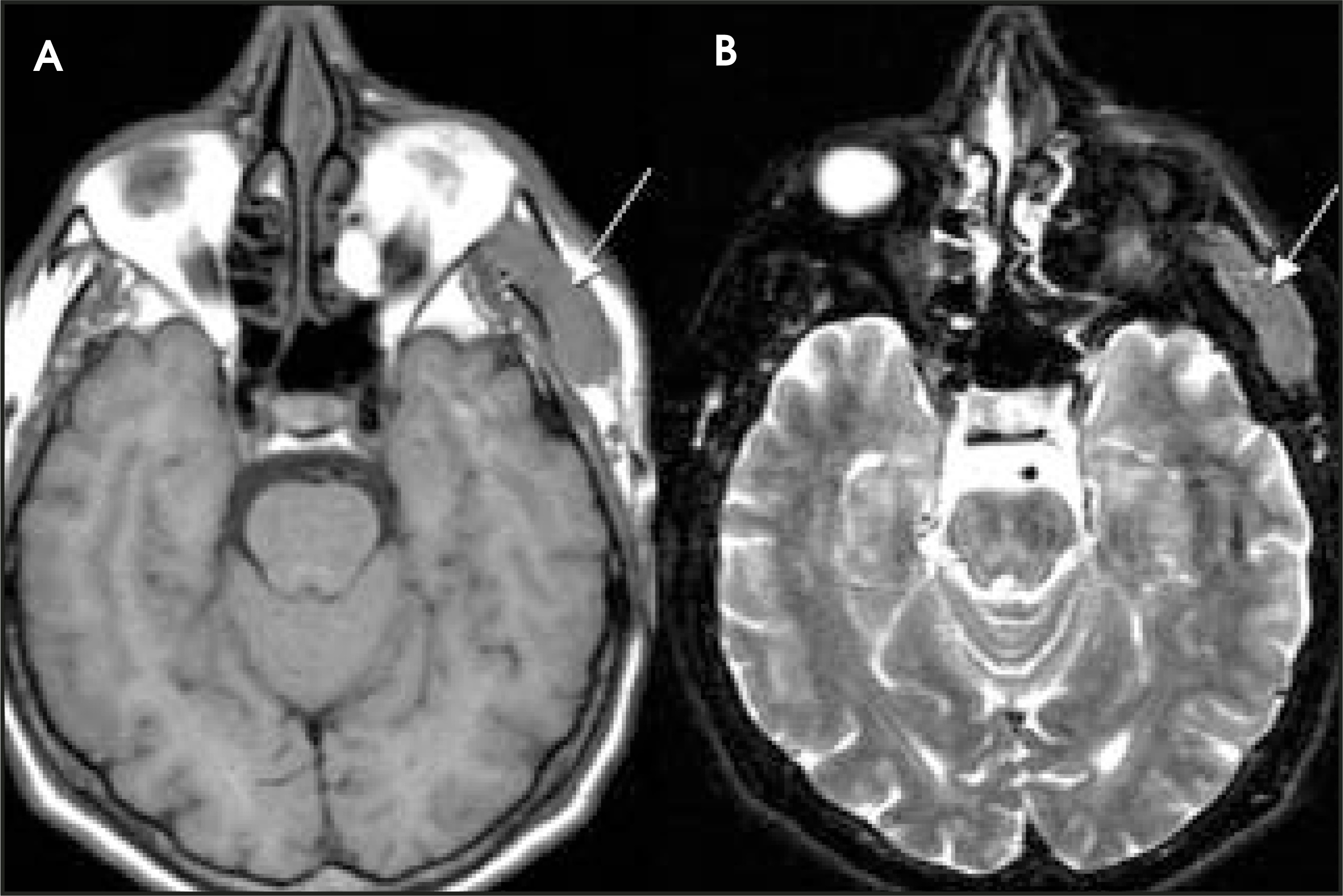
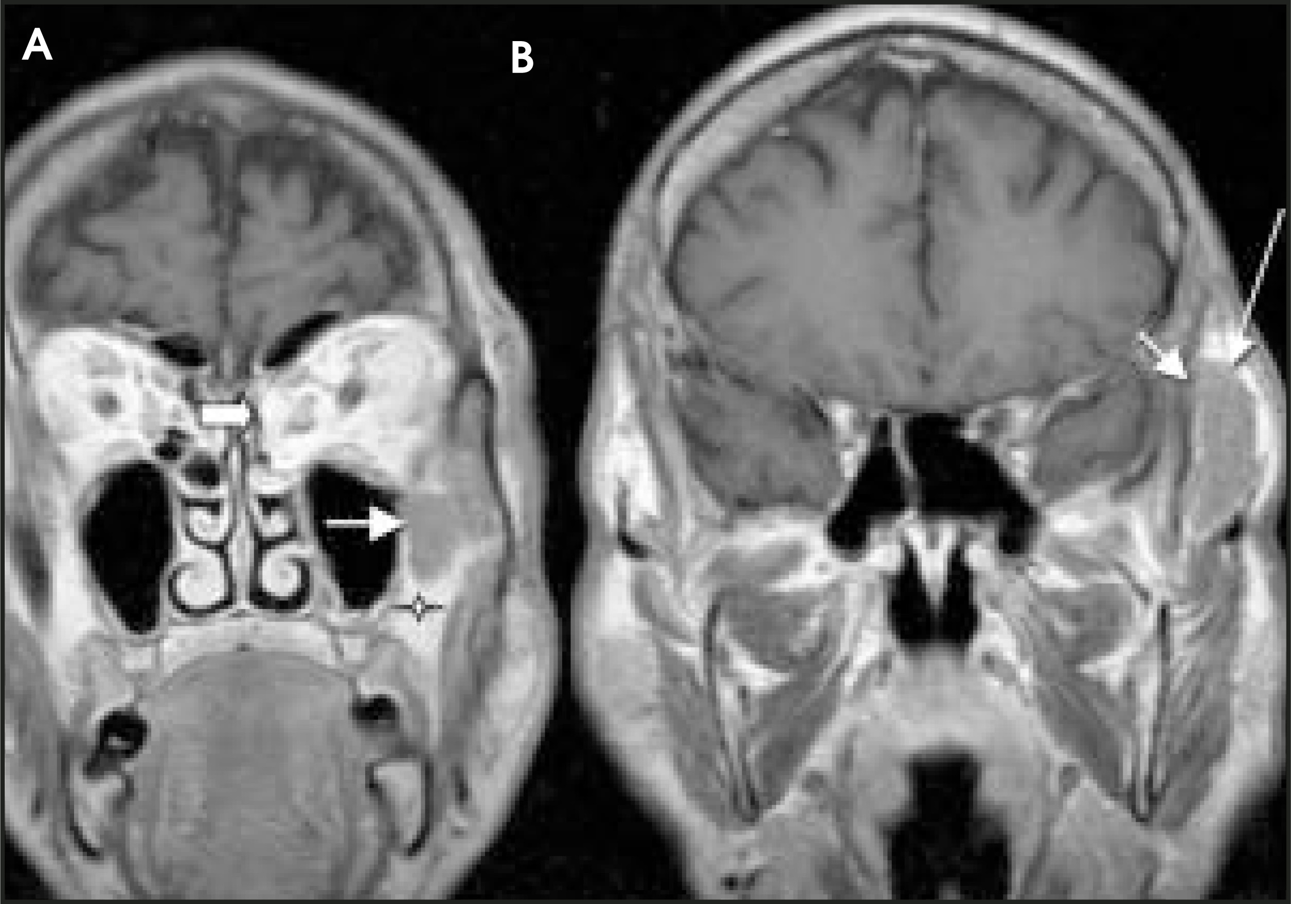
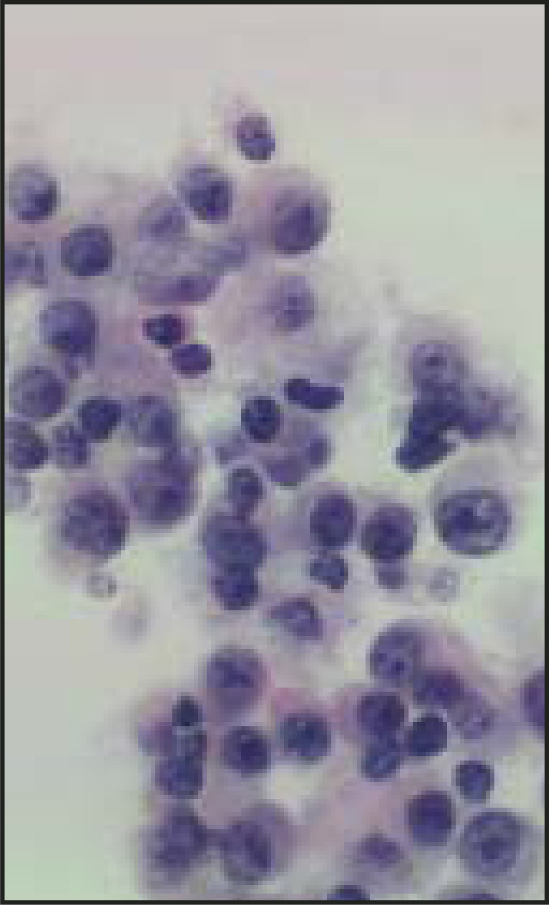
A fine-needle aspiration (FNA) of the left temporalis lesion was performed (Figure 1).
DISCUSSION
Extramedullary plasmacytoma (EMP) is a plasma-cell neoplasm in a class of tumors that includes multiple myeloma (MM) and solitary plasmacytoma of bone. Although EMPs comprise <1% of head and neck cancers, 85% of EMPs are found in the head and neck. Most commonly, they are found in the nasal cavity, nasopharynx, or paranasal sinuses but are also frequently seen in the larynx, tonsillar fossa, and at the base of the tongue. Symptoms usually include epistaxis, nasal obstruction, nasal discharge, headache, or local pain.1 Men are 3 times more likely than women to have EMP. Less than 30% of EMPs progress to MM, which is more often associated with morbidity and mortality.2 Histologically, plasma-cell neoplasms are composed of monotonous sheets of plasma cells that can appear very similar to each other. Therefore, when evaluating a plasma-cell neoplasm, it is critical to rule out multiple myeloma, which is done with a normal radiographic skeletal survey, negative serum and urine immunoelectrophoresis, and negative bone marrow biopsy (<5% plasma cells). Determination of clonality of the lesion is felt to be important, as polyclonal EMP tends not to progress, whereas monoclonal EMP can be followed immunologically.3
Histopathologically, high nuclear-to-cytoplasmic ratios, prominent nucleoli, more reticular (less clumped, “clock-face”–appearing chromatin), and the presence of cytoplasmic inclusions are signs of cytologic irregularity indicative of malignancy rather than plasma-cell reactivity.4 Diagnosis can be made by FNA or, proving insufficient for diagnosis, by excisional biopsy. This can generally be performed under local anesthesia.5
Treatment typically involves radiotherapy, as plasma-cell neoplasms are quite radiosensitive. In 1988, Abemayor et al6 reported a low rate of recurrence after the use of at least 4000 cGy to treat EMP. A small monoclonal serum level may be detectable but should disappear after local treatment but, if persistent, is worrisome for dissemination. In 26 patients with 31 EMPs, locoregional tumor control, with radiation alone, was limited by poor performance status, treatment fields (from previous radiation therapy), or concurrent illness in patients with bulky disease.7 Analysis of the dose-effect relationship of radiation to these tumors indicates that 40 to 50 Gy is likely adequate for macroscopic disease. Between 7.5% and 15% of patients have regional lymph node involvement at presentation. For node-negative necks, radiation therapy (XRT) is elective because of high locoregional control with treatment of primary tumor alone, and node-positive necks are appropriately treated with XRT following neck dissection or larger doses of XRT alone.7,8 A literature search pooling 609 patients with upper aerodigestive tract EMPs found that surgery alone resulted in longer median survival time than did XRT alone (156 versus 144 months).4 This may be due to the self-selection for surgery of less locally aggressive tumors (good resectability) and, therefore, better survival characteristics. Still, only 6.6% of those with surgical intervention alone went on to develop MM, compared with 17.5% and 15.5% with XRT alone or surgery plus XRT, respectively.
CONCLUSION
In our patient, given the proximity to the frontal branch of the seventh cranial nerve and patient preference, radiation therapy was initiated. He will require lifelong monitoring for local or systemic recurrence, or development of multiple myeloma, although overall prognosis is good (>70% 10-year survival).5
ACKNOWLEDGMENT
The authors appreciate the contributions of Dr. Robert Pistey in the Department of Pathology, Boston University School of Medicine, Boston, MA, in the preparation of this manuscript.
References
- Castro E, Lewis J, Strong E. Plasmacytoma of paranasal sinuses and nasal cavity. Arch Otolaryngol. 1973;97:326-329.
- Nofsfinger Y, Mirza N, Rowan P. Head and neck manifestations of plasma cell neoplasms. Laryngoscope. 1997;107:741-746.
- Hotz M, Schwaab G, Bosq J, Munck J. Extramedullary solitary plasmacytomas of the head and neck: A clinicopathological study. Ann Otol Rhinol Laryngol. 1999;108:495-500.
- Alexiou C, Kau R, Dietzfelbinger H. Extramedullary plasmacytoma: Tumor occurrence and therapeutic concepts. Cancer. 1999;85:2305-2314.
- Miller F, Lavertu P, Wanamaker J. Plasmacytomas of the head and neck. Otolaryngol Head Neck Surg. 1998;119:614-618.
- Abemayor E, Canalis R, Greenberg P. Plasma cell tumors of the head and neck. J Otolaryngol. 1988;17:376-381.
- Strojan P, Soba E, Lamovec J, Munda A. Extramedullary plasmacytoma: Clinical and histopathologic study. Int J Radiat Oncol Biol Phys. 2002;53:692-701.
- Bush S, Goffinet D, Bagshaw M. Extramedullary plasmacytoma of the head and neck. Radiology. 1981;140:801-805.
Citation
. RCOM RADIOLOGICAL CASE OF THE MONTH. Applied Radiology. 2005;34(6):40-43. doi:10.37549/AR1347.