RCOM RADIOLOGICAL CASE OF THE MONTH
Applied Radiology — Vol. 34 , Issue 6 , pp. 44 -46
DOI: 10.37549/AR1349
Published: June 1, 2005
Categories
CASE SUMMARY
A 35-year-old woman with no significant medical history presented to her primary-care doctor after experiencing an episode of severe dizziness and headache, which she claimed never happened to her before. Upon physical examination, the doctor could not feel the left carotid pulse. The right carotid pulse was normal.
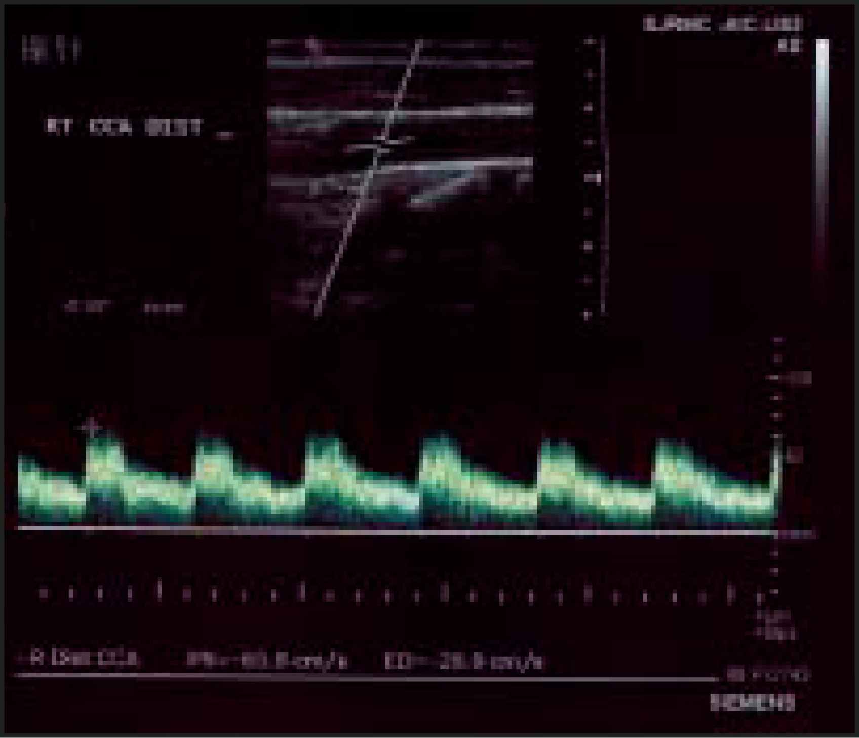
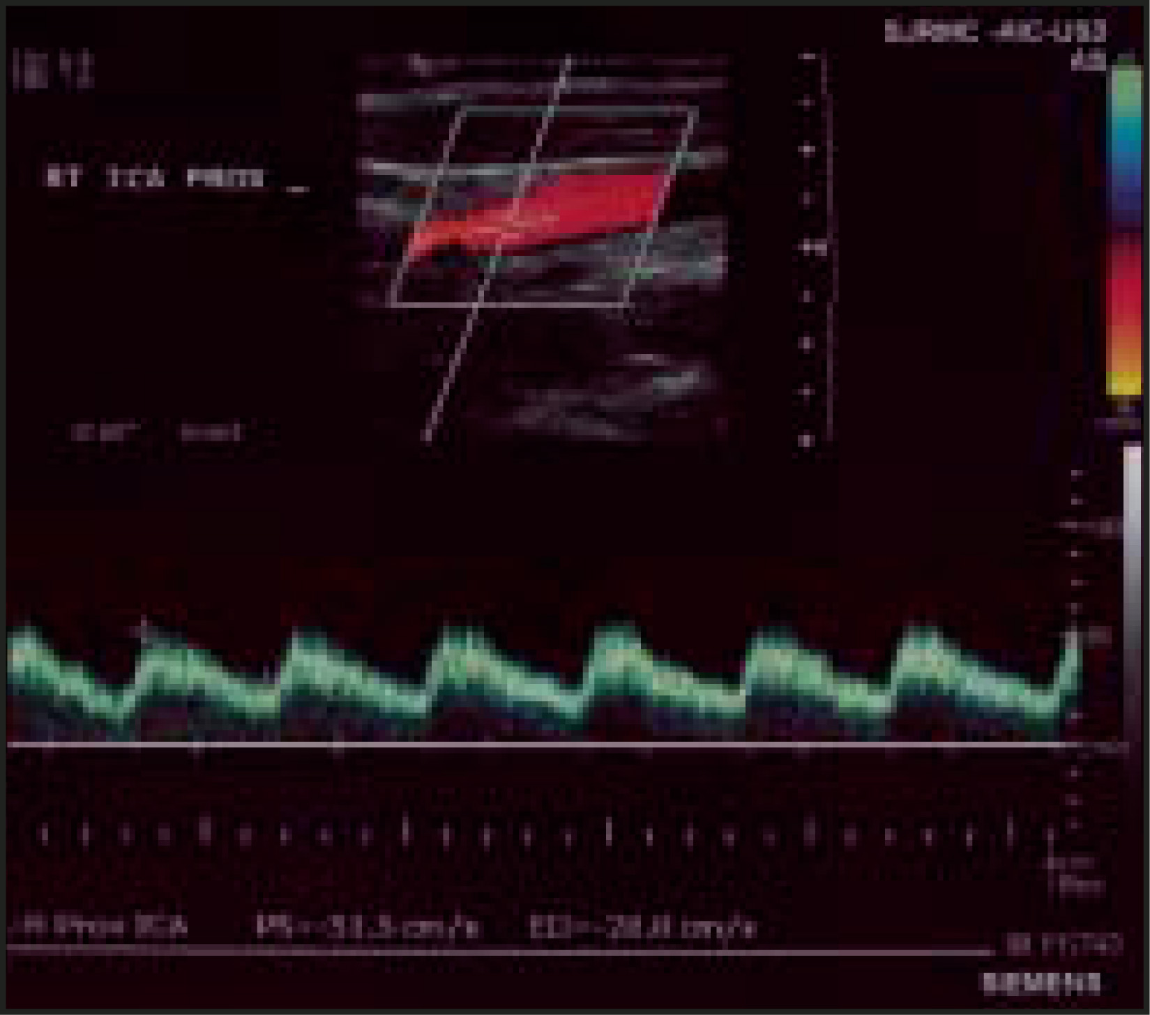
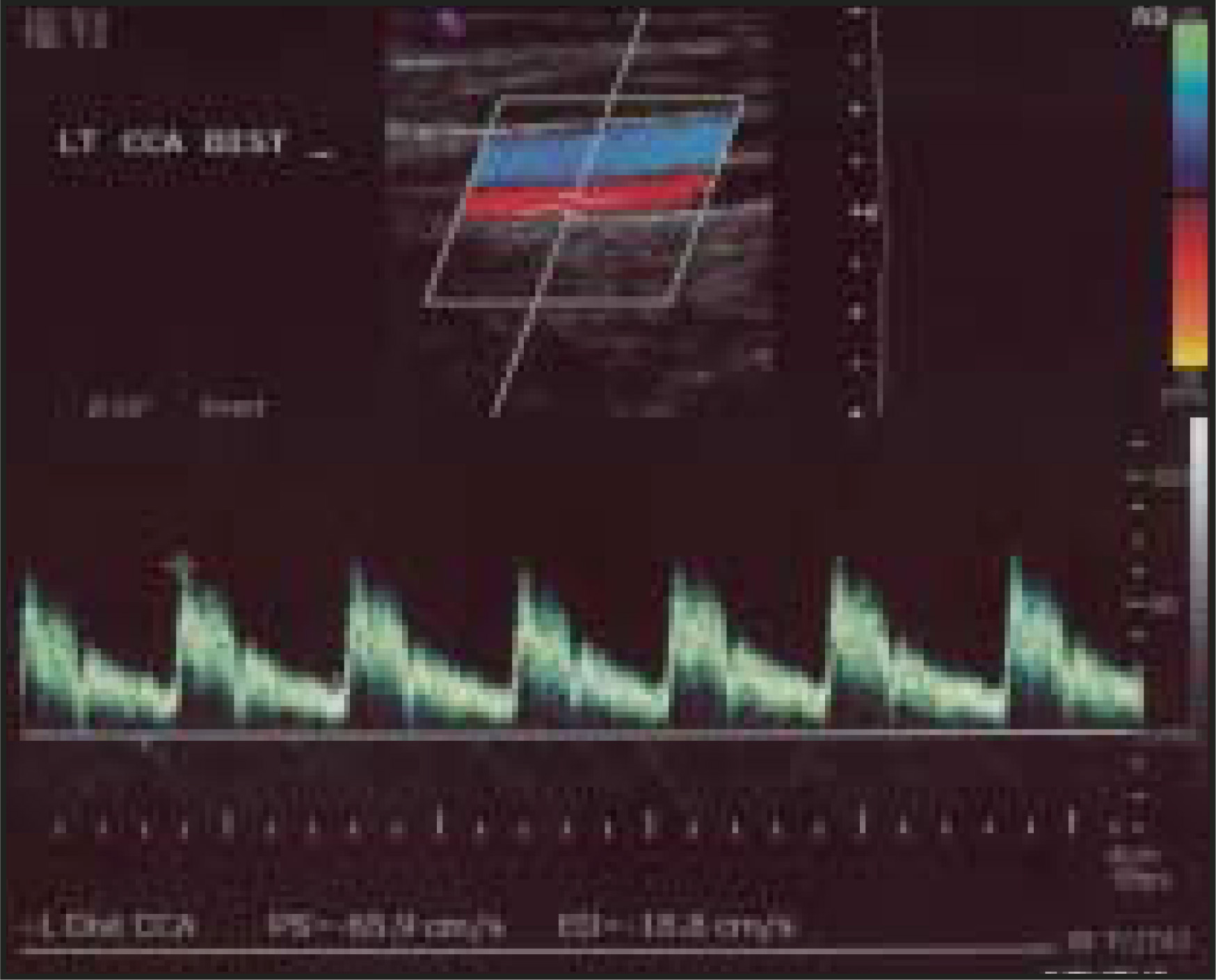
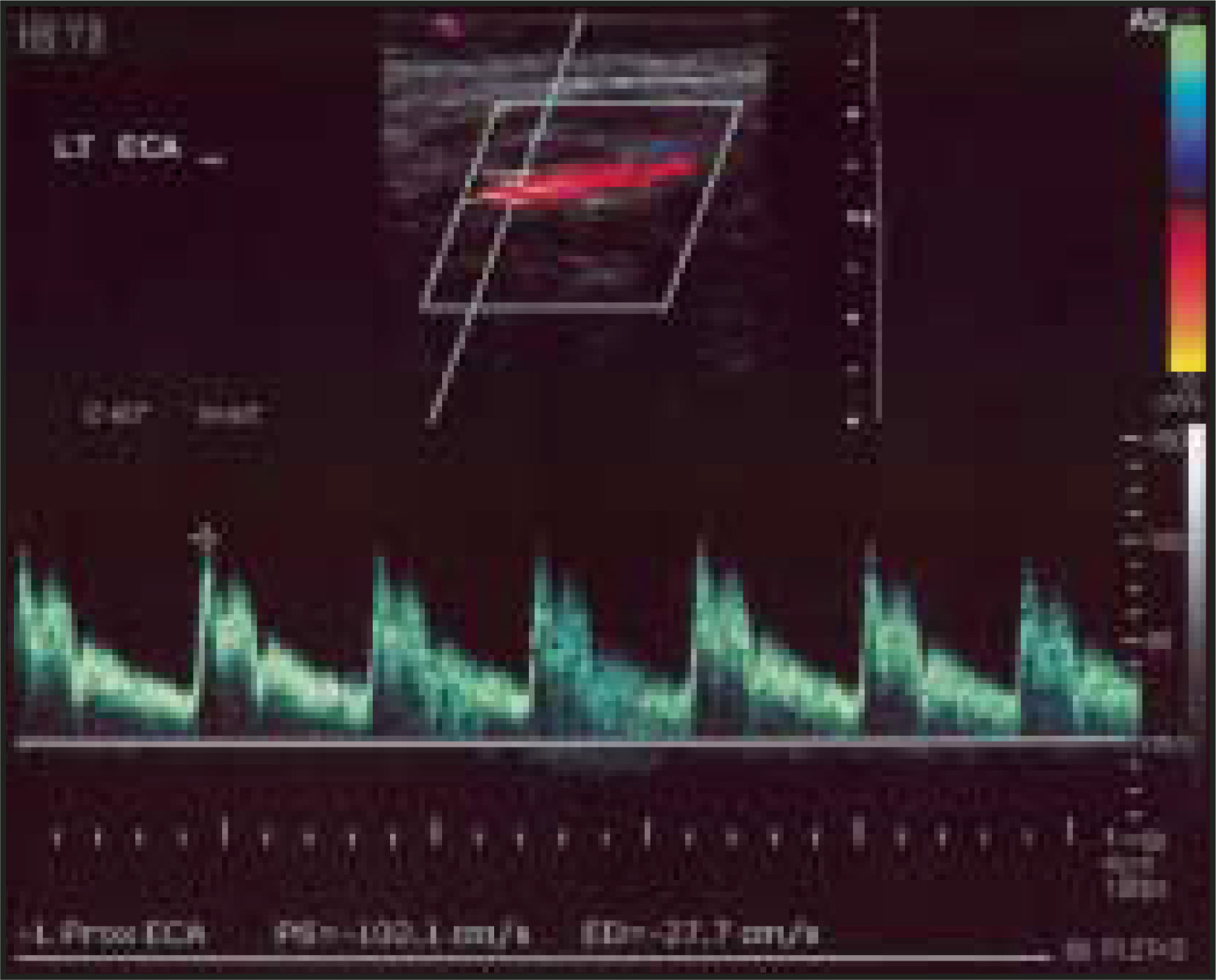
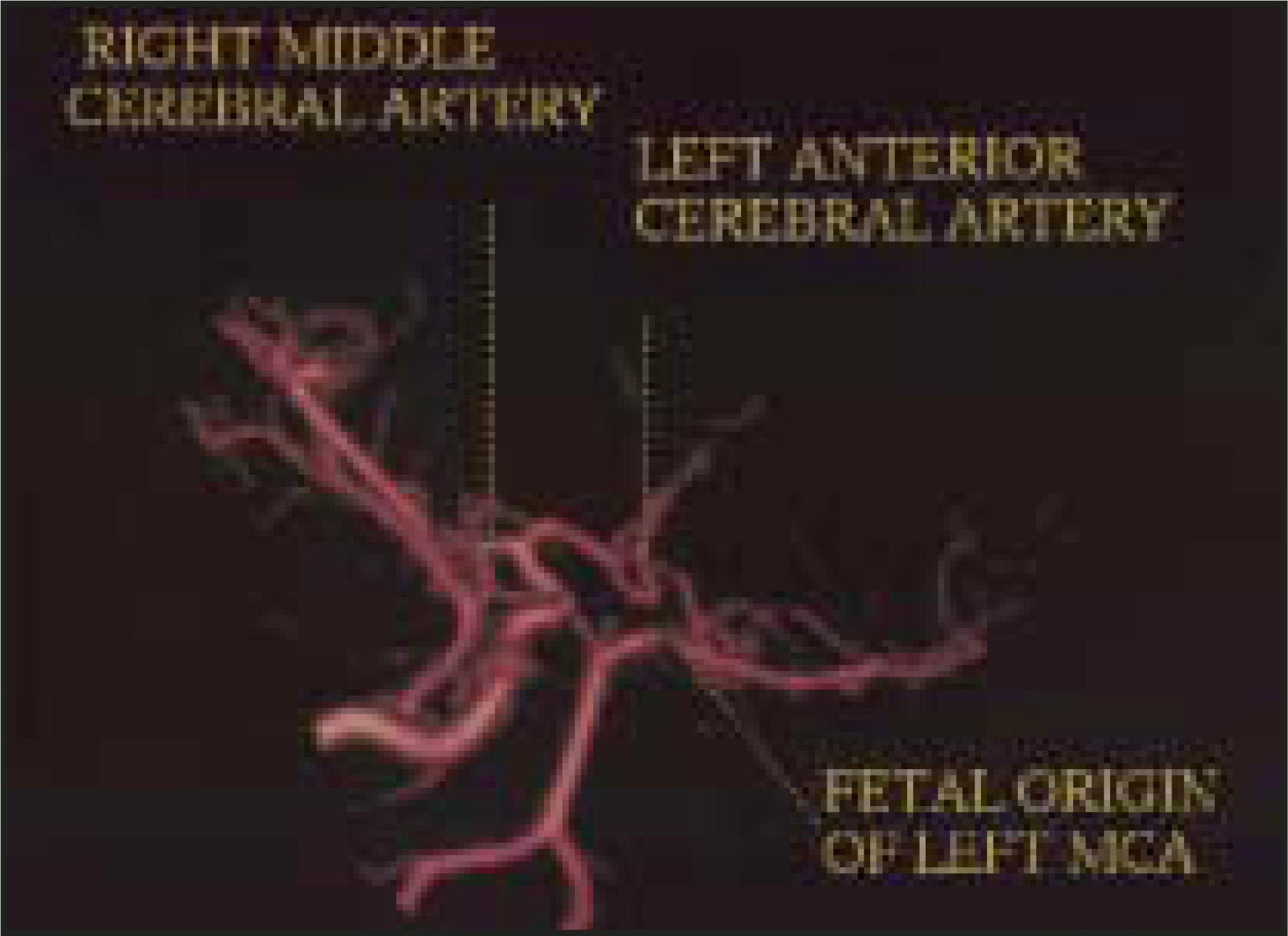
A subsequent carotid ultrasound/Doppler study showed normal right common (Figure 1), internal (Figure 2), and external carotid arteries. The left common (Figure 3) and external (Figure 4) carotid arteries were markedly small, but with expected normal Doppler waveforms. The left internal carotid artery (ICA) was not clearly visualized, or defined, by either gray-scale ultrasound, color Doppler, or Doppler waveform. A 3-dimensional (3D) magnetic resonance angiography (MRA) was then performed (Figures 5 and 6), which showed an absence of the left internal carotid artery and aberrant intracranial arteries.

DIAGNOSIS
Congenital absence of left internal carotid artery associated with aberrant intracranial arterial circulation
IMAGING FINDINGS
The carotid ultrasound/Doppler study showed normal right common carotid artery (Figure 1), right internal carotid artery (ICA; Figure 2), and external carotid artery, with normal color flows and Doppler waveforms. The left common carotid artery (Figure 3) and external carotid artery (Figure 4) were diffusely small, but with normal Doppler waveforms. The left internal carotid artery was neither visualized nor detected.
Three-dimensional gradient-echo postgadolinium MRA (Figures 5 and 6) showed complete absence of the left ICA. The left external carotid artery and common carotid artery were diffusely small in caliber (Figure 5). The right ICA and external carotid artery were normal (Figure 6). Bilateral vertebral arteries were unremarkable.
Intracranially, the 3D MRA showed the circle of Willis to be incomplete. The right middle cerebral artery and anterior cerebral artery arose from the right ICA, the left anterior cerebral artery was fed by the contralateral anterior cerebral artery via anterior communicating artery. The left A1 segment was absent.
The left middle cerebral artery had a fetal origin from the left posterior cerebral artery. The right posterior cerebral artery and posterior communicating artery were normal. The left posterior cerebral artery was normal; the left posterior communicating artery was absent.
The above findings are consistent with congenital absence of the left internal carotid artery with aberrant intracranial arterial circulation.
DISCUSSION
Hypoplasia or aplasia of bilateral ICAs is a rare congenital malformation1; only 17 cases had been reported through 1989. Hypoplasia is distinguished from aplasia by the presence of a patent but very reduced vascular lumen, while aplasia is associated with vestiges of nonpatent vessels. Agenesis of bilateral ICAs is even rarer; only 10 cases had been reported before 1986.2 The mechanism of the development of such anomalies is not completely clear. It is believed that secondary regression following a normal initial phase of development or arrest of development at a certain early embryonic stage is likely.
These anomalies are important on cerebral hemodynamic and embryology. In addition, they are associated with other central nervous system vascular or parenchymal abnormalities. Unilateral or bilateral aplasia or agenesis is compatible with life for an indefinite period of time because of the development of extensive collateral flows. However, the newly created vascularity is threatened by rupture with subarachnoid or meningeal hemorrhage and by ischemia.1
The patterns of compensatory collateral flow secondary to agenesis of the ICA are variable.1–6 Ruptured aneurysm of anterior and posterior communicating arteries in patients with agenesis of the ICA has been reported.1,7
An association between unilateral agenesis of the ICA and congenital hypopituitarism has been reported in several papers.3-5,8,9 Although the exact mechanism behind it is unproven, assumptions are that the development of the ICA and the pituitary gland occur at approximately the 4th embryogenic week, they, therefore, tend to be related. The hypopituitarism related to agenesis of aplasia of ICA usually occurs at birth.3,5,8,9 However, late onset hypopituitarism has also been reported in a female patient who was first diagnosed in her 20s.4
Other associations include Horner’s syndrome from absence of ipsilateral ICA,10 ischemic optic neuropathy,11 and spasmodic torticollis secondary to a dilated vertebral artery related to agenesis of the ICA.12
Conventional angiography, CT angiography, MRI, MRA, and single-photon–emission computed tomography (SPECT) can provide excellent depiction of extra- and intracranial vascular abnormalities.13-15 In most of the reported cases of ICA agenesis, no evidence of brain lesions or cerebral perfusion defects were shown with MRA or SPECT,15 despite the markedly altered vascular anatomy.
CONCLUSION
The authors have presented a case of complete agenesis of the left ICA with variation of intracranial arterial circulation and no other parenchymal neuroanatomic or functional abnormalities. Agenesis of the ICA is a rare but important entity due to the associated hemodynamic changes and other related congenital and developmental disorders. Recognizing this condition is important so that further evaluation, prevention, and treatment of the associated vascular and neurologic abnormalities can be conducted promptly.
References
- el Khamlichi A, Amrani F, el Azzusi M. Bilateral hypoplasia of the internal carotid arteries associated with aneurysm of the right posterior communicating artery. Apropos of a case. Neurochirurgie. 1989;35(1):23-30.
- Sunami N, Kunishio K, Yamamoto Y. Bilateral agenesis of internal carotid arteries. No Shinkei Geka. 1986;14:809-814.
- Inamo Y, Harada K. Agenesis of the internal carotid artery and congenital pituitary hypoplasia: Proposal of a cause of congenital hypopituitarism. Eur J Pediatr. 2003;162:610-612.
- Mellado J, Merino X, Ramos A. Agenesis of the internal carotid artery with a trans-sellar anastomosis: CT and MRI findings in late-onset congenital hypopituitarism. Neuroradiology. 2001;43:237-241.
- Kjellin I, Kaiserman K, Curran J, Geffner M. Aplasia of right internal carotid artery and hypopituitarism. Pediatr Radiol. 1999;29:586-588.
- Kidooka M, Okada T, Handa J. Agenesis of the internal carotid artery-report of a case combined with arachnoid cyst in a child. No To Shinkei. 1992;44:371-375.
- Nakai H, Kawata Y, Aizawa S. Unilateral agenesis of the internal carotid artery in a patient with ruptured aneurysm of the anterior communicating artery; a case report. No Shinkei Geka. 1992;20:893-898.
- Shulman D, Martinez C. Association of ectopic posterior pituitary and anterior pituitary hypoplasia with absence of the left internal carotid. J Pediatr Endocrinal Metab. 1996;9:539-542.
- Moon W, Porto L, Lanfermann H. Agenesis of internal carotid artery associated with congenital anterior hypopituitarism. Neuroradiology. 2002;44:138-142.
- Ryan F, Kline L, Gomez C. Congenital Horner’s syndrome resulting from agenesis of the internal carotid artery. Ophthalmology. 2000;107:185-188.
- Horowitz J, Melamud A, Sela L. Internal carotid artery hypoplasia presenting as anterior ischemic optic neuropathy. Am J Ophthalmol. 2001;131:673-674.
- Kikuchi K, Kowada M, Kojima H. Hypoplasia of the internal carotid artery associated with spasmodic torticollis: The possible role of altered vertebrobasilar haemodynamics. Neuroradiology. 1995;37:362-364.
- Worthington C, Olivier A, Melanson D. Internal carotid artery agenesis: Correlation by conventional and digital subtraction angiography, and by computed tomography. Surg Neurol. 1984;22:295-300.
- Martinez-Granero M, Martinez-Bermejo A, Arcas J. Unilateral agenesis of the internal carotid artery in childhood: Description of a case. Rev Neurol. 1997;25:1207-1209.
- Iaccarino V, Tedeschi E, Brunetti A. Congenital agenesis/aplasia of the internal carotid arteries: MRA and SPECT findings. J Comput Assist Tomogr. 1995;19:987-990.
Citation
. RCOM RADIOLOGICAL CASE OF THE MONTH. Applied Radiology. 2005;34(6):44-46. doi:10.37549/AR1349.