RCOM RADIOLOGICAL CASE OF THE MONTH
Applied Radiology — Vol. 34 , Issue 7 , pp. 43 -44
DOI: 10.37549/AR1352
Published: July 1, 2005
Categories
CASE SUMMARY
A 58-year-old white woman was transferred to the emergency department from a nearby facility. The patient complained of acute onset right-upper-quadrant abdominal pain and nausea. She denied fever, chills, and emesis. The patient was taken for exploratory laparotomy due to symptomatic anemia.
IMAGING FINDINGS
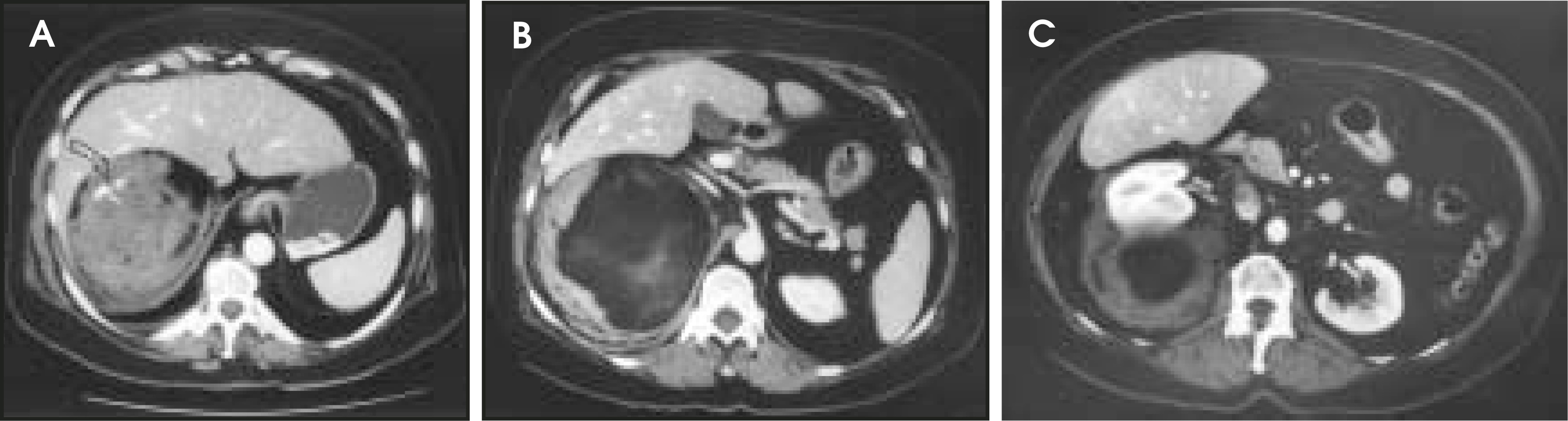
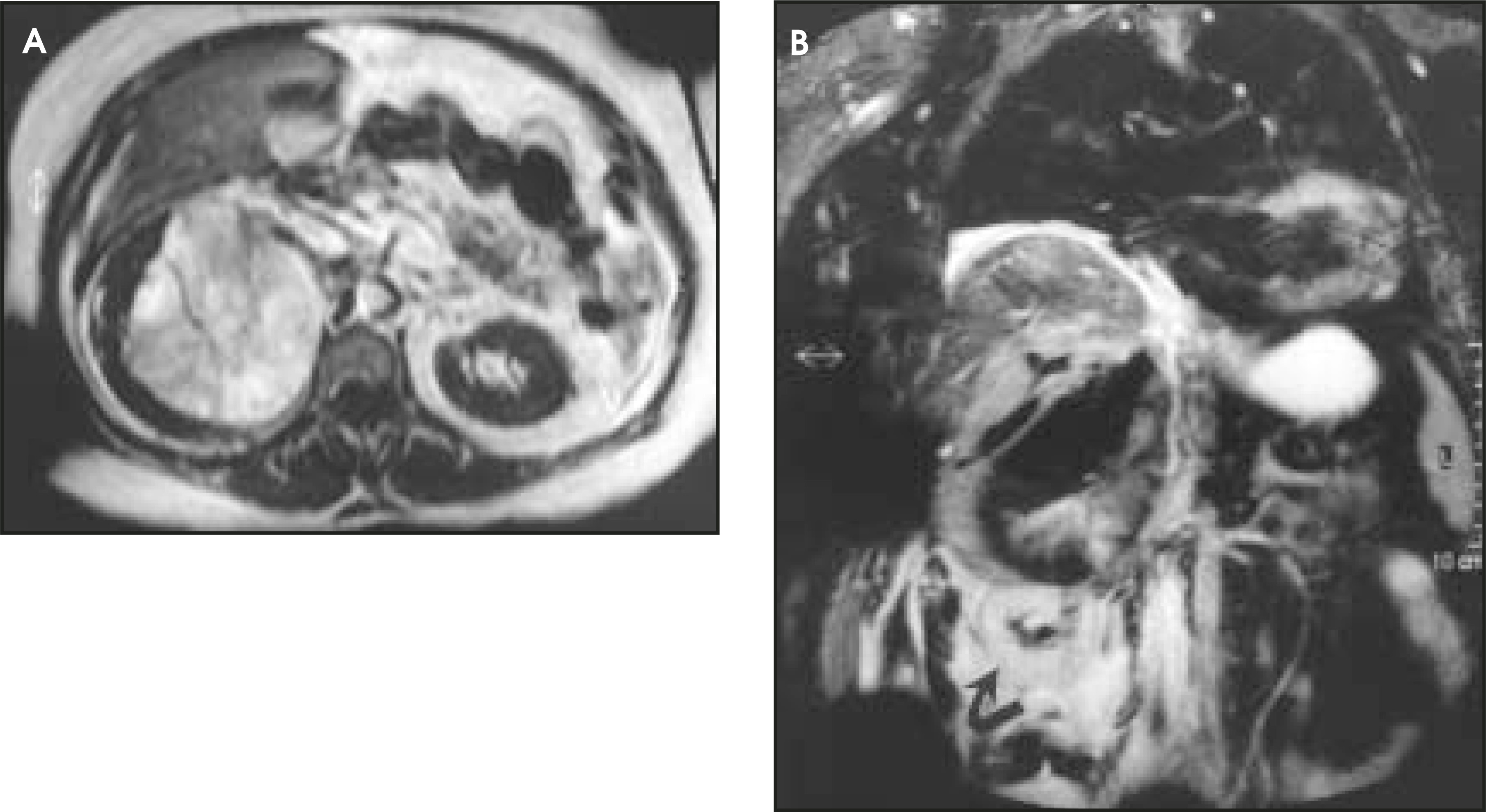
A contrast-enhanced computed tomography (CT) scan performed at an outside facility showed a large mixed-density mass measuring roughly 10 × 20 cm extending from a right suprarenal location inferiorly below the iliac crest (Figure 1). The retroperitoneal mass contained soft-tissue and fatty elements and displaced the right kidney anteriorly and inferiorly and exhibited mass effect upon the right lobe of the liver. Focal high attenuation seen in the superior aspect of the mass is consistent with active extravasation of contrast. Magnetic resonance imaging (MRI) of the abdomen revealed a right suprarenal 10-× 18-cm mixed-signal mass containing heme, fat, and soft-tissue/solid components (Figure 2). Significant mass effect upon the right kidney and to a lesser extent on the liver was again noted. The right adrenal gland was not identified.
DIAGNOSIS
Adrenal myelolipoma with rupture into the retroperitoneum
DISCUSSION
At surgery, the mass extended superiorly from the kidney and posterior to the liver to the level of the diaphragm without hepatic involvement. Clot (approximately 3 units) and some solid tumor components were manually debrided. The mass appeared to originate from the adrenal gland. The retroperitoneal mass and the right kidney were removed. The right nephrectomy was performed because inspection of the right kidney revealed some adherent tumor. Histologic examination revealed a 9- × 14-cm hemorrhagic, friable fibrofatty mass composed of mature fat with scattered foci of marrow elements. No normal adrenal gland was identified, but adrenal fragments were attached to the tumor. The kidney was unremarkable.
The incidence of adrenal myelolipoma is reported to be <0.5%.1,2 The majority of myelolipomas are asymptomatic, and almost all are adrenal in origin. Rupture with significant retroperitoneal hemorrhage is perhaps the most serious complication. Most adrenal myelolipomas contain enough bulk fat to permit their differentiation by CT or MRI from other adrenal masses such as adenomas, metastases, and pheochromocytomas.1,2 When a fat-containing retroperitoneal mass is seen in the perirenal area associated with retroperitoneal hemorrhage, renal angiomyolipoma is the first consideration. If the mass is centered in the adrenal area, the possibility of myelolipoma should be considered. Another consideration is retroperitoneal liposarcoma. The key to the differential diagnosis is proving intrarenal origin (angiomyolipoma), adrenal origin (myelolipoma), or retroperitoneal or renal capsular origin (liposarcoma).3
References
- Otal P, Escourrou G, Mazerolles C. Imaging features of uncommon adrenal masses with histopathologic correlation. RadioGraphics. 1999;19:569-581.
- Adusumilli S, Ramchandani P. Adrenal myelolipoma. eMedicine J. 2005.
- Khan A, Boylan C, Macdonald S. Kidney angiomyolipoma. eMedicine J. 2005.
Citation
. RCOM RADIOLOGICAL CASE OF THE MONTH. Applied Radiology. 2005;34(7):43-44. doi:10.37549/AR1352.