RCOM RADIOLOGICAL CASE OF THE MONTH
Applied Radiology — Vol. 34 , Issue 7 , pp. 40 -42
DOI: 10.37549/AR1353
Published: July 1, 2005
Categories
CASE SUMMARY
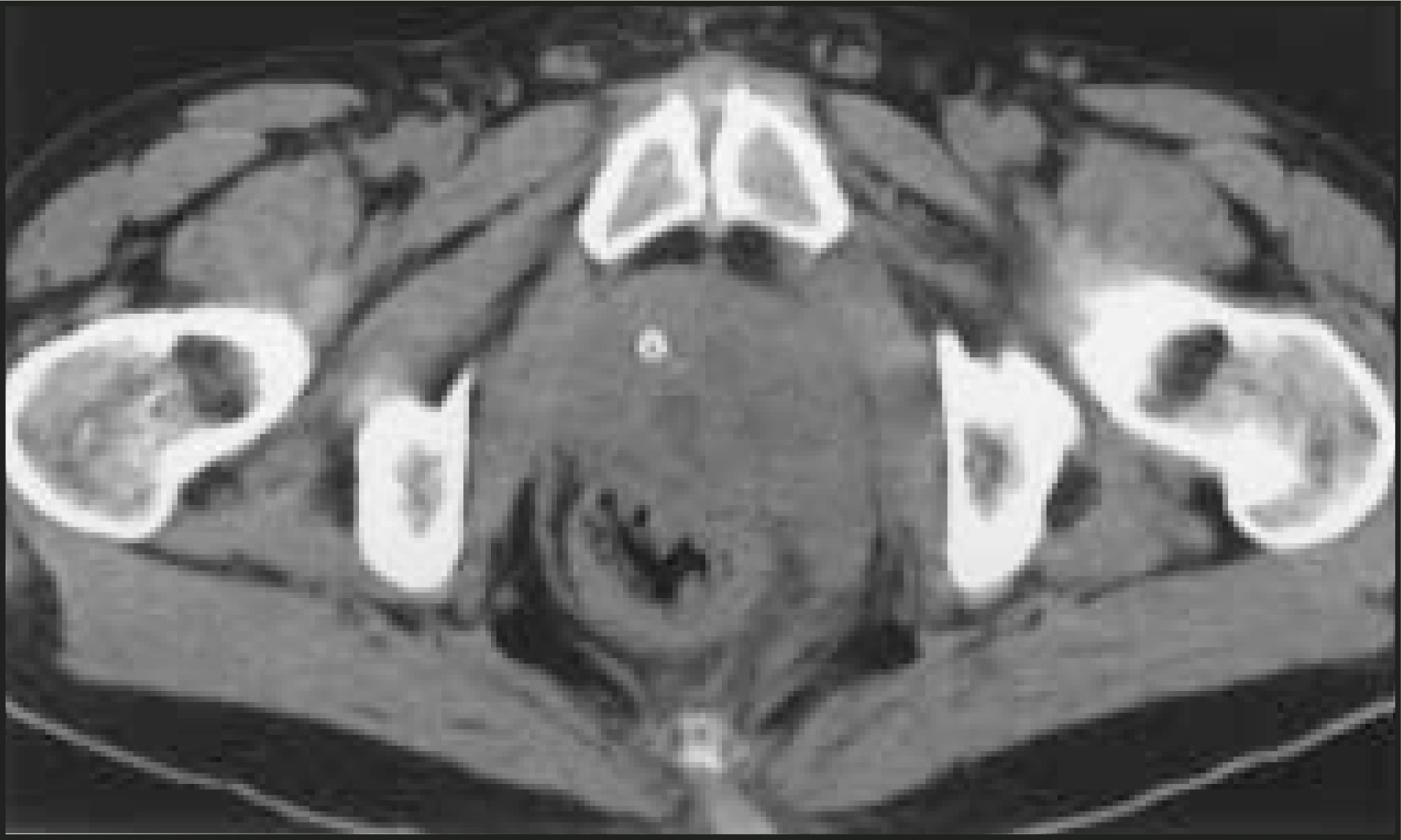
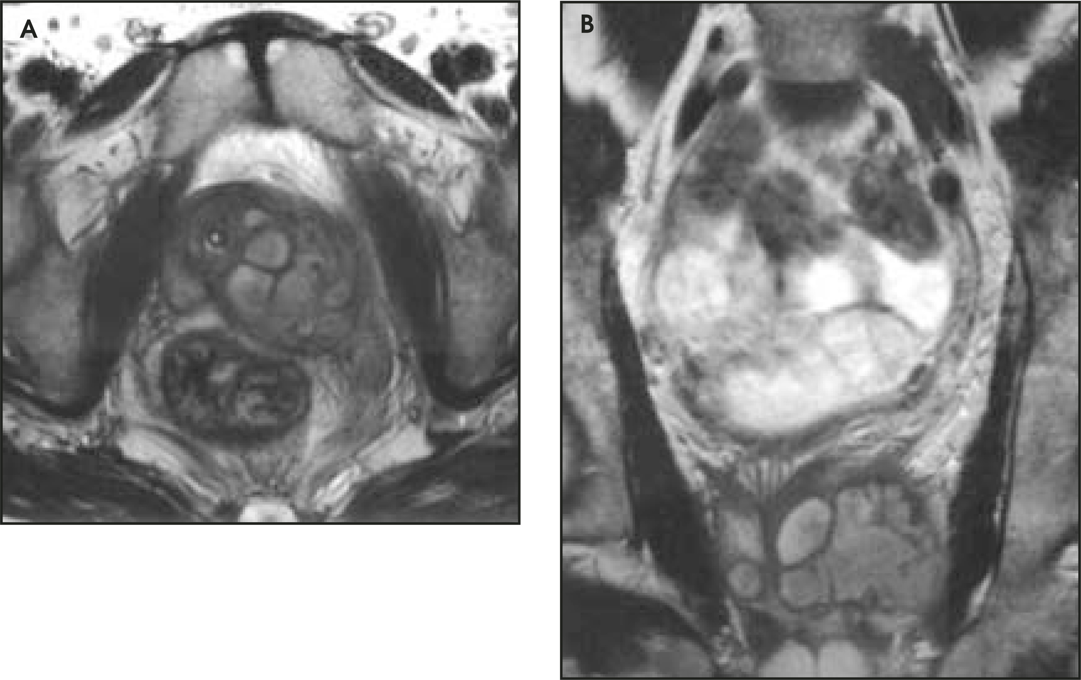
A 53-year-old African American man presented to the emergency department complaining of left-sided pelvic pain, which radiated to the left gluteal region and scrotum. He described increased difficulty with urination, and pain with defecation. His medical history included type 2 diabetes mellitus and chronic renal insufficiency. The prostate gland was exquisitely tender to palpation on digital rectal examination. Several days prior to presentation, he had been started on a course of ciprofloxacin for presumptive prostatitis. At presentation, his blood glucose was poorly controlled, elevated to 232 mg/dL. White blood cell (WBC) count was within normal limits at 8.4 × 10⁹/L; however, the urinalysis was abnormal, with +1 protein, +3 glucose, +1 leukocyte esterase, 9 WBC/high-powered field, and + bacteria. Urine cultures would later grow Escherichia coli and Klebsiella pneumoniae in >100,000 colony-forming units. A noncontrast computed tomography (CT) scan was performed (Figure 1). No intravenous contrast was administered for the CT scan because of the patient’s history of renal insufficiency. A contrast-enhanced magnetic resonance imaging (MRI) study was subsequently performed (Figure 2) to further characterize the abnormalities seen on the CT scan.
IMAGING FINDINGS
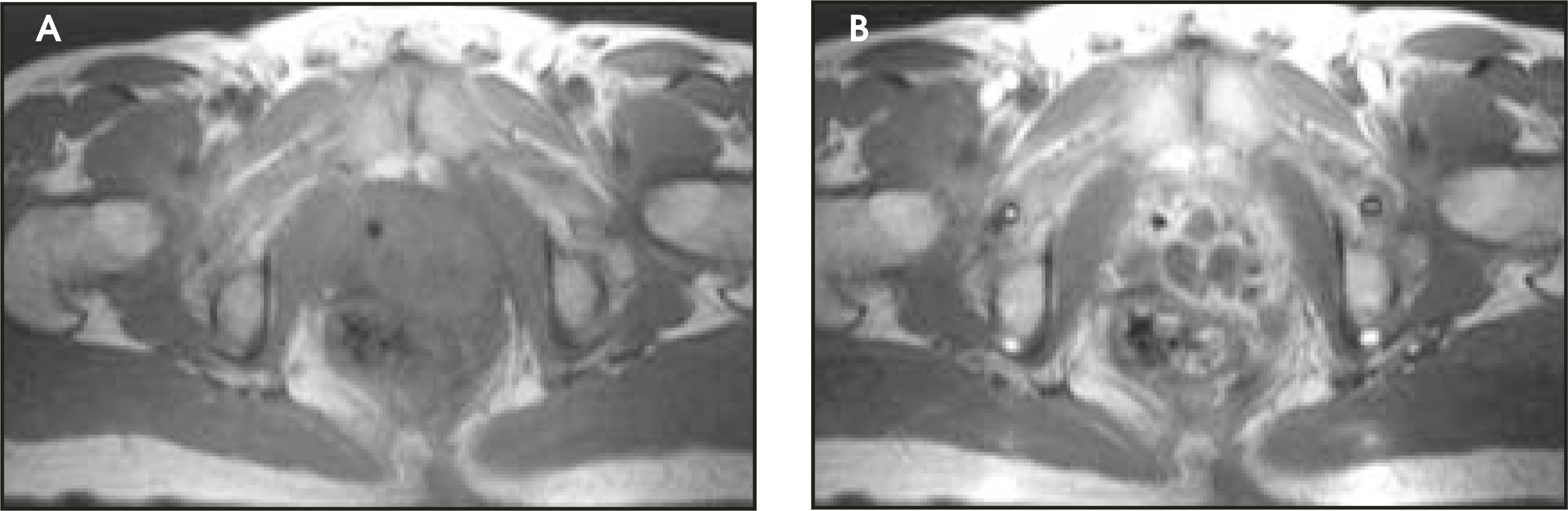
The noncontrast CT scan of the abdomen and pelvis showed abnormal enlargement of the left side of the prostate gland to approximately 6 × 4 cm, with focal areas of relative low density. Additionally, there was an abnormal soft-tissue density extending posteriorly toward the rectum, with diffuse inflammatory stranding surrounding the area (Figure 1); however, there was no gas within the area, and no definite fluid collection was identified. To further characterize the pathology suggested by noncontrast CT, and to more accurately delineate the anatomy of the involved area, a gadolinium- enhanced MRI of the pelvis was performed. The MRI showed deviation of the existing Foley catheter to the right by a multiloculated cystic lesion in the left lobe of the prostate gland. A single cystic lesion measuring 1 cm in diameter was present in the right lobe. These lesions had slightly increased signal intensity in T2-weighted sequences (Figure 2), were relatively isointense in T1-weighted sequences (Figure 3A), and had bright peripheral enhancement upon administration of gadolinium, consistent with abscess (Figure 3B). Of note, the seminal vesicles were identified and appeared to be normal.
DIAGNOSIS
Prostatic abscess
OPERATIVE FINDINGS AND MANAGEMENT
In the operating room, a suprapubic catheter was placed, and the abscess was drained via transurethral resection of prostate with unroofing, during which multiple pockets of pus were entered in serial resections. The resected prostate fragments were sent for pathologic examination, which revealed patchy acute and chronic inflammation, with focal cavitary purulent debris, consistent with prostatic abscess. The patient had an uneventful postoperative course, and was discharged home on postoperative day 3. On routine outpatient follow-up, the patient was voiding without difficulty, and had no prostate abnormality on digital rectal examination.
DISCUSSION
Prostatic abscess is an uncommon condition, often difficult to clinically discern from acute bacterial prostatitis.1 Historically, the common infecting organisms were Neisseria gonorrhoeae, Staphylococcus aureus, and Mycobacterium tuberculosis; however, more recently, gram-negative bacteria, such as Escherichia coli, are the causative species.1,2 Patients with diabetes mellitus (DM) are predisposed to prostatic abscess.1 As in the case described here, a common comorbidity of DM is renal insufficiency. MRI has several advantages for these patients. In cases in which patients have significant risk factors for contrast-induced nephropathy (albumin <3.5 g/L, diabetes mellitus, serum sodium <135 mmol/L, and serum creatinine >1.5 mg/dL), MRI has the ability to provide contrast enhancement with gadolinium without the potentially nephrotoxic effects of iodinated intravenous CT contrast.3 Additionally, the patient is spared the dose of ionizing radiation provided by CT. Like CT, MRI also saves the patient from the additional discomfort and potential bacteremic consequences, sometimes leading to septic shock, which may be encountered from a transrectal ultrasound examination.
In the case discussed by Papanicolaou et al,4 the prostatic abscess was described as a “well defined high-signal-intensity abnormality” in the T2-weighted images. In this case, the abscess was only slightly hyperintense relative to the adjacent prostatic tissue in T2-weighted sequences, and was not well defined in the precontrast T1-weighted images. This was thought to be secondary to a difference in protein content in the abscess fluid. Peripheral enhancement was evident following administration of intravenous gadolinium. A clear delineation of the extent of disease and the involved structures provided by a contrast-enhanced study was helpful in determination of surgical management versus transrectal or transperineal drainage.
Our institution uses a protocol for imaging the prostate gland, which is normally indicated for assessing local invasion of prostate cancer, but was also found to be beneficial in this case. The protocol includes multiple pre- and postgadolinium sequences, including high-resolution T2 turbo spin echo, and two-dimensional fast low-angle shot (FLASH) sequences with and without fat saturation, in multiple planes. This protocol takes advantage of smaller fields of view to yield greater anatomic detail.
CONCLUSION
Prostatic abscesses are rare in the modern antibiotic era.1,2 A misdiagnosis can have serious repercussions, including urethrorectal fistula formation from rupture involving surrounding structures, sepsis, and, possibly, death.1,2,5 Traditional radiologic studies in the clinical workup of a prostate abscess have included CT or trans-rectal sonography.2,5,6 Diagnosis of prostatic abscess with MRI has been described previously4 but may be underutilized. With continued improvement in technique, equipment, and experience, this may become the preferred imaging modality for this entity.
References
- Jacobsen J, Kvist E. Prostatic abscess: A review of literature and a presentation of 5 cases. Scand J Urol Nephrol. 1993;27:281-284.
- Ludwig M, Schroeder-Printzen I, Schiefer H, Weidner W. Diagnosis and therapeutic management of 18 patients with prostatic abscess. Urology. 1999;53:340-345.
- Rich M, Crecelius C. Incidence, risk factors, and clinical course of acute renal insufficiency after cardiac catheterization in patients 70 years of age or older. A prospective study. Arch Intern Med. 1990;150:1237-1242.
- Papanicolaou N, Pfister R, Stafford S, Parkhurst E. Prostatic abscess: Imaging with transrectal sonography and MR. AJR Am J Roentgenol. 1987;149:981-982.
- Washecka R, Rumancik W. Prostatic abscess evaluated by serial computed tomography. Urol Radiol. 1985;7:54-56.
- Thornhill B, Morehouse H, Coleman P, Hoffman-Tretin J. Prostatic abscess: CT and sonographic findings. AJR Am J Roentgenol. 1987;148:899-900.
Citation
. RCOM RADIOLOGICAL CASE OF THE MONTH. Applied Radiology. 2005;34(7):40-42. doi:10.37549/AR1353.