RCOM RADIOLOGICAL CASE OF THE MONTH
Applied Radiology — Vol. 34 , Issue 8 , pp. 43 -44
DOI: 10.37549/AR1360
Published: August 1, 2005
Categories
CASE SUMMARY
An 8-year-old previously healthy boy presented to the emergency department with a 1-day history of 2 episodes of grossly bloody stool. The patient had been involved in a go-cart accident 2 days prior to presentation but denied any pain or rectal bleeding at that time. On clinical examination, the patient was afebrile and complained of minimal diffuse abdominal pain on palpation. A rectal examination revealed gross blood. His laboratory results were normal, with the exception of a low hematocrit of 29%.
IMAGING FINDINGS
Initial radiographic evaluation of the bilateral hips, thoracic spine, and cervical spine was performed, and the resulting images were unremarkable.
Multiple postcontrast axial computed tomographic (CT) images of the abdomen and pelvis were obtained to rule out posttraumatic soft-tis-sue injury. A midline serpiginous, cystic, wall-enhancing structure containing a punctate gaseous collection was seen within the pelvis. There were no associated inflammatory changes, and the lumen did not opacify with contrast (Figure 1). The remainder of the study was unremarkable and without evidence of free air or fluid.
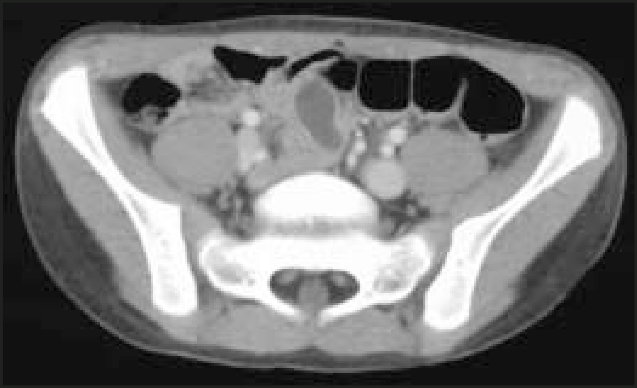
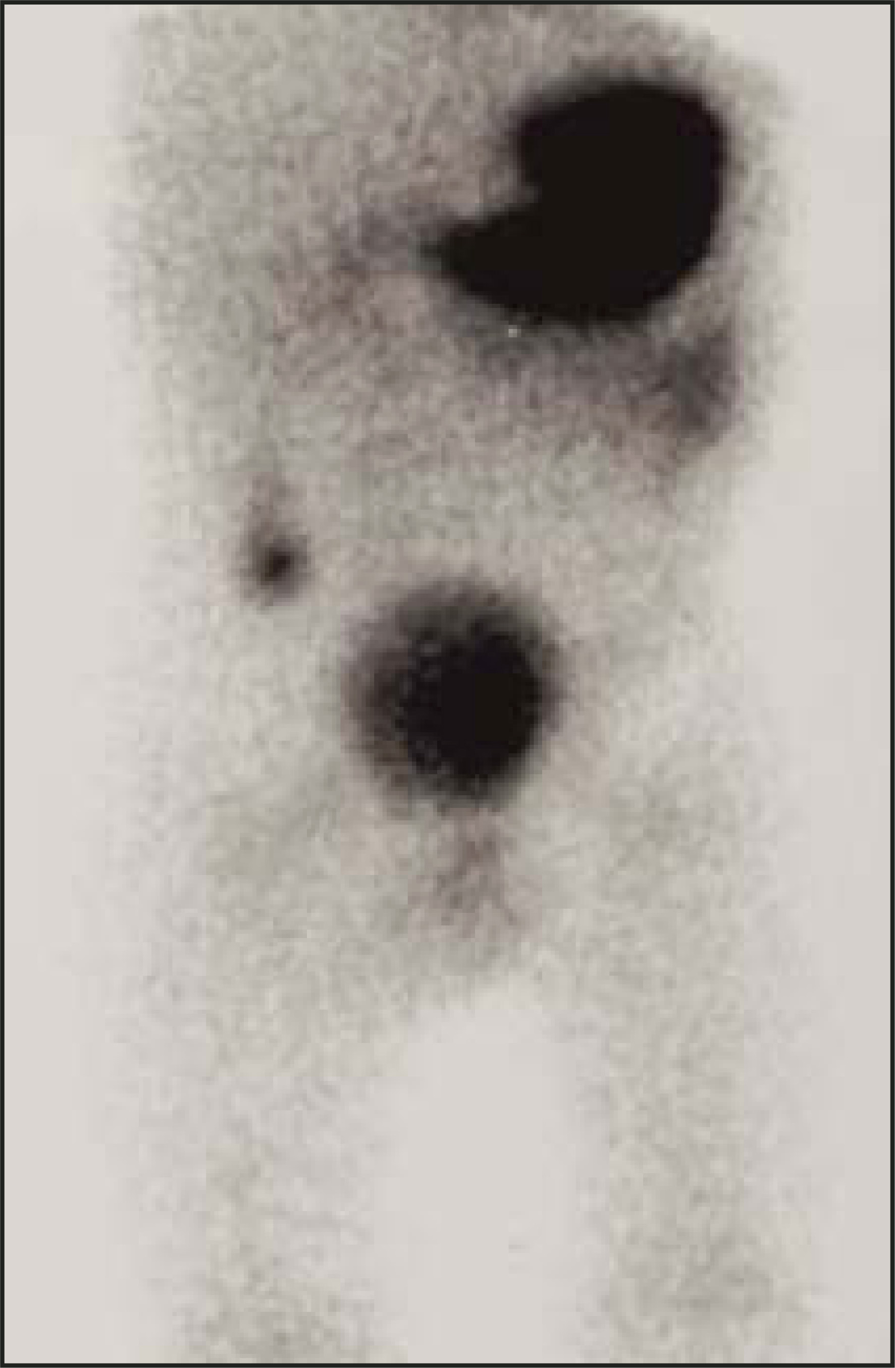
This CT finding is nonspecific, and the differential diagnosis included enteric duplication cyst, ileal diverticulum, nonopacified small bowel, ruptured viscus, and noninflamed Meckel’s diverticulum. The consulting radiologist believed the mass was most suggestive of a Meckel’s diverticulum. A Meckel’s scan was performed using 5.39 mCi of technetium (Tc)-99m pertechnetate intravenously. Sequential anterior and lateral images of the abdomen were obtained. A prominent and somewhat fusiform focus of radiotracer uptake was noted in the right mid-abdomen (Figure 2), which was consistent with a Meckel’s diverticulum.
DIAGNOSIS
Noninflamed Meckel’s diverticulum
DISCUSSION
The patient was managed surgically due to continued rectal bleeding associated with anemia. A nonperforated, noninflamed Meckel’s diverticulum was removed by laparotomy. The histopathologic specimen confirmed the diagnosis of a noninflamed Meckel’s diverticulum containing gastric mucosa.
Meckel’s diverticulum is a remnant of the vitelline duct that is usually located 45 to 60 cm proximal to the ileocecal valve on the antimesenteric border of the ileum.1 It is the most common congenital anomaly of the small intestine, occurring in 2% of the population. Meckel’s diverticulum is a common cause of rectal bleeding in children. The usual source of the bleeding is a chronic acid-induced ulcer in the ileum adjacent to the Meckel’s diverticulum that contains gastric, or less commonly, pancreatic mucosa.1 Diagnosis by radiography, CT, and ultrasound are rarely helpful,2 despite the findings in this case. In children, scintigraphy has been found to be the most accurate diagnostic test.
The findings seen with an obstructed, necrotic, inflamed, torsed, infarcted, or intussuscepted Meckel’s diverticulum diagnosed on CT have been described previously.3-7 We present a case of a noninflamed, nonobstructed, intact Meckel’s diverticulum diagnosed by CT, and confirmed with scintigraphy and histopathology as an uncommon presentation of a common condition.
References
- Townsend C, Beauchamp D, Evers M. Sabiston Textbook of Surgery. 2001:907-909.
- Hughes J, Hatrick A, Rankin S. Computed tomography findings in an inflamed meckel diverticulum. Br J Radiol. 1998;71:882-883.
- Prall R, Bannon M, Bharucha A. Meckel’s diverticulum causing intestinal obstruction. Am J Gastroenterol. 2001;96:3426-3427.
- Macari M, Panicek D. CT findings in acute necrotizing Meckel diverticulitis due to obstructing enterolith. J Comput Assist Tomogr.. 1995;19:808-810.
- Nigogosyan M, Dolinskas C. Demonstration of inflamed Meckel diverticulum. J Comput Assist Tomogr.. 1990;14:140-142.
- Farris S, Fernbach S. Axial torsion of Meckel’s diverticulum presenting as a pelvic mass. Pediatr Radiol. 2001;31:886-888.
- Russ P, Friefeld G, Nauck C, Wilmouth R. Infarcted Meckel diverticulum detected by CT. AJR Am J Roentgenol. 1988;150:299-300.
Citation
. RCOM RADIOLOGICAL CASE OF THE MONTH. Applied Radiology. 2005;34(8):43-44. doi:10.37549/AR1360.