RCOM RADIOLOGICAL CASE OF THE MONTH
Applied Radiology — Vol. 34 , Issue 8 , pp. 38 -41
DOI: 10.37549/AR1361
Published: August 1, 2005
Categories
CASE SUMMARY
An 80-year-old woman presented to the emergency department approximately 8 hours after a routine barium enema with complaints of shortness of breath and swelling of the neck and face. She denied any abdominal pain, fever, vomiting, or any similar previous episodes. Her medical history was significant only for hypertension and diverticulosis. The patient did appear to be in moderate respiratory distress; however, a clinical examination revealed a soft, nontender, and nondistended abdomen, with positive bowel sounds. She was afebrile at 36.7°C, her blood pressure was 208/84 mm Hg, pulse was 88 bpm, and respirations were 26/min. Laboratory data revealed a mild leukocytosis of 12,600 per mm3, with the remaining chemistry and coagulation panel within normal limits. Plain radiographic films of the chest and abdomen were performed, followed by a computed tomographic (CT) scan of the chest, abdomen, and pelvis.
IMAGING FINDINGS
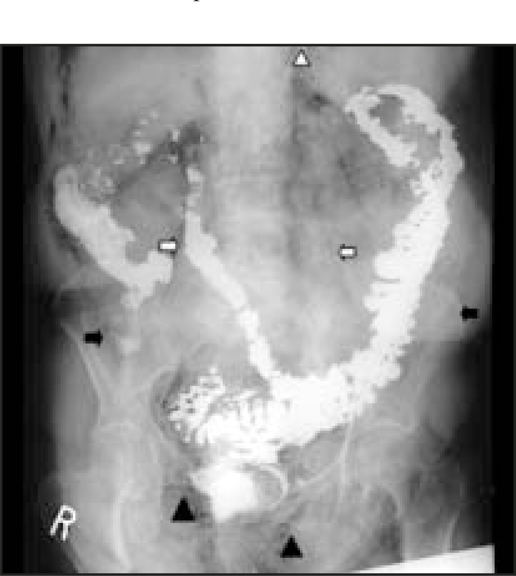
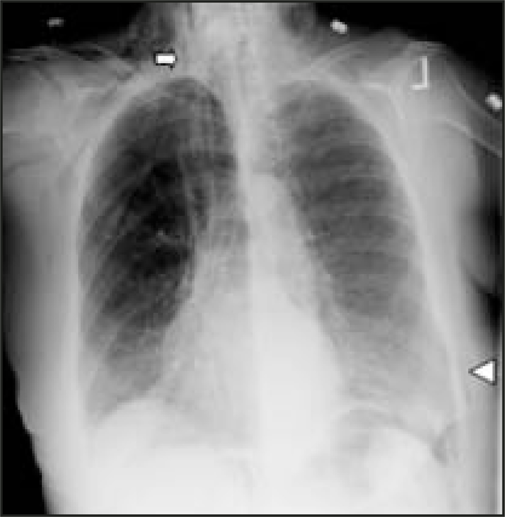
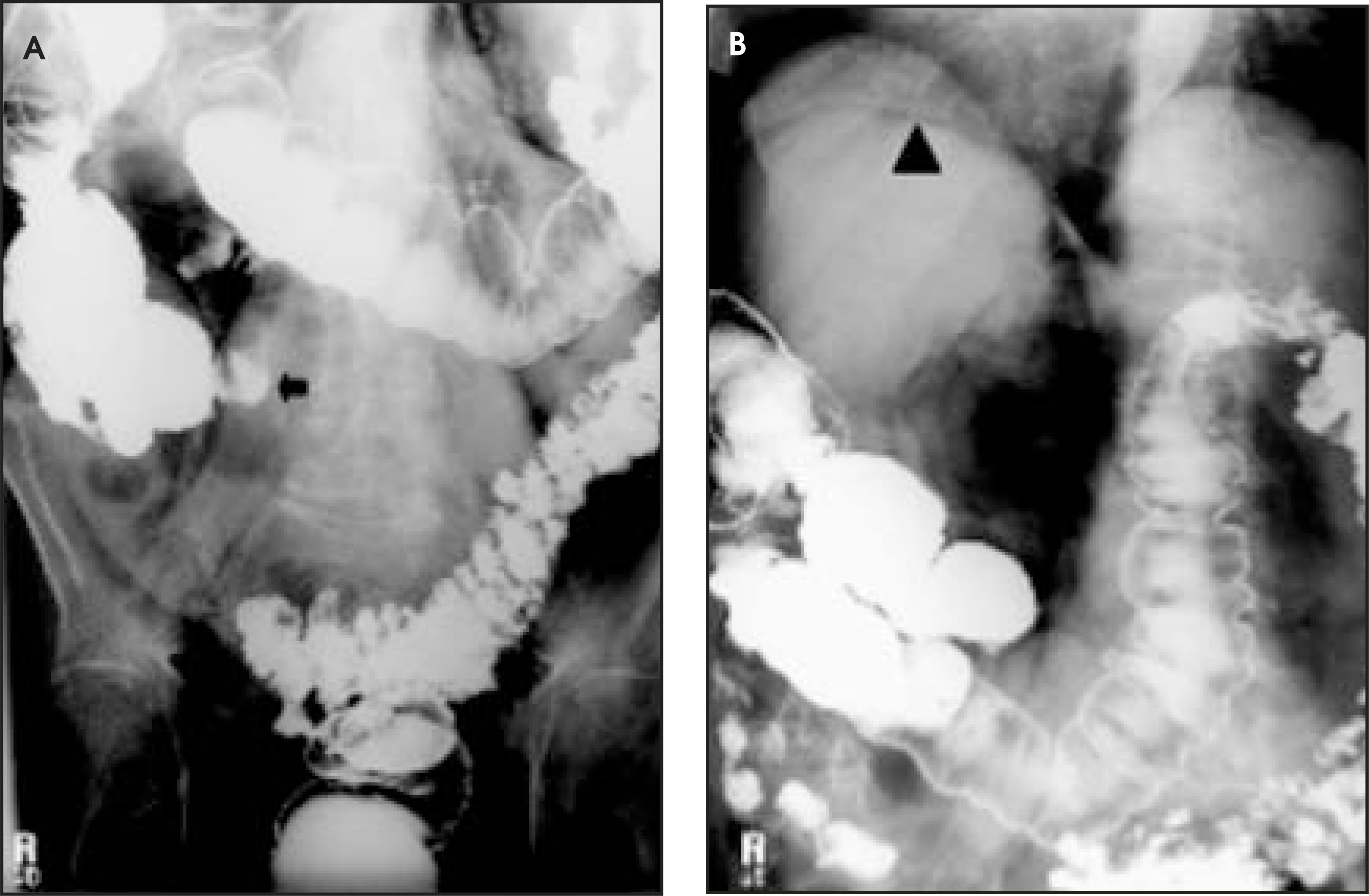
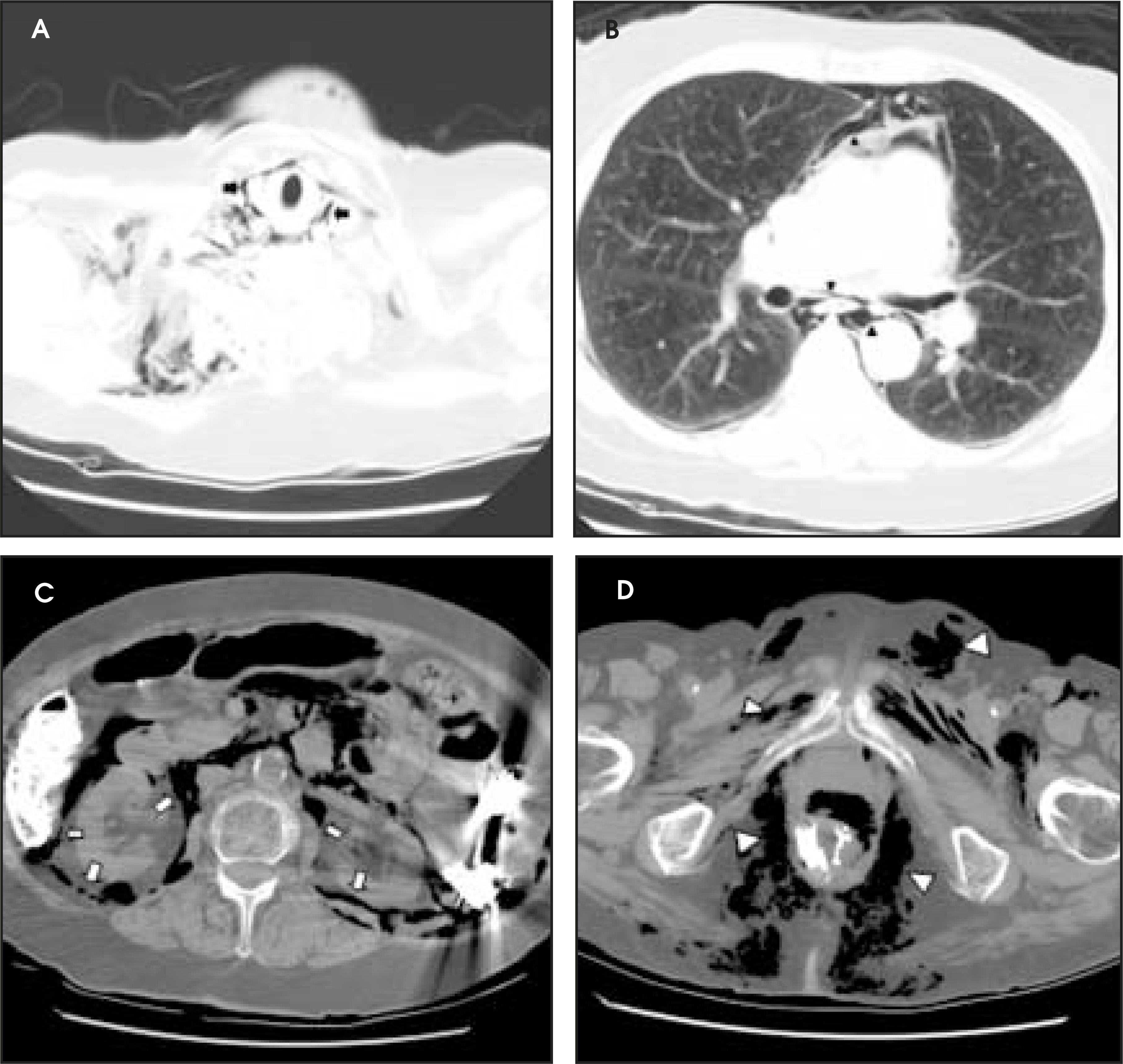
A plain film of the abdomen revealed extensive retroperitoneal air outlining both psoas margins and tracking superiorly below the hemidiaphragms and up into the chest. Extensive subcutaneous emphysema was seen adjacent to both flank stripes (Figure 1). The chest X-ray depicted the significant pneumomediastinum with extension superiorly into the soft tissues of the neck, where marked bilateral subcutaneous air was noted (Figure 2). The barium enema examination performed approximately 8 hours prior to this presentation revealed severe sigmoid diverticulosis without frank barium extravasation (Figure 3). A CT scan of the chest, abdomen, and pelvis depicted a large quantitiy of air extending from the subcutaneous soft tissues of the tracheal region all the way down to the perineum. Air was noted around the vocal cords and great vessels of the neck, within the mediastinum surrounding the esophagus and aorta, and in between the parietal pleura and the chest wall. Neither pneumopericardium nor pneumothorax were appreciated. Abdominal slices revealed air below both hemidiaphragms with a plethora of retroperitoneal air surrounding the perirenal space. A large amount of perirectal air was noted. Intraluminal barium was present without extravasation (Figure 4).
DIAGNOSIS
Massive retroperitoneal air, pneumomediastinum, and subcutaneous air secondary to bowel perforation after barium enema
DISCUSSION
Bowel perforation following barium enema examination is recognized as a rare but potentially devastating complication of what is generally considered to be a safe procedure. The incidence of bowel perforation following colonoscopy has been reported to be in the vicinity 0.075%, according to a Mayo Clinic retrospective study of 57,028 colonoscopic procedures performed between 1980 and 1995.1 This incidence is likely even lower for barium enema since it entails less insufflation and no endoscopic manipulation. Many mechanisms have been proposed as the cause for perforation, including an overdistended balloon, overzealous catheter tip positioning, hydrostatic pressure, and recent colonic instrumentation or biopsy. At the same time, certain patient populations are at increased risk for a perforation whether from underlying diverticulosis, rectal mucosal disease, cancer, inflammatory bowel disease, or stricture.2 The patient in this specific case was likely at increased risk due to her marked sigmoid diverticulosis (Figure 3). Perforation can be identified on plain films as gas under the hemidiaphragms, gas tracking along the psoas margins, or the classic presentation of gas outlining the falciform ligament or both sides of the bowel wall (Rigler sign). Having correctly identified the presence of a bowel perforation, the focus changes to that of patient management.
Some authors contend that all patients with colonic perforation should get prompt surgical laparotomy, and these advocates defend the traditional premise that early aggressive management decreases morbidity and mortality. Armed with the knowledge that barium sulfate can cause a progressive and lethal peritonitis, the prompt and thorough removal of all intraperitoneal barium is seen as an essential measure to avoid peritonitis or even subsequent adhesion formation. Irrigation and a gentle debriding of peritoneal surfaces is performed. Colostomy, resection, or even a Hartmann’s pouch are reserved for more severe cases, including those with an obstruction distal to the perforation.2
More recently, conservative management has been heralded for asymptomatic perforations, perforations without extravasation, and for mild or localized symptoms diagnosed within 4 to 8 hours of injury. These patients are admitted to the hospital to receive bowel rest, fluids, and intravenous (IV) antibiotics. Single therapy cefoxitin will suffice in an immunocompetent patient.3 They are closely followed for 5 to 7 days and monitored for resolution of abdominal pain; reduction in pulse, temperature, or white blood cells; and return of bowel function. If peritonitis or clinical deterioration ensues, then the patient receives a laparotomy.4
While the patient in this specific case was successfully managed conservatively and discharged uneventfully with close follow-up, the question remains as to why the perforation resulted in such an extensive pattern of massive retroperitoneal air, pneumomediastinum, and subcutaneous emphysema. The pattern can be explained by a knowledge of the various fascial compartments interconnecting one anatomic structure to another. Specifically, the visceral space is a fascial compartment that extends from the neck, through the mediastinum, and eventually to the retroperitoneum, forming an anatomic connection between all three areas. Gas in any one of these areas may spread to the other via a pressure gradient from the lumen of the bowel to the adjacent soft tissues.5 Gas from pneumatosis cystoides coli can dissect along the mesentery and into the retroperitoneum. Gas extending down into the scrotal region suggests a retroperitoneal mechanism that will likely be treated conservatively. Finally, subcutaneous emphysema occurs from dissection of air from the confounds of the visceral space to the periphery, such as is the case with a pneumomediastinum tracking cephalad around the great vessels of the neck (Figure 4). Gas can also escape into the soft tissues of the abdominal wall via the anatomic connection between the posterolateral retroperitoneum and the preperitoneal flank fat.6 Ultimately, this understanding of the close anatomic fascial connections allows one to avoid becoming prematurely alarmed about the impressive imaging appearance of retroperitoneal air, pneumomediastinum, and subcutaneous emphysema without first evaluating the patient clinically.
CONCLUSION
Plain radiography is a useful initial imaging modality to make the diagnosis of bowel perforation following barium enema examination, and CT is a useful tool to differentiate retroperitoneal air from air in other compartments. An understanding of anatomic fascial compartments explains how bowel perforations can result in such a profound imaging appearance as massive retroperitoneal air, pneumomediastinum, and subcutaneous emphysema. Nonetheless, the patient’s clinical examination should be followed in the postperforation period and used as a guideline for when to incorporate conservative or surgical management. Patients without intraperitoneal extravasation of barium (such as the patient in this case) who remain stable or continue to improve clinically without signs of peritonitis can be successfully managed with a conservative course of bowel rest, fluids, and IV antibiotics.
References
- Farley D, Bannon M, Zietlow S. Management of colonoscopic perforations. Mayo Clin Proc. 1997;72:729-733.
- Wang T, Tu H. Colorectal perforation with barium enema in the elderly: Case analysis with the POSSUM scoring system. J Gastroenterol. 1998;33:201-205.
- Kavin H, Sinicrope F, Esker A. Management of perforation of the colon at colonoscopy. Am J Gastroenterol. 1992;87:161-167.
- Damore L, Rantis P, Vernava A, Longo W. Colonoscopic perforations: Etiology, diagnosis, and management. Dis Colon Rectum. 1996;39:1308-1314.
- Ho H, Burchell S, Morris P, Yu M. Colon perforation, bilateral pneumothoraces, pneumopericardium, pneumomediastinum, and subcutaneous emphysema complicating endoscopic polypectomy: Anatomic and management considerations. Am Surg. 1996;62:770-774.
- Humphreys F, Hewetson K, Dellipiani A. Massive subcutaneous emphysema following colonoscopy. Endoscopy. 1984;16:160-161.
Citation
. RCOM RADIOLOGICAL CASE OF THE MONTH. Applied Radiology. 2005;34(8):38-41. doi:10.37549/AR1361.