RCOM RADIOLOGICAL CASE OF THE MONTH
Applied Radiology — Vol. 34 , Issue 9 , pp. 44 -47
DOI: 10.37549/AR1370
Published: September 1, 2005
Categories
CASE SUMMARY
A 22-year-old woman presented to our hospital with complaints of primary amenorrhea and intermittent lower abdominal pain. She also complained of cyclical hematuria. She reported normal development of secondary sexual characteristics by 13 years of age. By the age of 14, she noticed irregular abdominal cramping, which gradually progressed over several years to severe pelvic pain occurring at monthly intervals with associated cyclical hematuria. Her mother, sister, and other female relatives had normal onset of menses without dysmenorrhea.
The patient was a normally developed woman: 5 feet 4 inches tall and weighing 58 kg. External genitalia were normally developed. The vagina was well cornified and had adequate depth but ended in a blind pouch without a palpable cervix. Her blood count and urinalysis were normal. Intravenous pyelography showed a pelvic functioning kidney. A buccal smear was positive for sex chromatin. Her recorded 1-month basal body temperature showed a biphasic curve, indicative of ovulation. Cystoscopy confirmed a fistulous opening in the urinary bladder. Laparoscopy confirmed a normal-sized uterus. Levels of serum follicle-stimulating hormone (FSH), luteinizing hormone (LH), and prolactin were within normal limits.
IMAGING FINDINGS
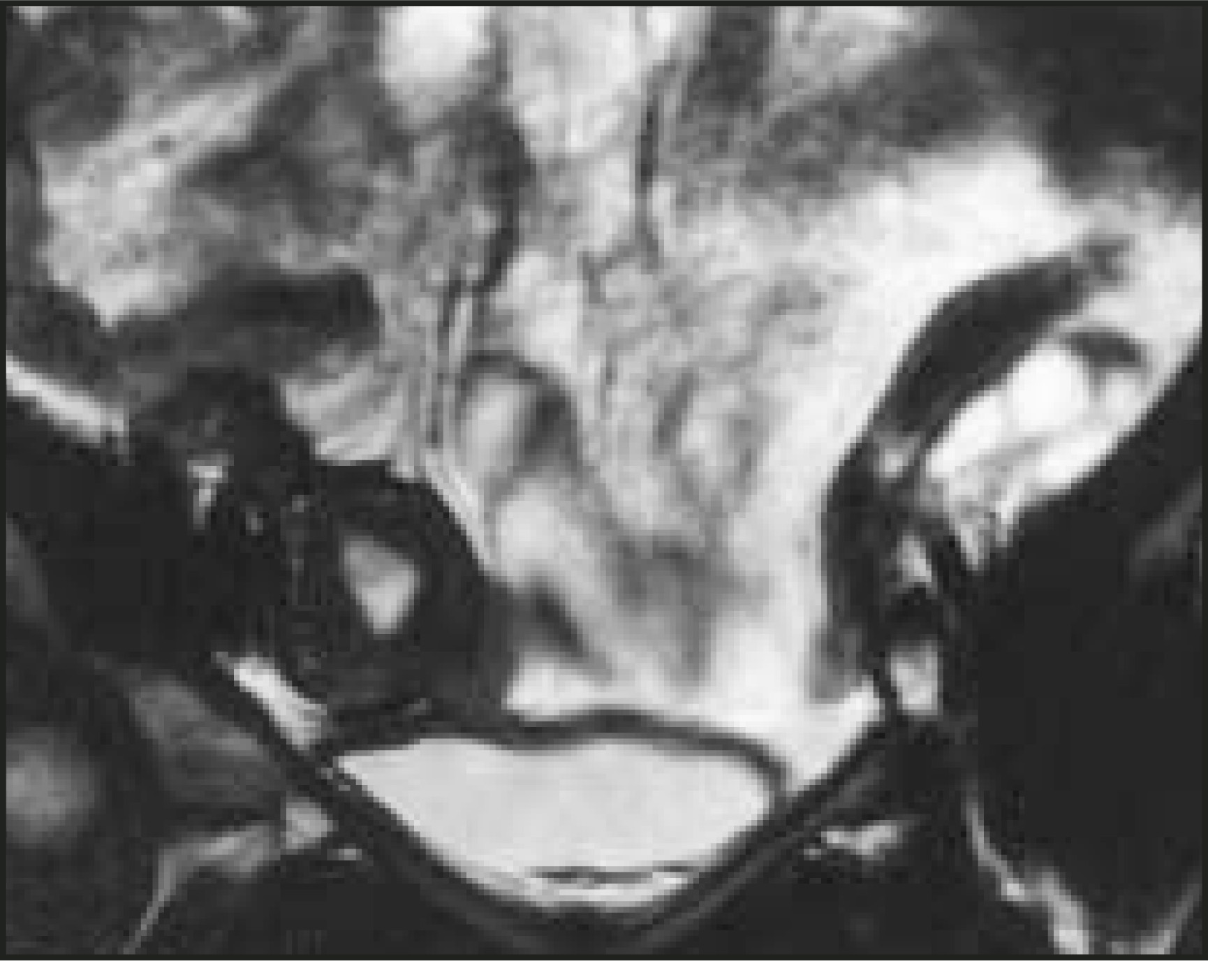
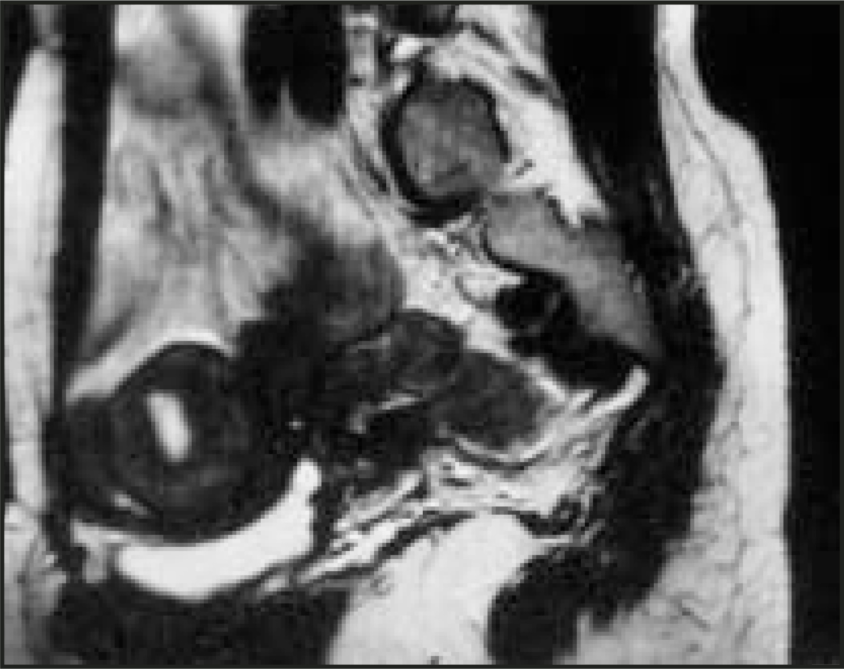
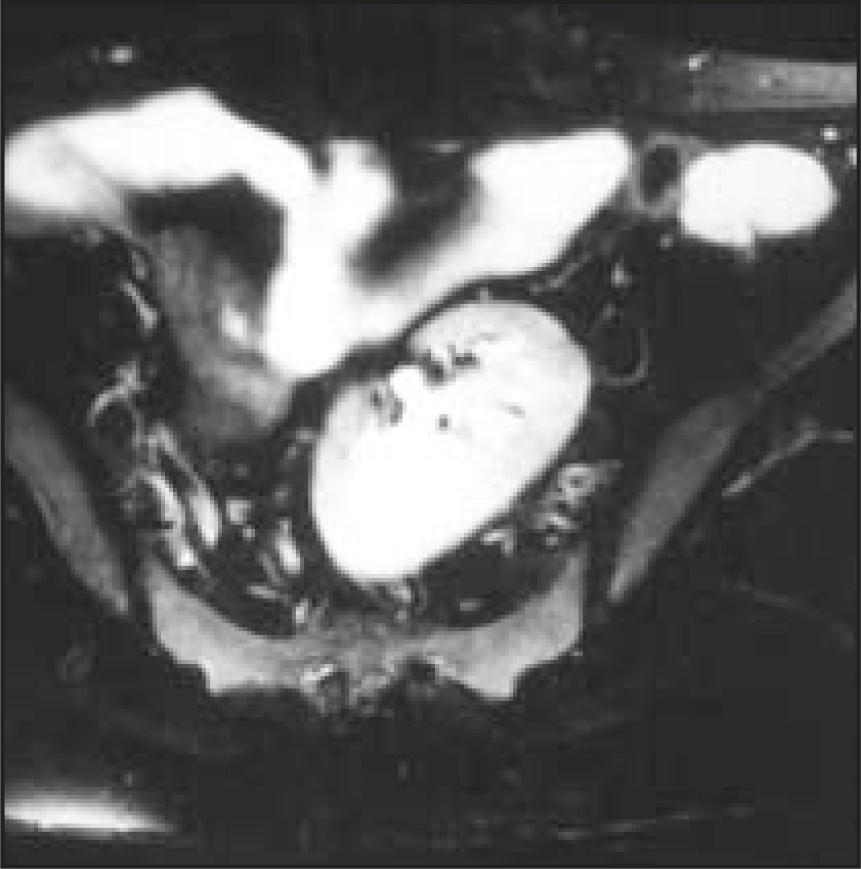
Magnetic resonance imaging (MRI) of the abdomen was performed using a phased-array body coil on a 1.5T scanner. In the coronal T2-weighted image, a normal-appearing uterus with a central high signal intensity due to presence of endometrium was seen superior to the urinary bladder on the right side. The left ovary appeared normal (Figure 1). On sagittal T2-weighted imaging, the uterus was identified superior to the urinary bladder. The cervix was not identified (Figure 2). An axial MR image revealed that the left kidney was located at level of the pelvic inlet (S1 to S3 level) with a vascular supply from the left iliac vessels (Figure 3).
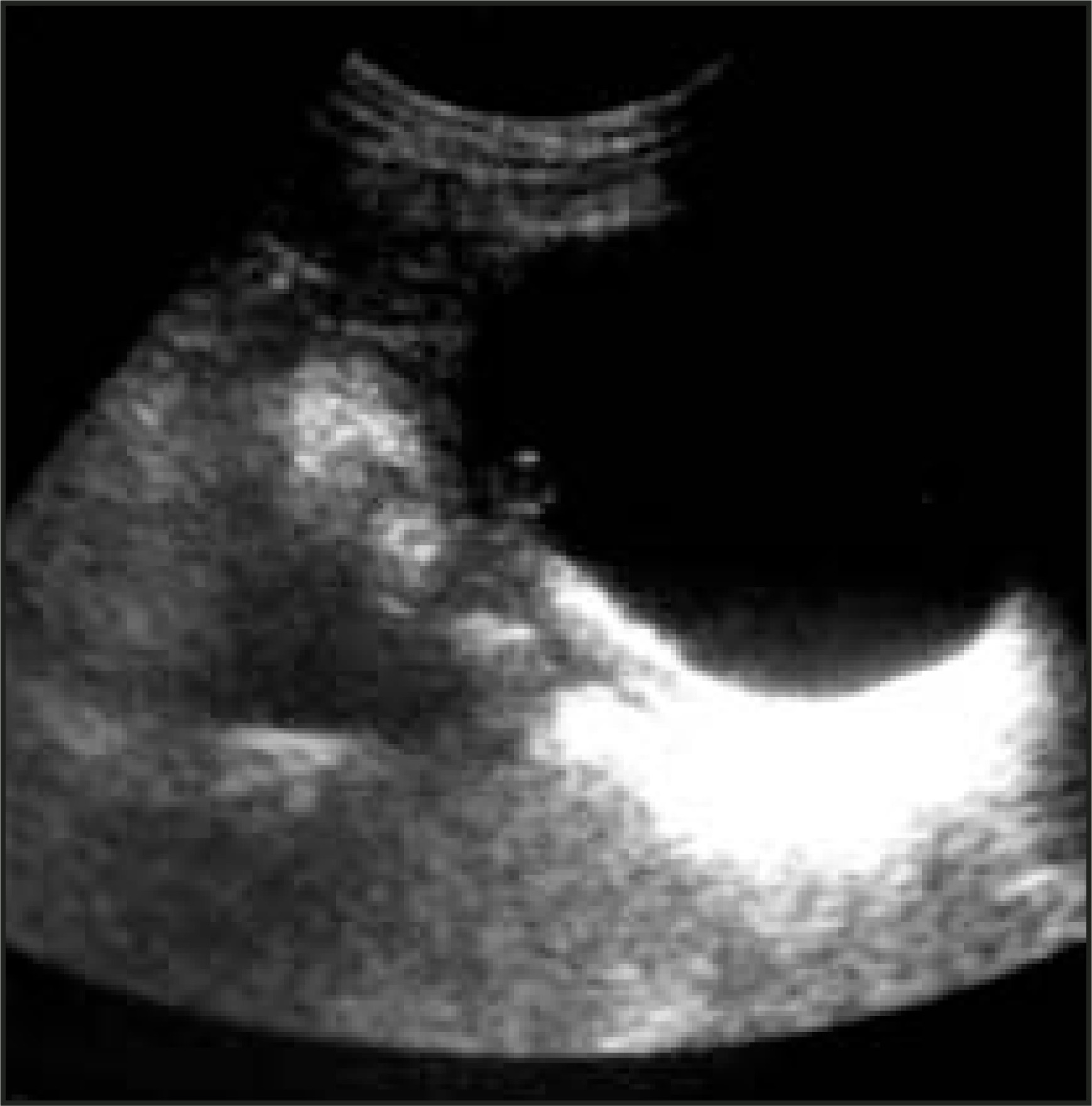
Transabdominal ultrasound revealed a normally shaped uterus superior to the urinary bladder. No cervix or endocervical canal was identified. A transverse sonographic image showed the site of communication of the uterus with the urinary bladder. This fistulous communication was confirmed on cytoscopy (Figure 4).
DIAGNOSIS
Cervical agenesis with uterovesical fistula and pelvic kidney
DISCUSSION
Although defects in Müllerian duct fusion produce a variety of congenital anomalies, the lack of communication between the uterus and the vagina is extremely rare. Our patient with this anomaly had the typical clinical picture consisting of primary amenorrhea and cyclic lower abdominal pain, but she was different from previously reported cases in that she had an uterovesical fistula and a pelvic kidney. Both ultrasound and MRI provided the significant information required to make the diagnosis.
The function of the uterine cervix encompasses providing a conduit for menstrual egress, maintenance of the intrauterine gestation, and mucus secretion, which, in turn, facilitates sperm transport and forms a barrier to the microflora of the vagina. Why the cervix alone would undergo atretic change remains unknown. Patten’s theory of local segmental atrophy is often quoted.1
Congenital cervical atresia is a rare class IB Müllerian anomaly. Cervical agenesis (class IB) has been reported in <20 patients in the English literature. Attempts to create an endometrial-vaginal fistula have been reported with short-term restoration of the menses and 1 pregnancy.2 However, these fistulous tracts may allow infection to ascend; a few deaths due to sepsis have been reported.3 Moreover, conception is highly unlikely because the cervical mucus is absent. Reports have suggested that the conservative approach to the establishment of uterovaginal patency has not resulted in the conservation of reproductive potential. For these reasons, heroic surgical efforts to preserve fertility is unwarranted in most instances. Hysterectomy remains the definitive therapeutic method.4,5
During childhood, most patients with Müllerian anomalies are asymptomatic. At puberty, when the uterus begins to menstruate, in the absence of a communicating channel between the uterus and vagina, the blood will remain hidden behind the obstruction and will cause a condition known as cryptomenorrhea. In patients with cervical agenesis, menstrual blood will collect inside the uterus and reflux along the fallopian tube into the peritoneal cavity; pelvic endometriosis is a possible complication.6
Diagnosis of cervical agenesis is quite difficult. In this situation, the clinical examination is of limited value; hysterosalpingography would not be possible, and even laparoscopy would be inconclusive. Both ultrasound and MRI are the most helpful in preoperative evaluation of Müllerian anomalies. Ultrasound can be utilized as an early imaging modality. MRI has the ability to delineate uterine, cervical, and vaginal anomalies. Recent reports suggest that MRI offers the best support in achieving a diagnosis of Müllerian anomaly. An intravenous pyelogram is often done because of known incidence of urinary tract anomalies, as was seen in the present case.7
MRI is a noninvasive diagnostic modality that is being applied with increasing frequency in obstetrics and gynecology. The preference for MRI over computed tomography (CT) scans is obvious. Fast scan times and higher resolution machines have improved image quality such that MRI is now the modality of choice for soft-tissue imaging in the pelvis. Its advantages over CT scanning include an absence of interference and artifact from pelvic bones and the ability to reconstruct images axially, sagittally, or coronally.8 Also, it is not operator-dependent and is not limited by bowel loops, which is also an advantage over sonography.9
MRI is also particularly adept at defining uterine anomalies because it highlights endometrial outlines. This allows MRI on T2weighted images to reveal uterine architecture in relation to the endometrial cavity. For this reason, MRI may be considered the method of choice for diagnosing uterine anomalies.8
CONCLUSION
Müllerian anomalies are not often encountered by the obstetrician-gynecologist; but when they are, the clinician is frequently frustrated in both diagnosis and treatment because so little is known about them. The present case is unique, as cervical agenesis per se is an extremely rare anomaly, and, to the best of our knowledge, association with uterovesical fistula and a pelvic kidney has never been reported in the literature. Our case highlights the role of sonography and MRI in the diagnosis of a rare Müllerian anomaly.
References
- Jacob J, Griffin W. Surgical reconstruction of the congenitally atretic cervix: Two cases. Obstet Gynecol Surv. 1989;44:556-569.
- Buttram V. Mullerian anomalies and their management. Fertil Steril. 1983;40:159-163.
- Casey A, Laufer M. Cervical agenesis: Septic death after surgery. Obstet Gynecol. 1997;90(4 Pt 2):706-707.
- Rock J, Schlaff W, Zacur H, Jones H. The clinical management of congenital absence of the uterine cervix. Int J Gynaecol Obstet. 1984;22:231-235.
- Geary W, Weed J. Congenital atresia of the uterine cervix. Obstet Gynecol. 1973;42:213-217.
- Vutyavanich T, Buddhirakkul P. Cervical and vaginal agenesis: A case report. J Med Assoc Thai. 1989;72:527-530.
- Chen C, Wu M, Ho H. Congenital atresia of the uterine cervix and vagina: Report of case. Acta Obstet Gynecol Scand. 1996;75:770-771.
- Kelley J. Magnetic resonance imaging to diagnose a mullerian anomaly during pregnancy. Obstet Gynecol. 1990;75(3 Pt 2):521-523.
- Vainright J, Fulp C, Schiebler M. MR imaging of vaginal agenesis with hematocolpos. J Comput Assist Tomogr. 1988;12:891-893.
Citation
. RCOM RADIOLOGICAL CASE OF THE MONTH. Applied Radiology. 2005;34(9):44-47. doi:10.37549/AR1370.