RCOM RADIOLOGICAL CASE OF THE MONTH
Applied Radiology — Vol. 34 , Issue 9 , pp. 38 -42
DOI: 10.37549/AR1372
Published: September 1, 2005
Categories
CASE SUMMARY
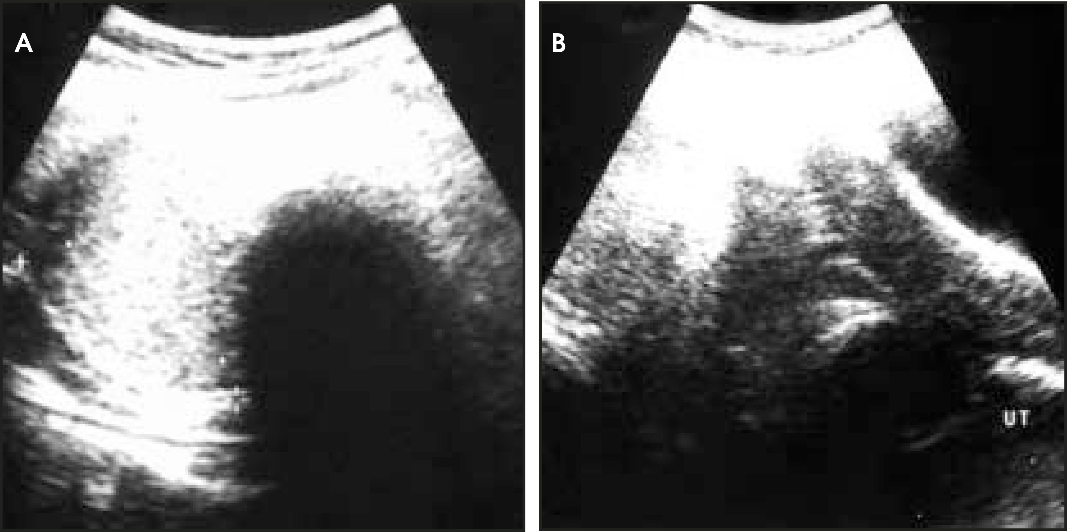
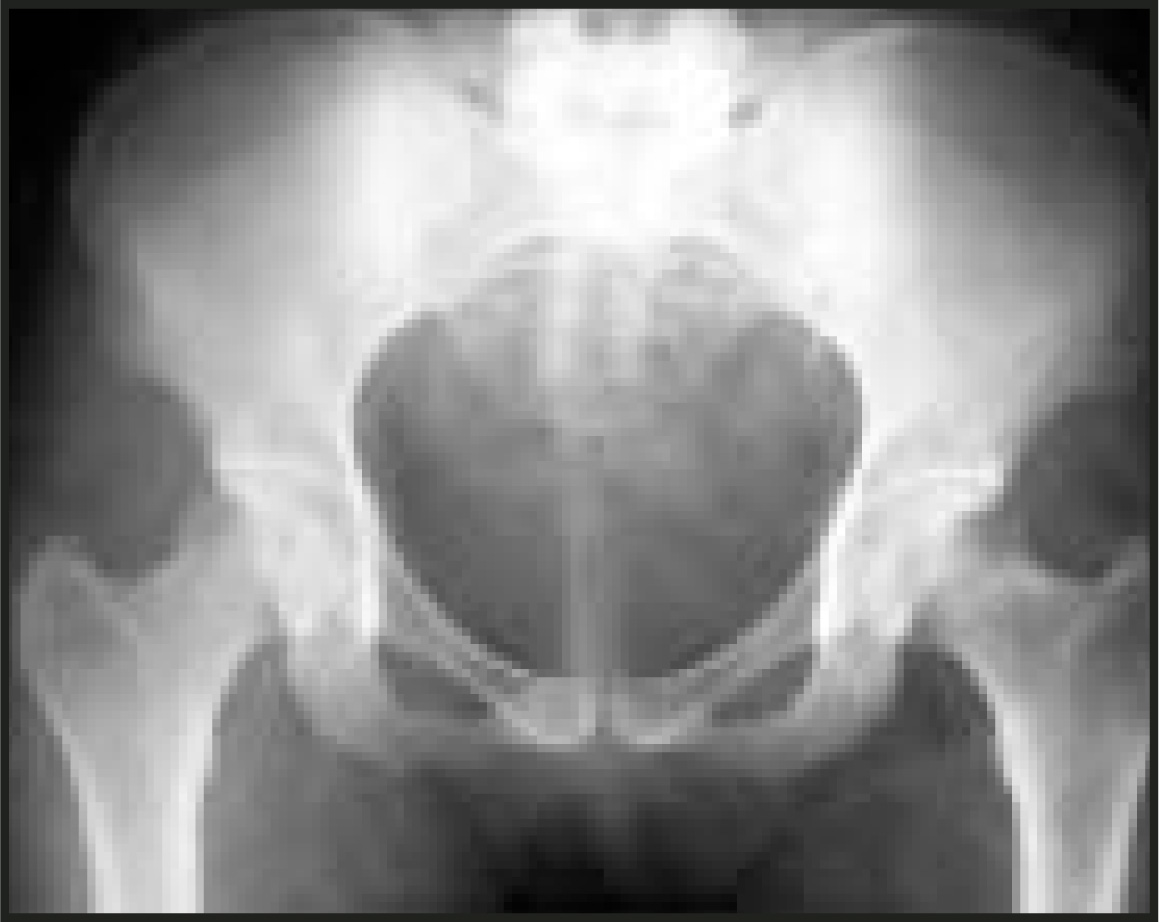
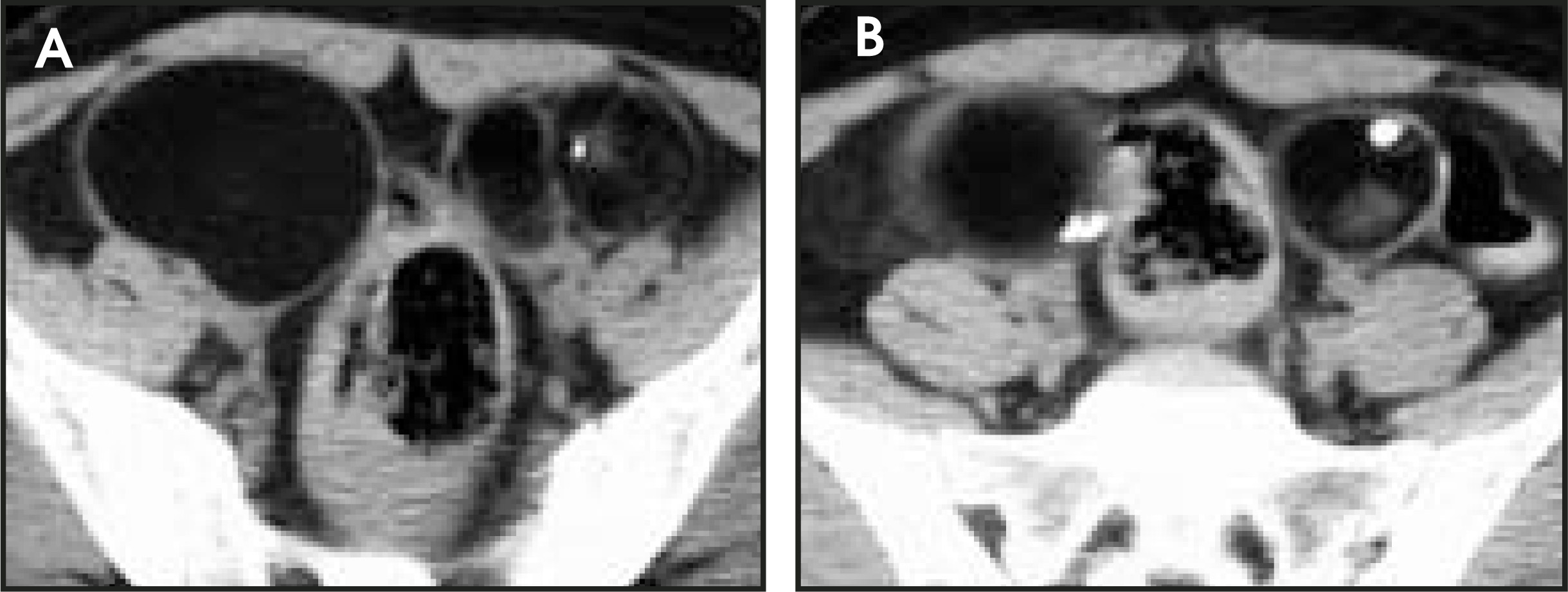
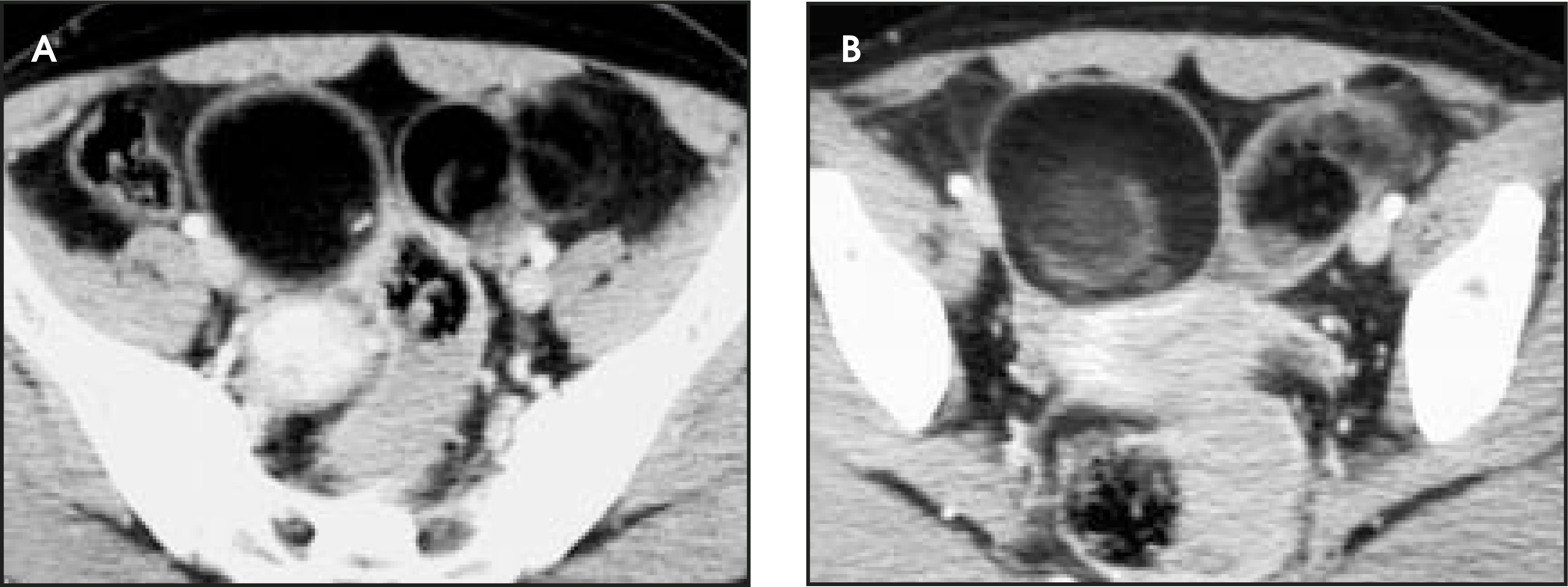
A 30-year-old woman presented to her primary care physician with abdominal discomfort and a palpable pelvic mass. Pelvic ultrasound revealed a bilateral ovarian mass (Figure 1). Pelvic radiography (Figure 2) and computed tomographic (CT) scans (Figures 3 and 4) were performed for further evaluation.
IMAGING FINDINGS
The pelvic ultrasound revealed a bilateral ovarian mass containing a solid hyperechoic component, suggesting the presence of fat (Figure 1). Some areas demonstrated acoustic shadowing, in keeping with a focus of calcification.
A pelvic X-ray showed tooth-like calcific densities (Figure 2). Noncontrast and contrast-enhanced CT scans of the abdomen showed a bilateral well-defined adnexal soft-tissue mass of fat density with two rounded soft-tissue densities within it and peripheral calcified areas (Figures 3 and 4). The appearances were typical for a dermoid cyst.
The tumors were resected. Histology confirmed the diagnosis of dermoid cyst.
DIAGNOSIS
Bilateral ovarian dermoid cyst
DISCUSSION
An ovarian dermoid cyst (DC) or a benign cystic teratoma is a benign tumor descending from germinal cells.1 In approximately 80% of the cases, this lesion occurs in young women (20 to 30 years of age) and represents 18% to 20% of benign ovarian tumors. In most cases, dermoid cysts are unilateral, but they are bilateral in 10% to 15% of cases.2,3
Dermoid cysts can be composed of elements descending from all three of the germinal layers, but in ovarian DCs, ectodermic differentiation frequently occurs. Typically, the diameter of an ovarian DC is <10 cm and is rarely >15 cm. On histologic examination, lipidic substance, hair, sebaceous secretions, hair follicles, and eggshell calcifications are seen in 50% of cases; real organoid structures (teeth, fragments of bone) in 30% of the cases.
Frequently, symptoms arise acutely with abdominal and pelvic pain, and, in 15% of cases, the symptoms are associated with menstrual abnormalities. Torsion is the most common complication, whereas rupture and suppuration are uncommon.4 Malignant degeneration occurs in 1% to 2% of cases, usually originating from squamous epithelial cells.1
At plain radiography of the abdomen, DCs can be easily detected if calcifications are present. Sometimes a typical radiographic finding of DC, “fat floating” appears (corresponding to the “fat-fluid level” on ultrasound and CT features); this radiographic sign is a horizontal line between 2 soft tissues of different opacities. It is caused by oily and sebaceous fluid floating over serous and over intracystic debris. Ultrasound appearances are often characteristic because of the presence of a highly reflective dermoid plug (a Rokitansky nodule), which is the solid element within the cyst that contains hair follicles, sebaceous glands, fat, and calcified or ossified elements. It usually forms an acute angle with the wall of the cyst and can produce acoustic shadowing due to the presence of hair, calcium, or bone.5 A fluid-fluid level may be detected due to sebum floating on an aqueous, more echogenic, layer.2 Echogenic lines and dots may be seen within the fluid, caused by strands of hair in the cyst, although fibrinous strands in hemorrhagic cysts can also cause this appearance.6
Tubo-ovarian abscesses can also contain fluid-fluid levels and echogenic pus and can produce acoustic shadowing due to gas. Ectopic pregnancies also demonstrate shadowing from bones and contain echogenic hemorrhage, which may separate to give fluid-fluid levels. Therefore, these conditions can mimic ovarian DC sonographically, although the clinical setting should allow for an accurate diagnosis in the majority of these cases.6 Hemorrhagic cysts can also cause diagnostic difficulty; however, the echogenic focus produced by fresh hemorrhage displays through transmission rather than acoustic shadowing.7 Ovarian DCs are the most commonly missed ovarian neoplasm on sonography, often due to the “tip of the iceberg” sign, in which the back wall of the cyst is obscured by acoustic shadowing, causing the echogenic Rokitansky nodule to be misinterpreted as bowel gas.7
CT and magnetic resonance imaging (MRI) are more sensitive to the presence of fat and calcium than is ultrasound, thus making the diagnosis using these modalities more straightforward.2
On CT, the fat content and the well-defined cystic appearance is highly suggestive of an ovarian DC.8 Whereas most DCs contain some soft-tissue components, the presence of a large (>10 cm), irregular soft-tissue mass within the tumor should raise the suspicion of malignant transformation. Contrast enhancement is also suggestive of malignant change.5
With MR, the intracystic sebum and the adipose tissue usually present in a Rokitansky nodule typically have high signal on T1-weighted images and intermediate signal on T2-weighted images. These findings are not diagnostic of ovarian DCs, as they can also occur in intracystic hemorrhage, eg, in endometriomas. Hemorrhagic products in an endometrioma can also produce a layered appearance on T2-weighted images or, sometimes, a gradated signal loss called “shading,” a finding that is not seen in DC. The loss of high signal on T1-weighted images with fat suppression is diagnostic of DC, allowing it to be differentiated from hemorrhagic lesions. Occasionally, nondependent spheres of intracyst lipid are seen within a DC.5 Areas of calcification and ossification are of low signal on all sequences on MR. A “salt-and-pepper” speckled appearance can sometimes be seen within a DC, which is thought to be due to multiple chemical shift artifacts at fat/water interfaces.9-11
The diagnosis of ovarian DC can often be made confidently on ultrasound; in one study, experienced observers were able to achieve 100% positive predictive value in cases in which ≥2 characteristic sonographic features were present.6 In uncertain cases, MR and CT will increase diagnostic accuracy because of their high sensitivity for fat and calcium content.9-11
Because of the potential for complications, such as torsion and rupture, DCs are usually resected. Therefore, gynecologic referral is recommended upon their diagnosis.
CONCLUSION
As shown in this case, ultrasound, X-rays, MRI, and CT are all useful in the diagnosis of a dermoid cyst.
References
- Bazot M, Cortez A, Sananes S. Imaging of dermoid cysts with foci of immature tissue. J Comput Assist Tomogr. 1999;23:703-706.
- Outwater E, Siegelman E, Hunt J. Ovarian teratomas: Tumor types and imaging characteristics. RadioGraphics. 2001;21:475-490.
- Caruso P, Marsh M, Minkowitz S, Karten G. An intense clinicopathologic study of 305 teratomas of the ovary. Cancer. 1971;27:343-348.
- Comerci J, Licciardi F, Bergh P. Mature cystic teratoma: A clinicopathologic evaluation of 517 cases and review of the literature. Obstet Gynecol. 1994;84:22-28.
- Quin S, Erickson S, Black W. Cystic ovarian teratomas: The sonographic appearances of the dermoid plug. Radiology. 1985;155:477-478.
- Patel M, Feldstein V, Lipson S. Cystic teratomas of the ovary: Diagnostic value of sonography. AJR Am J Roentgenol. 1998;171:1061-1065.
- Hertzberg B, Kliewer M. Sonography of benign cystic teratoma of the ovary: Pitfalls in diagnosis. AJR Am J Roentgenol. 1996;167:1127-1133.
- Occhipinti K, Frankel S, Hricak H. The ovary. Computed tomography and magnetic resonance imaging. Radiol Clin North Am. 1993;31:1115-1132.
- Buy J, Ghossain M, Moss A. Cystic teratoma of the ovary: CT detection. Radiology. 1989;171:697-701.
- Mitchell D. Benign disease of the uterus and ovaries. Applications of magnetic resonance imaging. Radiol Clin North Am. 1992;30:777-787.
- Troiano R, McCarthy S. Magnetic resonance imaging evaluation of adnexal masses. Semin Ultrasound CT MR. 1994;15:38-48.
Citation
. RCOM RADIOLOGICAL CASE OF THE MONTH. Applied Radiology. 2005;34(9):38-42. doi:10.37549/AR1372.