RCOM RADIOLOGICAL CASE OF THE MONTH
Applied Radiology — Vol. 34 , Issue 11 , pp. 66 -70
DOI: 10.37549/AR1384
Published: November 1, 2005
Categories
CASE SUMMARY
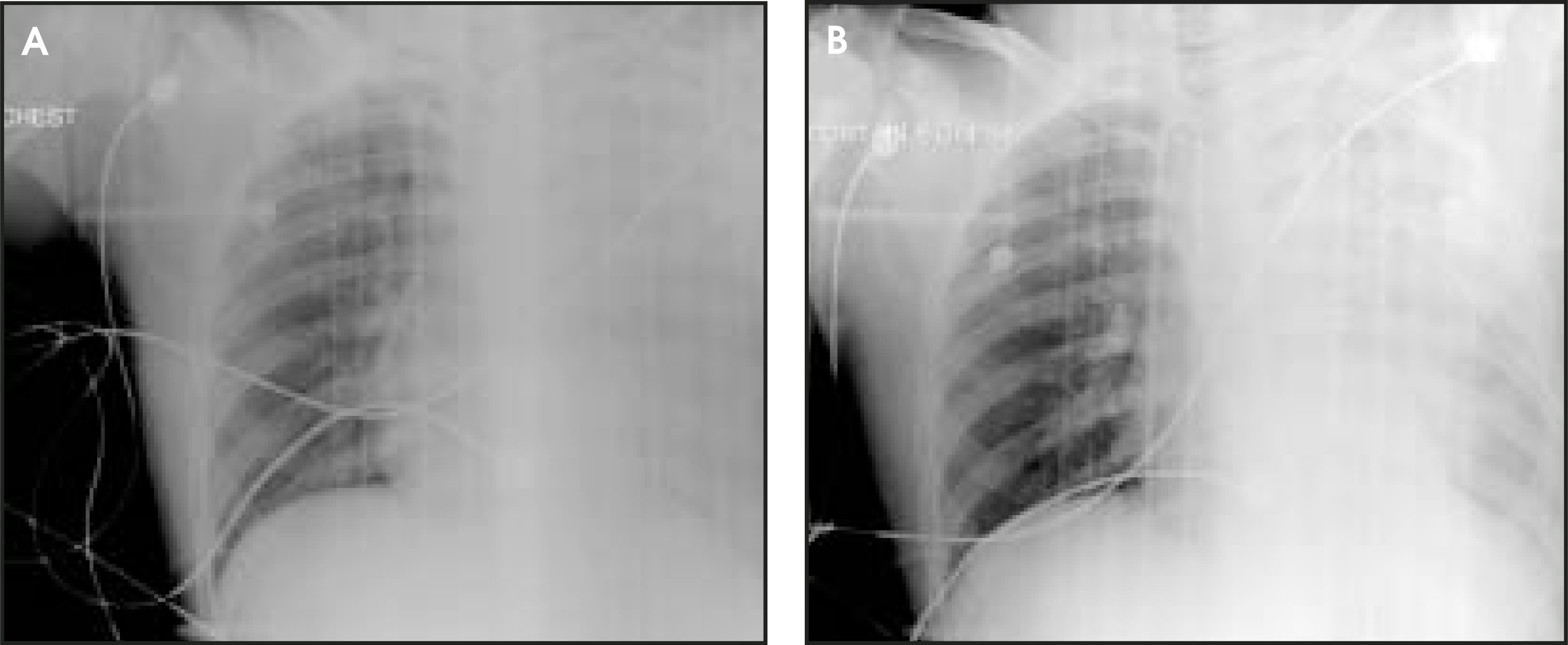
A 16-year-old otherwise healthy boy presented to the emergency department after being struck by a car and subsequently complaining of left-sided chest and back pain with shortness of breath. His initial vital signs were blood pressure of 110/62 mm Hg, pulse rate of 155, respiratory rate of 24, and oxygen saturation of 96% on a non-rebreather mask. The initial chest X-ray (CXR) showed complete opacification of the left hemithorax (Figure 1), which led to chest tube placement that drained approximately 1600 mL of blood. The patient was immediately taken to the operating room for exploratory thoracotomy, which revealed a large firm mass in the posterior chest wall. Bleeding was originating from a puncture site in the mass caused by a rib fracture. Intraoperative biopsy was suggestive of a nerve sheath tumor.
IMAGING FINDINGS
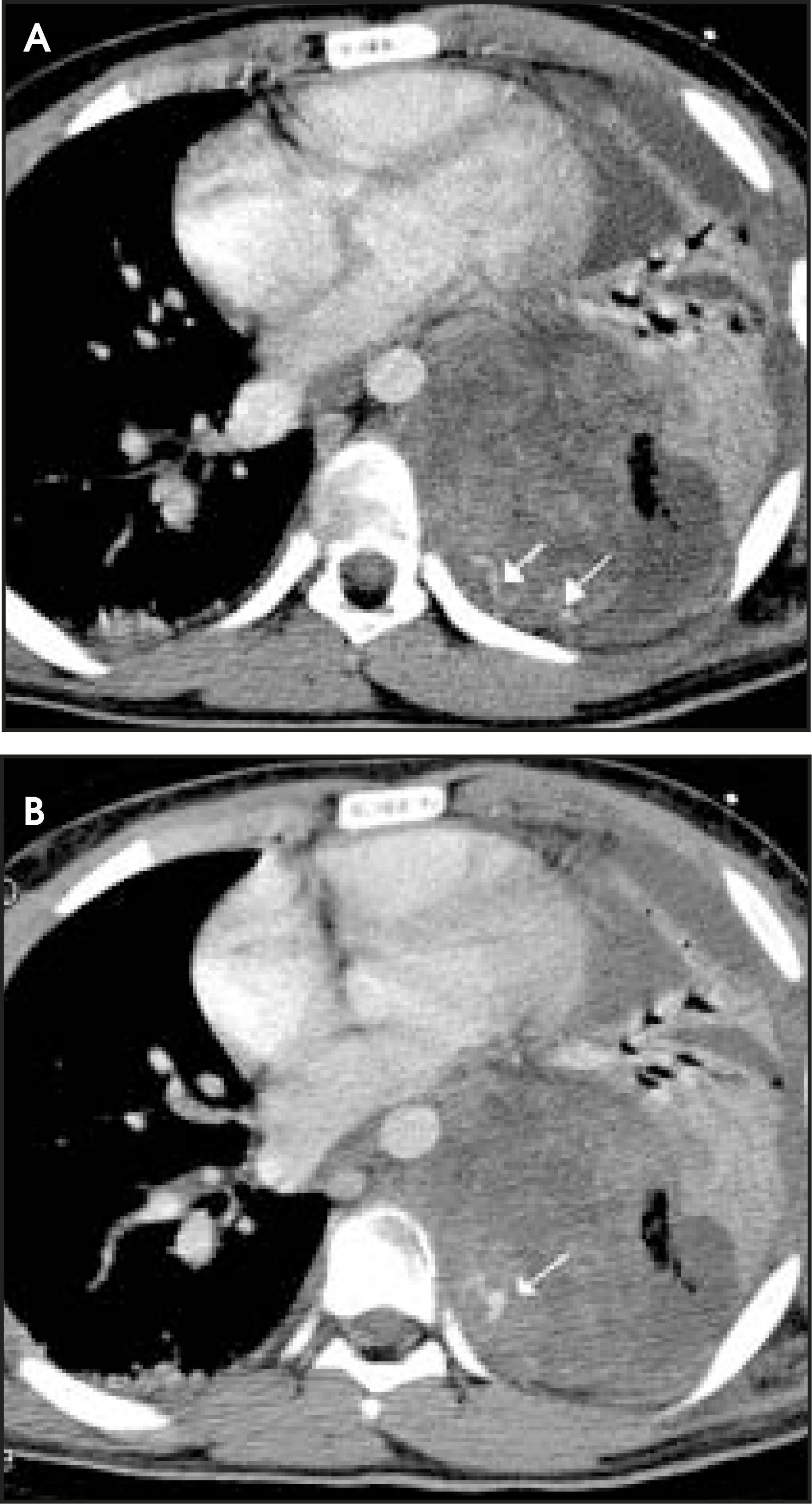
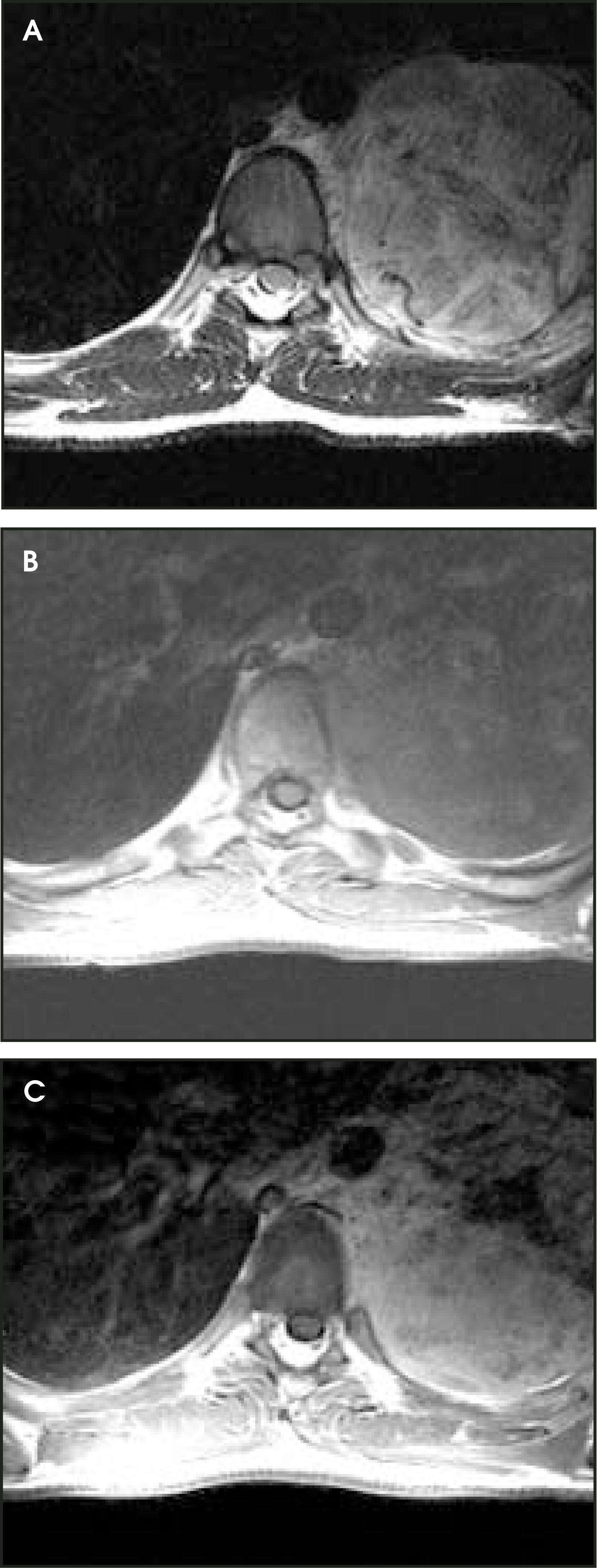
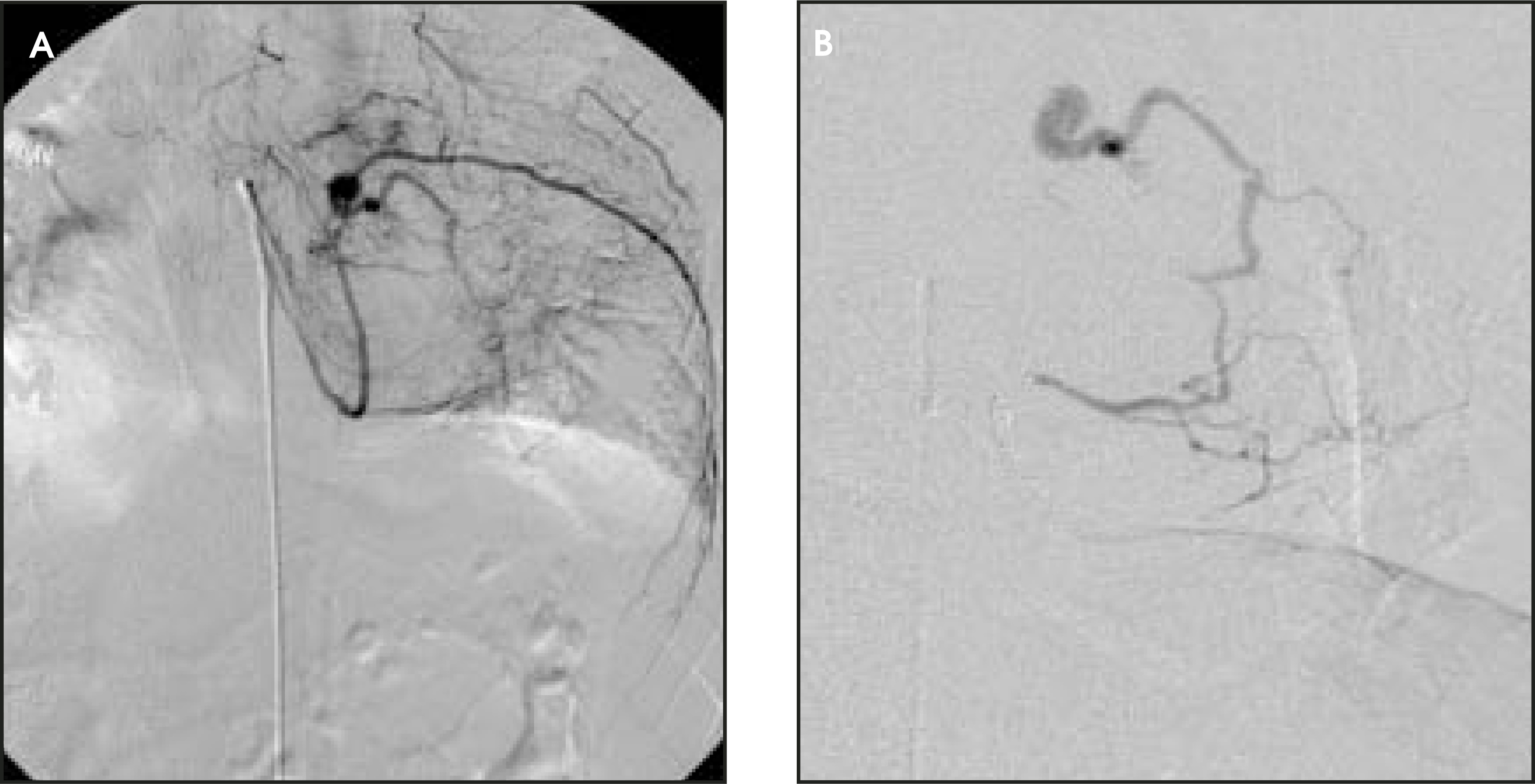
The initial CXR showed complete opacification of the left hemithorax (Figure 1A) with no bony abnormalities evident. The CXR taken after a chest tube was placed revealed the presence of pleural fluid in this patient, but the tumor was not yet visible (Figure 1B). Computed tomography (CT) scans showed a posterior mediastinal mass of heterogeneous density and a fluid collection in the left hemithorax with a prominent feeding vessel to the mass (Figure 2A). There was evidence of a small pseudoaneurysm in a vessel feeding the tumor, with no active contrast extravasation seen (Figure 2B). Magnetic resonance imaging (MRI) examination did not show any involvement of the intervertebral foramen by the tumor (Figure 3). The mass was inhomogeneous and of high intensity on T2-weighted images (Figure 3A), and it revealed significant enhancement on postcontrast images (Figure 3C). During angiography for preoperative embolization, vessels draping over the tumor mass without early filling of draining veins was seen (Figure 4). The pseudoaneurysm visible on the CT scan was seen more clearly on angiography without any evidence of active bleeding.
DIAGNOSIS
Thoracic schwannoma
DISCUSSION
Neurogenic tumors are the most common cause of a mass lesion in the thoracic paravertebral region in the general adult population. Schwannomas are rare in people <20 years of age and are largely asymptomatic.1 On gross pathologic analysis, they appear as sharply circumscribed, spherical soft-tissue masses.2 They are eccentric and encapsulated and do not have nerve fibers passing through them.1,3
There are no plain-film findings that are pathognomonic for intrathoracic schwannomas. Chest radiography usually shows a smoothly rounded or oval mass located in the paravertebral region.1 The mass is rarely calcified, and inferior and superior sulci are usually present.4 While bone changes on plain films are generally late manifestations of schwannomas,5 there are some findings that can help narrow the differential diagnosis. Bone changes, such as erosion or splaying of the ribs, may occur4 as well as neural foraminal enlargement and vertebral body erosion.4 Rib erosion with a sclerotic border is suggestive of a benign lesion; however, spreading to multiple ribs with erosion suggests malignancy.1
Schwannomas appear as well-circumscribed, round masses that are of homogenous soft-tissue density on plain CT images,2,3 with clear preservation of surrounding fat planes.6 The attenuation values of schwannomas tend to be lower than those of the surrounding muscle,2 which may be accounted for by their areas of low cellularity.3 Schwannomas may occasionally be seen as areas of very low attenuation on noncontrast CT exams if there is a high concentration of lipid-rich Schwann cells in these tumors.2 Schwannomas have even been described as presenting as cystic masses.6 Schwannomas show variable enhancement on contrast-enhanced CT scans.2,3,5 The heterogeneity is due to cystic degeneration, xanthomatous change, variabilities in hyper- and hypocellular areas, and areas of hemorrhage.3 A pattern of rim enhancement has also been described.7 Malignant schwannomas have CT findings of low-density areas, compression of adjacent structures, pleural abnormalities, such as pleural effusions or pleural nodules, and metastatic pulmonary nodules.5,8
In the more usual nonemergency setting, MRI is the preferred modality for imaging neurogenic tumors, as its multiplanar capability and high soft-tissue contrast resolution can best reveal the nature of lesions, intraspinal extension, and cranial-caudal extent.9 More specifically, MRI is more sensitive than CT for the identification of schwannoma.5 On MRI, schwannomas show low-to-intermediate signal intensity on T1-weighted images. On T2-weighted images, they show inhomogenously high intensity.5,10,11 Very high intensity regions seen on T2weighted images of schwannomas correspond to cystic degeneration with surrounding collagenous fibrous tissue.10 On MRI with gadolinium contrast, there will be dense enhancement of the tumor that will be heterogeneous if large and homogeneous if small. Most schwannomas have mild-to-markedly increased signal intensity on proton-density and T2-weighted sequences.5 Schwannomas do not always demonstrate the target appearance often associated with benign neurofibromas.11
There is little in the literature regarding the angiographic findings of extracranial schwannomas. This may be because angiography is used only in cases in which a highly vascular lesion is suspected, critical adjacent arterial structures need to be assessed prior to surgery, or when preoperative embolization is planned.12 In general, schwannomas are largely avascular or hypovascular lesions that are identified by focal displacement,13 stretching, and draping of adjacent vessels.5
CONCLUSION
This case illustrates an unusual presentation of a thoracic region schwannoma in a patient with trauma, and demonstrates the utility of CT, MRI, and angiographic imaging in providing critical information in trauma patients.
STATEMENT OF OWNERSHIP MANAGEMENT AND CIRCULATION (REQUIRED BY 39 U.S.C. 3685)
Title of Publication: APPLIED RADIOLOGY
Publication Number: 0160-9963
Filing Date: 9/30/05
Frequency: Monthly
Number of Issues Published Annually: 12
Annual Subscription Price: $95
Mailing Address and Business Headquarters:
APPLIED RADIOLOGY
Anderson Publishing, Ltd.
180 Glenside Avenue
Scotch Plains, NJ 07060-1518
Publisher: O. Oliver Anderson
Editor: Stuart E. Mirvis, MD, FACR
Managing Editor: Elizabeth A. McDonald
Owners: O. Oliver Anderson
Brenda M. Anderson
References
- Laurent F, Latrabe V, Lecesne R. Mediastinal masses: Diagnostic approach. Eur Radiol. 1998;8:1148-1159.
- Kumar A, Kuhajda F, Martinez C. Computed tomography of extracranial nerve sheath tumors with pathologic correlation. J Comput Assist Tomogr. 1983;7:857-865.
- Chui M, Bird B, Rogers J. Extracranial and extraspinal nerve sheath tumors: Computed tomographic evaluation. Neuroradiology. 1988;30:47-53.
- Reed J, Hallet K, Feigin D. Neural tumors of the thorax: Subject review from the AFIP. Radiology. 1978;126:9-17.
- Osborn A. Diagnostic Neuroradiology. 1994.
- Powers S, Norman D, Edwards M. Computerized tomography of peripheral nerve lesions. J Neurosurg. 1983;59:131-136.
- Ikezoe J, Sone S, Higashihara T. CT of intrathoracic neurogenic tumors. Eur J Radiol. 1986;6:266-269.
- Moon W, Im J, Han M. Malignant schwannomas of the thorax: CTfindings. J Comput Assist Tomogr. 1993;17:274-276.
- Erasmus J, McAdams H, Donnelly L, Spritzer C. MR imaging of mediastinal masses. Magn Reson Imaging Clin N Am. 2000;8:59-89.
- Sakai F, Sone S, Kiyono K. Intrathoracic neurogenic tumors: MR-pathologic correlation. AJR Am J Roentgenol. 1992;159:279-283.
- Suh J, Abenoza P, Galloway H. Peripheral (extracranial) nerve tumors: Correlation of MR imaging and histologic findings. Radiology. 1992;183:341-346.
- Abramowitz J, Dion J, Jensen M. Angiographic diagnosis and management of head and neck schwannomas. AJNR Am J Neuroradiol. 1991;12:977-984.
- Moscow N, Newton T. Angiographic features of hypervascular neurinomas of the head and neck. Radiology. 1975;114:635-640.
Citation
. RCOM RADIOLOGICAL CASE OF THE MONTH. Applied Radiology. 2005;34(11):66-70. doi:10.37549/AR1384.