RCOM RADIOLOGICAL CASE OF THE MONTH
Applied Radiology — Vol. 34 , Issue 12 , pp. 45 -47
DOI: 10.37549/AR1387
Published: December 1, 2005
Categories
CASE SUMMARY
A 19-year-old man presented with gross hematuria and urinary urgency. His medical history was notable for end-stage renal disease and hemodialysis. All other examinations and laboratory findings were unremarkable.
IMAGING FINDINGS
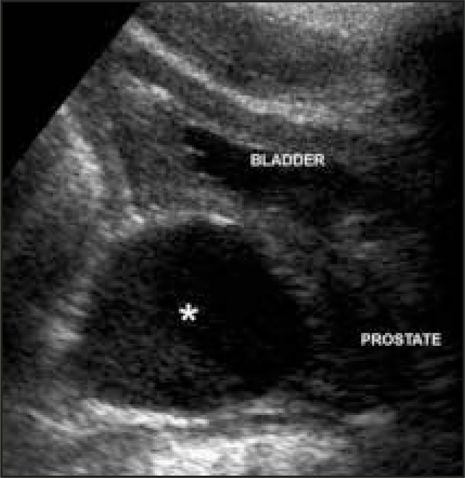
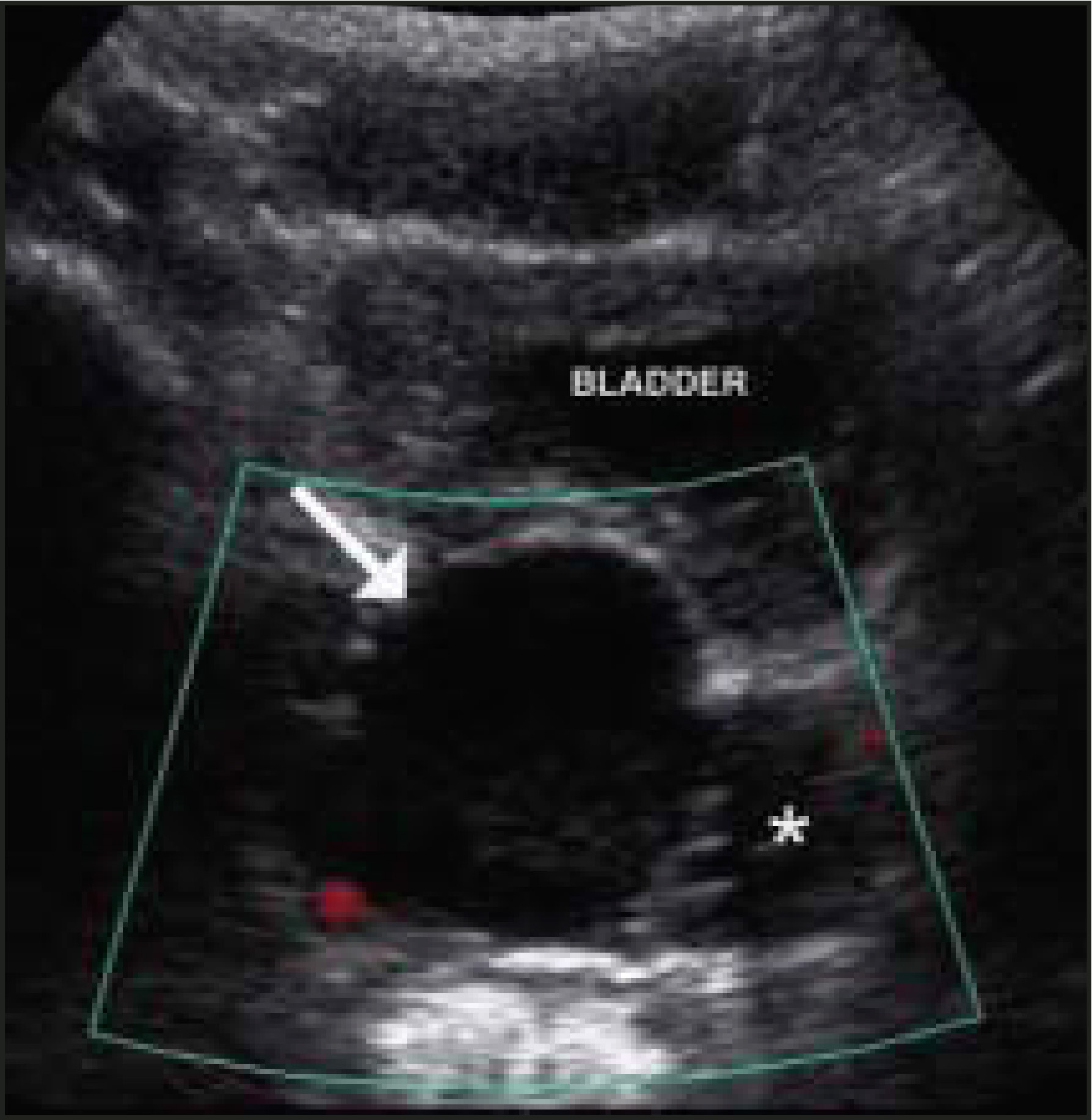
Images of the pelvis from the bladder ultrasound (Figures 1 and 2) revealed a 3.5-cm midline fluid collection posterior to the bladder and superior to the prostate gland. The cystic fluid collection appears contiguous with the prostate. Echoes are noted within the cyst fluid. No internal flow is seen on color Doppler imaging (Figure 2).
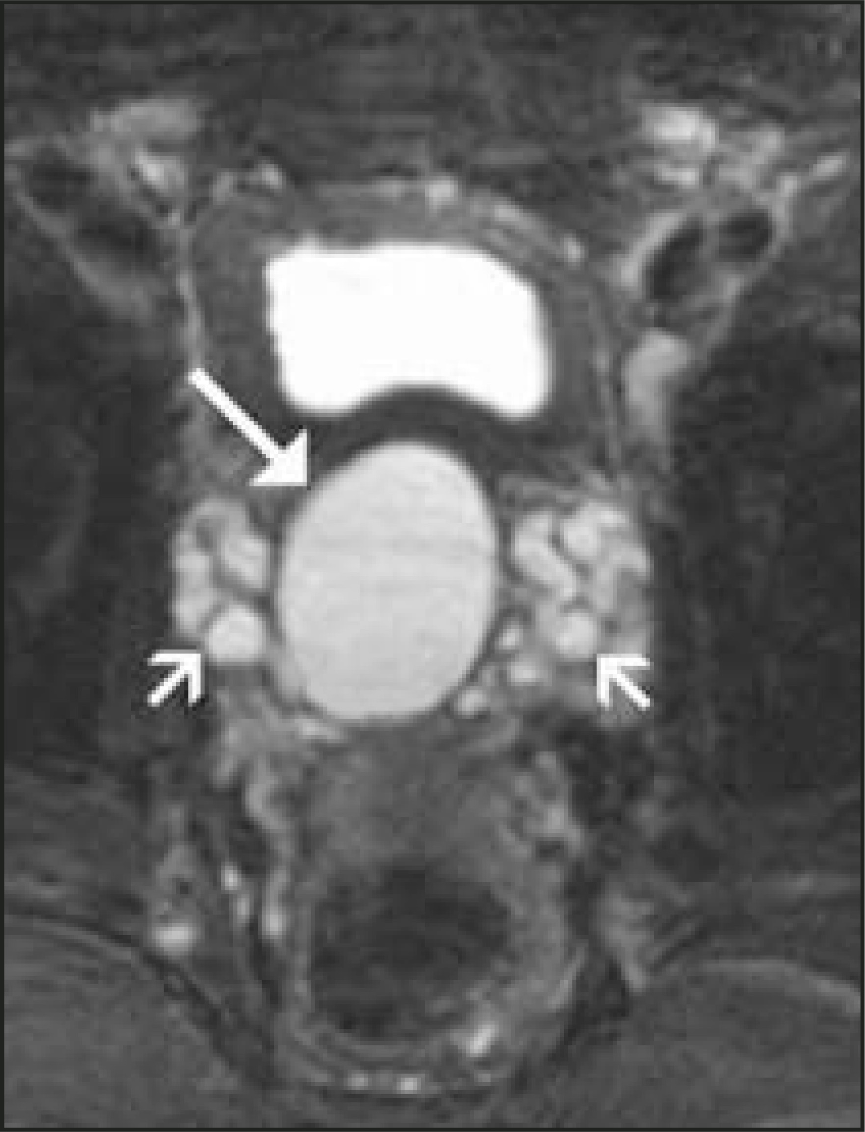
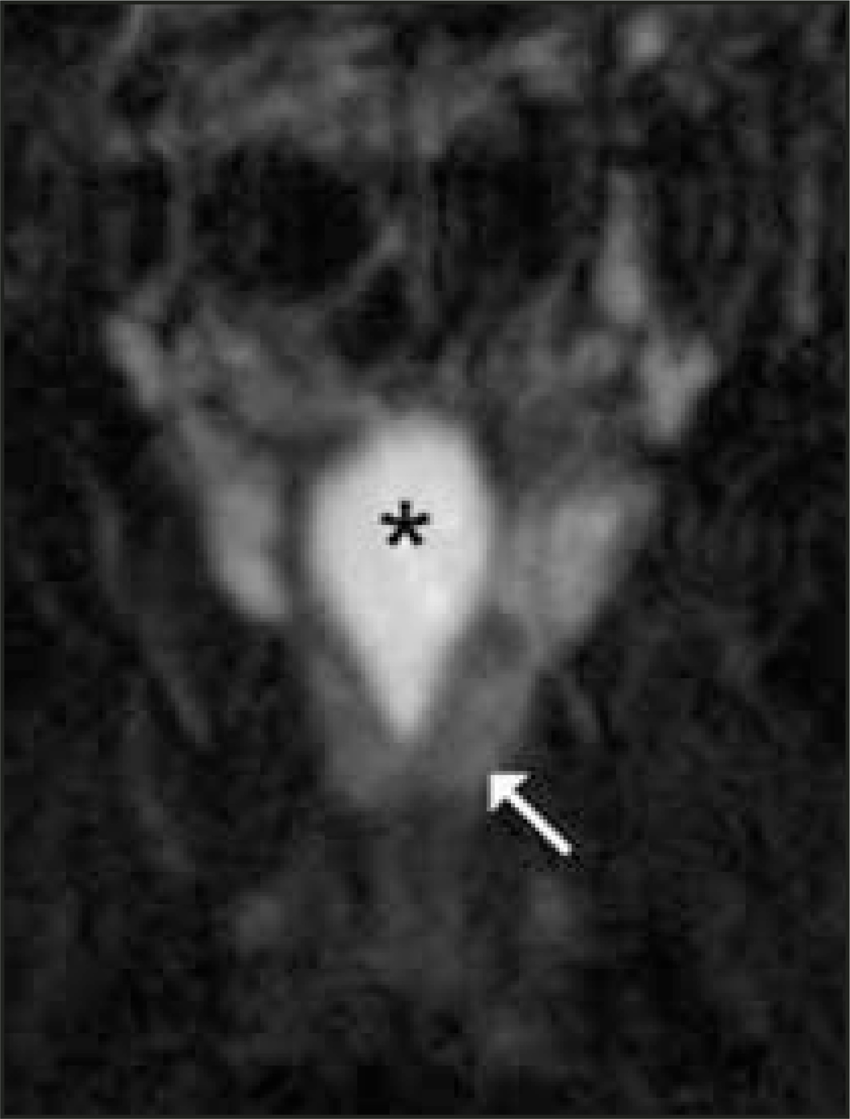
Subsequent magnetic resonance imaging (MRI) of the pelvis (Figures 3 and 4) confirmed the ultrasound findings, which depicted the midline position of the cyst, posterior to the bladder and extending superiorly from the verumontanum and the base of the prostate. No definite connection was seen to the posterior wall of the bladder or the urethra. No abnormal enhancement was identified on postcontrast MR images (not shown). Incidental note was made of bladder-wall thickening, likely due to coexisting cystitis.
DIAGNOSIS
Müllerian duct cyst
DISCUSSION
The müllerian duct cyst is a remnant of the caudal ends of the fused müllerian duct, a structure that typically regresses in utero. These cysts are typically located in the midline, posterior to the bladder, originating in the region of the verumontanum. Müllerian duct cysts do not communicate with the prostatic urethra, but are connected to the verumontanum by a thin stalk.1 Unlike utricular cysts, the müllerian duct cyst is not typically associated with other congenital abnormalities of the urinary tract and usually occurs as an isolated entity. Rarely, a müllerian duct cyst may be associated with renal agenesis.1 When aspirated, the cyst fluid is devoid of spermatozoa, and may be proteinaceous or hemorrhagic, accounting for the appearance of increased signal on T1weighted MR imaging. Rarely, calculi may occur within these cysts. The peak clinical incidence of müllerian duct cysts occurs in the age range of 20 to 40 years, as the cyst becomes larger and accumulates fluid.2 The clinical presentation is varied, including urinary frequency, urinary urgency, dysuria, urinary obstruction, hematuria, and pelvic pain. Digital rectal examination may reveal a midline mass superior to the prostate gland. Secondary evidence of a müllerian duct cyst can be noted on a voiding cystourethrogram or an intravenous urogram, manifesting as an impression along the posterior wall of the bladder.2 Pelvic ultrasound and transrectal ultrasound will reveal a fluid-filled cyst projecting superiorly from the prostate, extending posterior to the bladder.
MRI is a valuable imaging modality for the evaluation of müllerian duct cysts. The excellent soft-tissue contrast and multiplanar capability of MRI provide superb depiction of pelvic anatomy, allowing differentiation between entities that can mimic the müllerian duct cyst, such as posterior diverticuli of the bladder and urethra, utricle cyst, vas deferens cyst, and seminal vesicle cyst. Sagittal and coronal images are particularly useful in visualizing the location of the cyst in relation to the prostate and bladder. The müllerian duct cyst and utricle cyst both occur in the midline; however, the utricle cyst is typically smaller, confined to the base of the prostate, and communicates with the posterior urethra.1 Müllerian duct cysts are larger and extend above the base of the prostate. In the case presented, cystoscopy revealed no communication with the urethra or the bladder. Cysts of the vas deferens and seminal vesicles occur in a paramedian location; however, when large, they may project into the midline. Aspiration of the cyst fluid will reveal spermatozoa, thus differentiating these entities from the müllerian duct cyst.3
CONCLUSION
Müllerian duct cysts are rare, congenital lesions of the periprostatic region. This diagnosis should be considered in the presence of a midline cystic mass, posterior to the bladder and arising superiorly from the region of the verumontanum and base of the prostate.
References
- McDermott V, Meakem T, Stolpen A, Schnall M. Prostatic and periprostatic cysts: Findings on MR imaging. AJR Am J Roentgenol. 1995;164:123-127.
- Thurnher S, Hricak H, Tanagho E. Müllerian duct cyst: Diagnosis with MR imaging. Radiology. 1988;168:25-28.
- Noone T, Semelka R, Kubik-Huch R, Braga L, Semelka R. Abdominal-Pelvic MRI. 2002:983-984.
Citation
. RCOM RADIOLOGICAL CASE OF THE MONTH. Applied Radiology. 2005;34(12):45-47. doi:10.37549/AR1387.