RCOM RADIOLOGICAL CASE OF THE MONTH
Applied Radiology — Vol. 34 , Issue 12 , pp. 38 -40
DOI: 10.37549/AR1388
Published: December 1, 2005
Categories
CASE SUMMARY
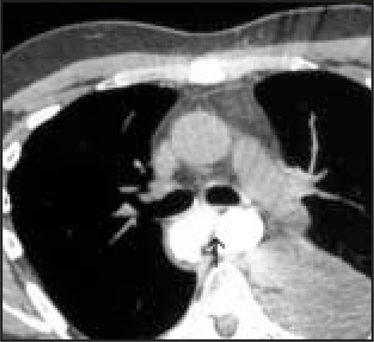
A 55-year-old man with moderately differentiated adenocarcinoma of the esophagus underwent transhiatal esophagectomy with gastric pull-up. The postoperative course was complicated by persistent low-grade fevers. Subsequent cine-esophagram showed a small amount of extravasation from the anastomosis. Because of the complex postoperative anatomy, the patient was referred for a thoracic computed tomography (CT) to evaluate for other complications.
IMAGING FINDINGS
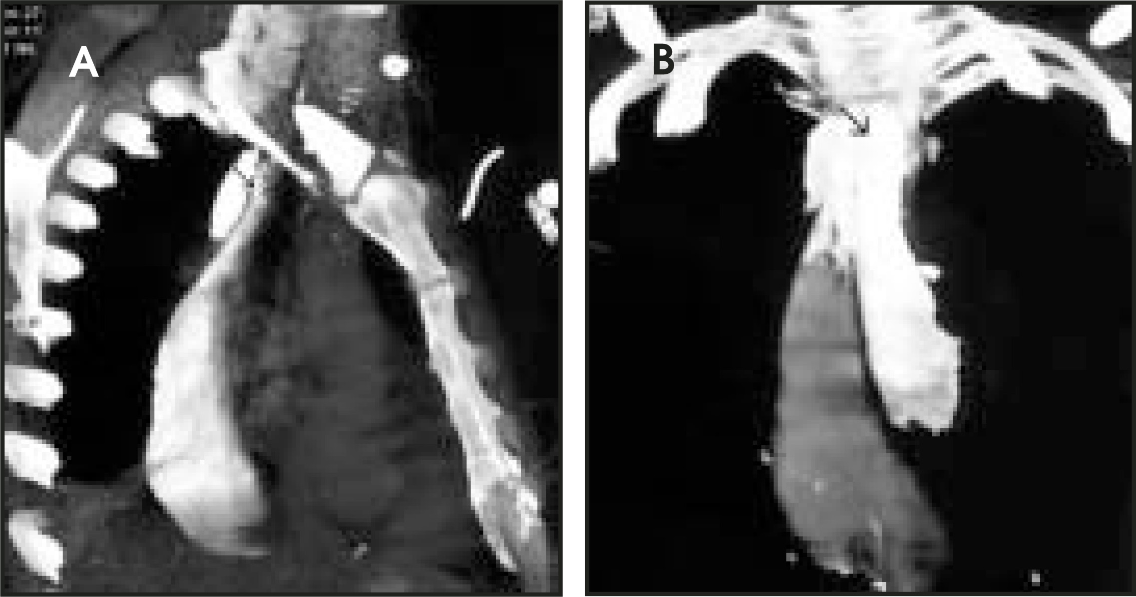
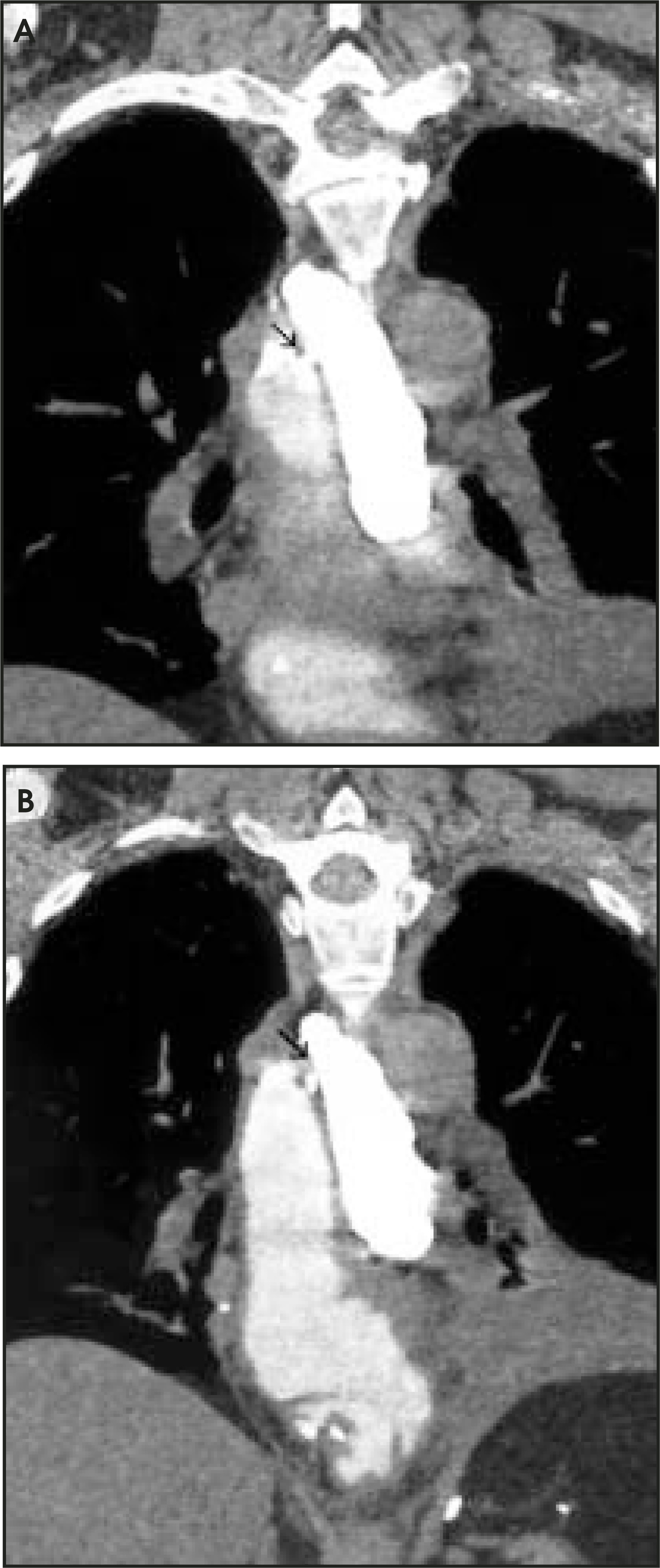
Helical CT of the chest (Figure 1) with 3-dimensional (3D) reconstructions (Figures 2 and 3) was performed with only oral contrast. The CT also depicts the anastomotic leak. A dense collection of contrast was present to the left of the gastric pull-up and anterior to the spine, extending from the thoracic inlet to the level of the left atrium, because of a leak at the anastomosis.
DIAGNOSIS
Anastomotic leak status post partial esophagectomy and gastric pull-up
DISCUSSION
Traditionally, cine-esophagrams have been used to evaluate postoperative anatomy and complications following esophagectomy and gastric pull-up. The development of multidetector computed tomography (MDCT) and 3D software now allows new methods for visualization and evaluation of this complex postoperative anatomy.
Cine-esophagram is widely used in the analysis of esophagectomy patients to evaluate for postoperative complications, such as leaking from the anastomotic site. CT has also been used to look for subtle leaks or postoperative complications because of better spatial anatomy.1,2
With the advent of 3D imaging, this complex postoperative anatomy becomes even clearer. Reconstructions can be performed in any plane. Our surgeons have found this more useful and helpful for planning repeat surgeries, as images can be reproduced in a familiar format. Three-dimensional imaging can manipulate the data set to resolve issues that are confusing in the axial plane alone, such as determining the site of the leak or the extent of fluid collection. As demonstrated in this case, the site of leak, the pooling of contrast, and their relationship to the gastric pull-up are easier to visualize on the coronal and sagital reconstructed images than on the axial plane alone.
CONCLUSION
Three-dimensional CT can provide additional information and can be a problem-solving tool in complicated postesophagectomy patients. Reconstructed images often make it easier for the referring surgeon to visualize the anatomy and pathology in order to plan necessary therapy.
References
- Heiken J, Balfe D, Roper C. CT evaluation after esophagogastrectomy. AJR Am J Roentgenol. 1984;143:555-560.
- Gimenez A, Franquet T, Erasmus J. Thoracic complications of esophageal disorders. RadioGraphics. 2002;22:S247-258.
Citation
. RCOM RADIOLOGICAL CASE OF THE MONTH. Applied Radiology. 2005;34(12):38-40. doi:10.37549/AR1388.