RCOM RADIOLOGICAL CASE OF THE MONTH
Applied Radiology — Vol. 35 , Issue 2 , pp. 41 -43
DOI: 10.37549/AR1403
Published: February 1, 2006
Categories
CASE SUMMARY
A 27-year-old white woman presented with symptoms of hyperglycemia, weakness, rash, and facial swelling, suggestive of Cushing’s syndrome. An ectopic focus of adrenocorticotropic hormone (ACTH) secretion was suspected from subsequent studies. Computed tomography (CT) of the chest, abdomen, and pelvis was unremarkable at her initial presentation. Although no specific therapy was initiated, the patient’s symptoms resolved over a period of several months.
Two years later, she presented with recurrent similar symptoms. Magnetic resonance imaging (MRI) of the thorax showed a mediastinal mass (Figure 1). As part of the staging evaluation, MRI and CT of the abdomen were also performed and revealed symmetric adrenal gland enlargement (Figure 2). The mass was removed via median sternotomy and was found to be a carcinoid tumor of the thymus. Postoperatively, the adrenal glands returned to normal size and shape.
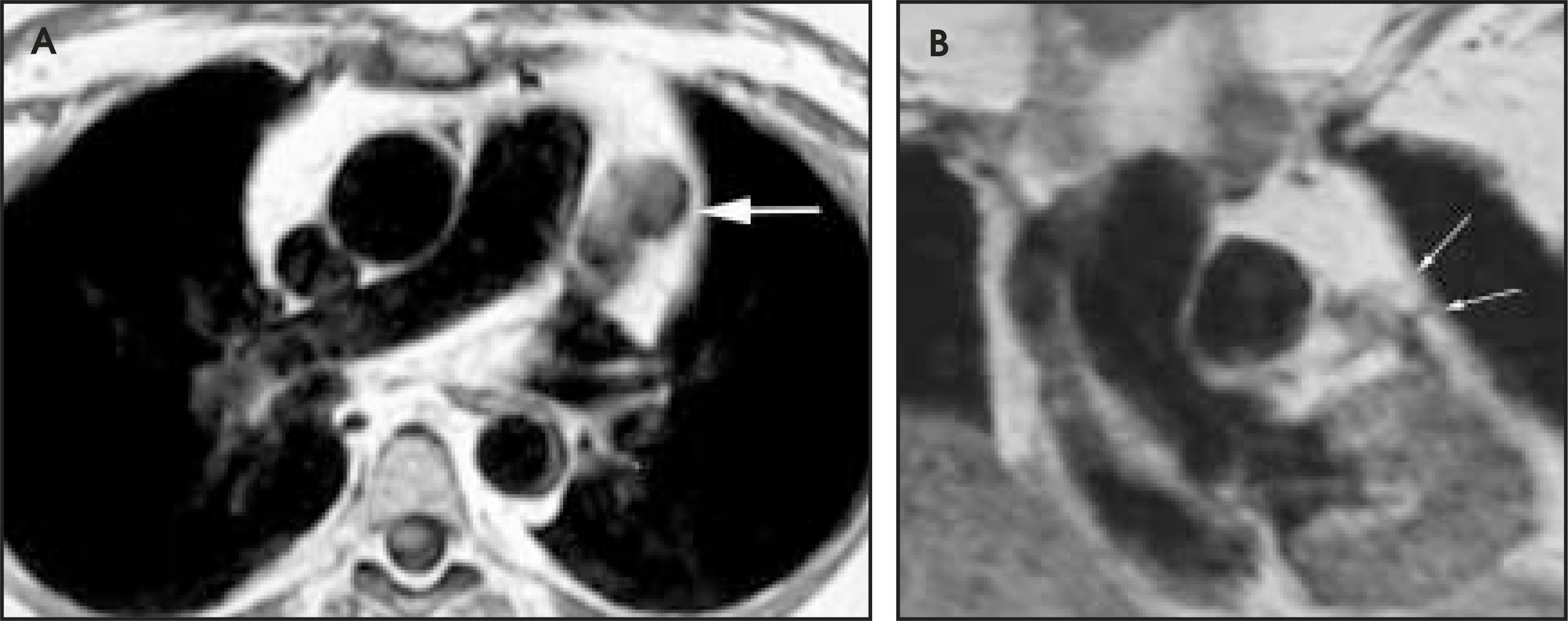
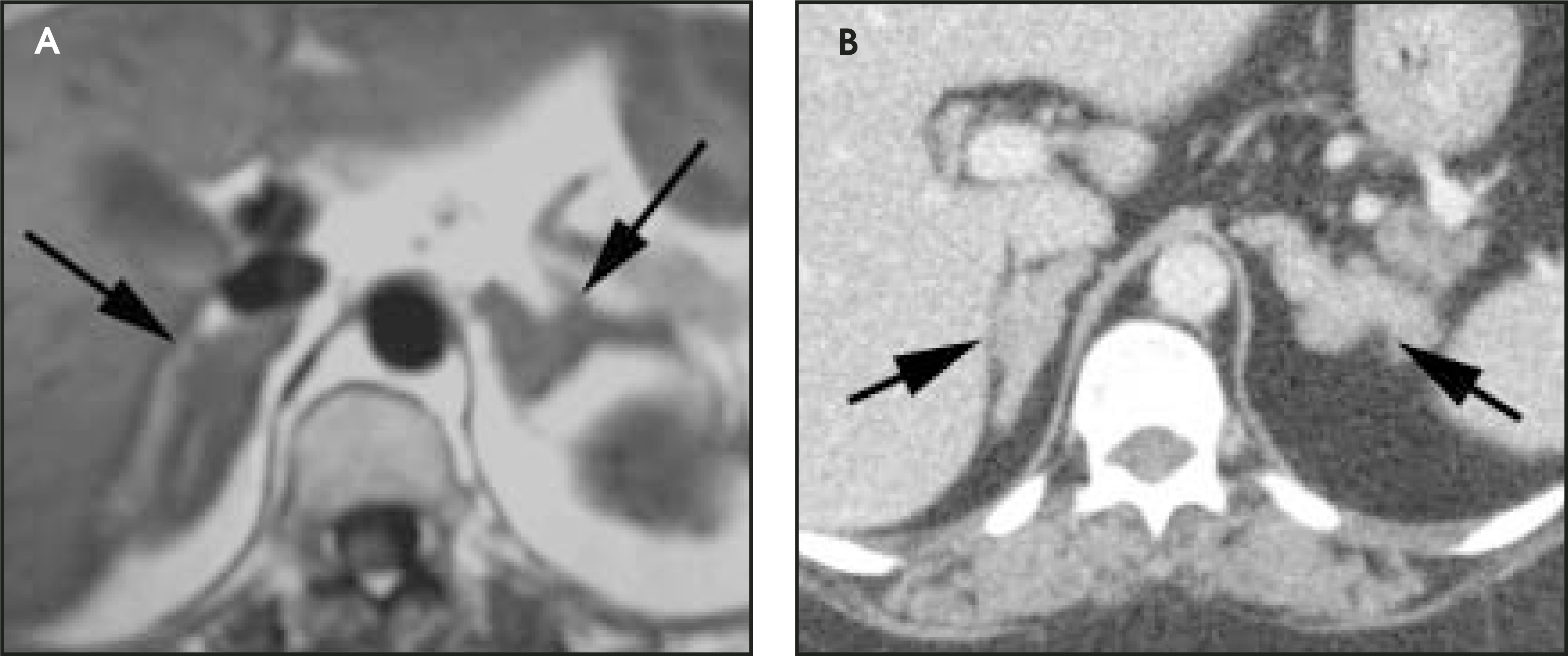
IMAGING FINDINGS
DIAGNOSIS
Thymic carcinoid with ectopic adrenocorticotropic hormone (ACTH) production
DISCUSSION
The term “karzinoide” (now carcinoid) was coined in 1907 by Oberndorfer in order to separate these tumors that have more indolent behavior from adenocarcinomas.1 Derived from Kulchitsky cells, these tumors can arise anywhere along the gastrointestinal tract or bronchial mucosa. The appendix is the most common site, but other relatively common areas of involvement include the rectum, ileum, lungs, and bronchi.1 Release of serotonin is thought to produce the flushing, diarrhea, and wheezing associated with carcinoid syndrome.1 Carcinoid tumors have been associated with other paraneoplastic syndromes, including the ectopic production of adrenocorticotropic hormone (Cushing’s syndrome) and growth-hormone–releasing factor (acromegaly). This usually heralds the presence of metastatic disease.
Thymic carcinoids are rare malignant tumors that account for approximately 2% to 4% of anterior mediastinal neoplasms, with just >200 cases reported in the literature.2,3 Clinically, patients may be asymptomatic or may present with symptoms of compression of the anterior mediastinal structures, endocrine abnormalities, or metastatic disease. Approximately half of thymic carcinoid patients have functionally active tumors, with 33% to 40% of these producing ACTH with associated Cushing’s syndrome.4 Multiple endocrine neoplasia type 1 (MEN-1) is also seen in 25% of patients with thymic carcinoid.4,5 There is a male preponderance, and the tumors tend to be more aggressive in patients with thymic carcinoid who also have MEN-1. Carcinoid of the thymus can range from well differentiated to very poorly differentiated. About 80% of cases show aggressive behavior, and both local recurrence and distant metastasis are common.3,4 As a result, surgical excision is necessary for treatment of the primary tumor or subsequent recurrence.4 This patient had multiple lymph node recurrences heralded by the return of Cushing’s-type symptoms. Although radiation therapy to the lymph nodes resulted in palliation of symptoms, 6 years later a bilateral adrenalectomy with adrenal hormone replacement was undertaken as definitive therapy for recurrent symptoms. The prognosis for this disease is poor, with a 5-year survival rate of 27%.2 It should be noted that carcinoid syndrome has not been associated with thymic carcinoids.6
Radiographically, these lesions are seen as large anterior mediastinal masses. CT and MRI of thymic carcinoid reveal a large, heterogeneous, lobulated mass, occasionally with areas of hemorrhage and central necrosis.2 Calcifications may be present. The differential diagnosis for thymic carcinoid is that of the “anterior mediastinal mass”: thymoma, thymic carcinoma, lymphoma, and germ cell neoplasms.2 Clinically, thymic carcinoid can be best differentiated from other diagnoses by the presence of symptoms that suggest a functional endocrine tumor. In the absence of a paraneoplastic process, it is much more difficult to establish the diagnosis. Thymoma, lymphoma, and germ cell neoplasms can all have a similar appearance either as a well-defined lobular mass (thymoma and thymic lymphoma) or heterogeneous attenuation and calcification (thymoma and germ cell neoplasm). The presence of adenopathy elsewhere in the mediastinum will favor a diagnosis of lymphoma, but thymic carcinoids do metastasize to regional lymph nodes. Thymic carcinomas tend to be poorly defined and infiltrative, giving a more aggressive appearance than is usually seen with a thymic carcinoid.
CONCLUSION
Thymic carcinoid is a rare, malignant disease with a poor prognosis. This condition should be suspected in individuals who have symp
toms of endocrine abnormalities with an anterior mediastinal mass. Even with adequate surgical excision of the primary tumor, there should be close follow-up for recurrence because of the aggressive nature of the disease.
References
- Kulke M, Mayer R. Carcinoid tumors. N Engl J Med. 1999;18.
- Chaer R, Massad M, Evans A. Primary neuroendocrine tumors of the thymus. Ann Thorac Surg. 2002;74:1733-1740.
- Moran C, Suster S. Neuroendocrine carcinomas (carcinoid tumor) of the thymus. A clinicopathologic analysis of 80 cases. Am J Clin Pathol. 2000;114:100-110.
- Rosado de Christenson M, Abbott G, Kirejczyk W. Thoracic carcinoids: Radiologic-pathologic correlation. RadioGraphics. 1999;19:707-736.
- Tiffet O, Nicholson A, Ladas G. A clinicopathologic study of 12 neuroendocrine tumors arising in the thymus. Chest. 2003;124:141-146.
- Dusmet M, McKneally M. Pulmonary and thymic carcinoid tumors. World J Surg. 1996;20:189-195.
Citation
. RCOM RADIOLOGICAL CASE OF THE MONTH. Applied Radiology. 2006;35(2):41-43. doi:10.37549/AR1403.