RCOM RADIOLOGICAL CASE OF THE MONTH
Applied Radiology — Vol. 35 , Issue 3 , pp. 44 -47
DOI: 10.37549/AR1409
Published: March 1, 2006
Categories
CASE SUMMARY
A 35-year-old man, who was suspected of having Hansen’s disease, presented to the dermatology outpatient department with a swelling on the lateral aspect of the right leg, just below the knee. The swelling was well defined and cystic and was approximately 3 × 2 cm in size. There was evidence of sensory deficit on the lateral aspect of the lower leg and dorsum of the foot. The results of routine investigations, including blood laboratory values, were normal.
IMAGING FINDINGS
Ultrasonography of the swelling was performed using a 7.5-MHz transducer. A well-defined elliptical lesion was seen on the lateral aspect of the leg just below the knee joint. The central portion of this lesion was clearly anechoic, whereas the walls were thickened, irregular, and echogenic (Figures 1 and 2). On the basis of the finding of a tubular, linear lesion along the course of a nerve, the differential diagnosis included organized hematoma, abscess, or mass/tumor. With the clinical picture, a diagnosis of leprotic nerve abscess was made.
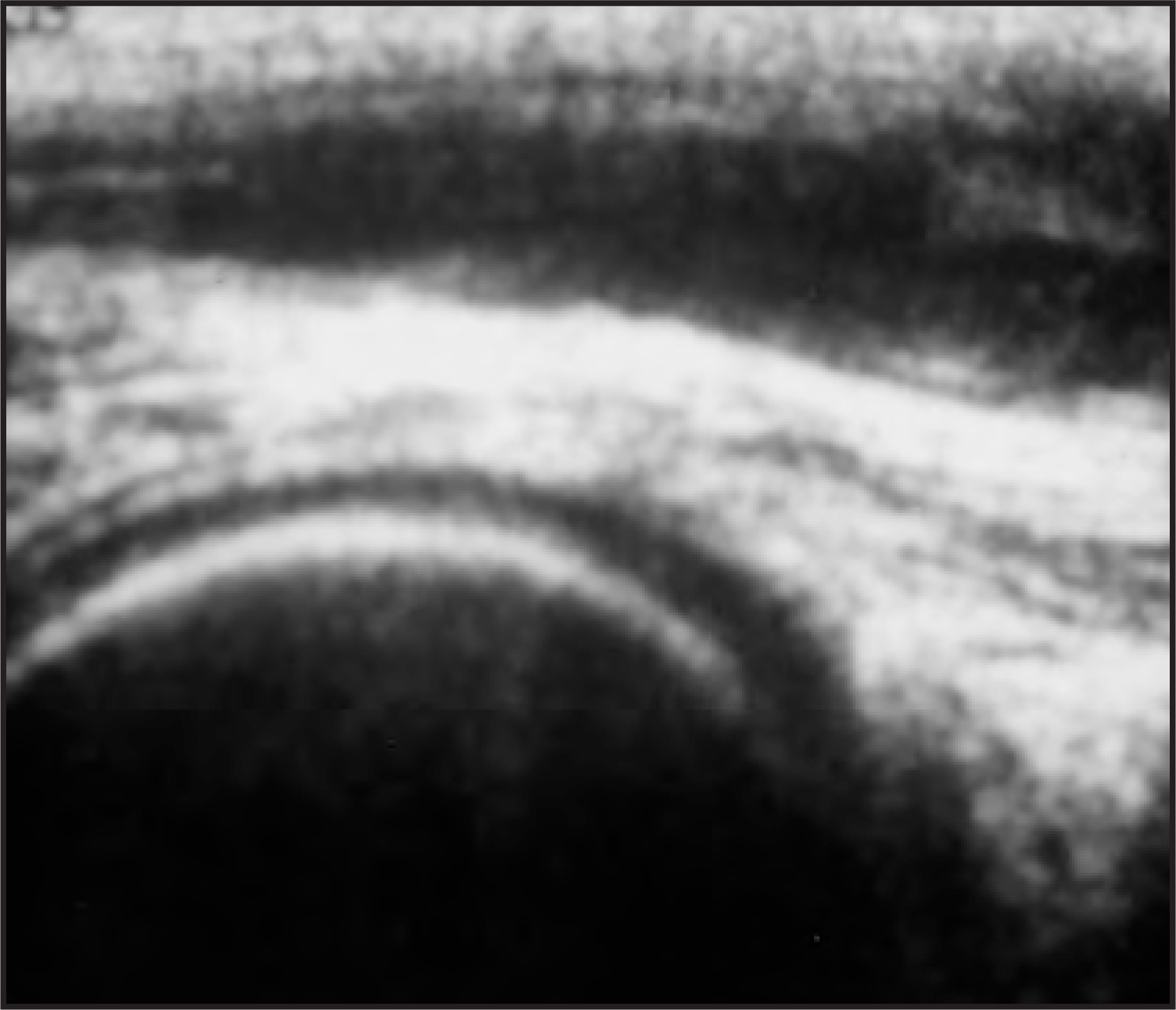
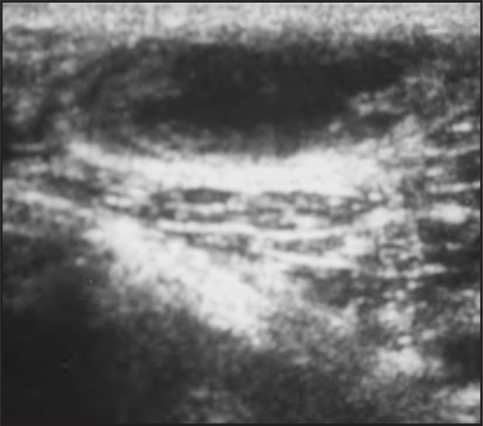
At surgery, this swelling was identified as an enlarged lateral popliteal nerve. On incision, the walls of the abscess were thick and irregular, and dirty yellow pus was drained. The pus smear was positive for Mycobacterium leprae.
DIAGNOSIS
Leprotic nerve abscess
DISCUSSION
Bacterial parasitization of peripheral nerves is a unique feature that is characteristic of leprosy. In most instances, the resulting neural lesion remains as a granuloma, but in a few cases the granuloma may soften and develop into an “abscess.” Progression to abscess formation is most commonly seen in patients with tuberculoid leprosy. Rarely, however, nerve abscess may also develop in other types of leprosy.1
Mycobacterium leprae has a predilection for peripheral nerves. The bacterial dissemination may be hematogenous or by contiguous spread from skin. The usual habitat in the nerve is the Schwann cell but occasionally the ensheathed axon becomes involved. The Schwann cells assume a phagocytic function and evolve into macrophages or epitheloid cells, resulting in the formation of a granuloma. Invasion of the endoneurium may follow, and the whole endoneurial zone may appear to be occupied by epitheloid cells with or without the presence of bacilli. Caseation may occur in microscopic foci within the granulomas, or areas of necrosis may coalesce, forming a cold abscess, particularly when the immunity is high. Cold abscesses occur more frequently in the tuberculoid form, especially in India.2,3
High-resolution ultrasound can effectively assess the preoperative 3-dimensional localization and the internal echotexture of masses arising from peripheral nerves. High-resolution sonography has identified normal peripheral nerves, all having echogenic fibrillar echotexture. A tubular/linear hypoechoic lesion along the course of a nerve should suggest an organized hematoma, an abscess, or a mass/tumor. In the appropriate clinical settings, a diagnosis of nerve abscess can be considered. In this case, the central anechoic area was frank pus, which was positive for lepra bacilli, surrounded by a thickened nerve sheath. This appearance may be typical of a cold abscess of a superficial nerve in leprosy.4
Once the nerve abscess has been detected, it has to be incised and drained; otherwise, it may rupture through skin onto the skin’s surface. Surgical incision and drainage without disturbing the intact nerve fibers is the method of choice. It has been suggested that an operation must be undertaken to rid the patient of the deleterious complications that may result because of persistent pressure on the nerve fibers that is caused by the tension within the nerve sheath.5,6
CONCLUSION
The case reported here illustrates the sonographic appearance and management of nerve abscess, a rare complication exclusive to leprosy. Leprotic nerve abscess is an extremely rare manifestation of leprosy. However, in the appropriate clinical settings and with the characteristic sonographic appearance, a diagnosis of nerve abscess should be considered, especially in endemic areas.
References
- Kumar P, Saxena R, Mohan L. Peripheral nerve abscess in leprosy: Report of twenty cases. Indian J Lepr. 1997;69:143-147.
- Char G, Cross J. Ulnar nerve abscess in Hansen’s disease. West Indian Med J. 1986;35(1):66-68.
- Singh G, Ojha D. Leprotic nerve abscesses. Dermatologica. 1969;139:409-412.
- Taneja K, Sethi A, Shiv V. Diagnosis of nerve abscess in leprosy by sonography. Indian J Lepr. 1992;64:105-107.
- Sehgal V, Tuli S. Leprotic nerve abscess: A case report. Indian J Dermatol. 1967;13(1):19-20.
- Browne S. Leprous nerve abscess: Report of two cases. Lepr Rev. 1957;28(1):20-24.
Citation
. RCOM RADIOLOGICAL CASE OF THE MONTH. Applied Radiology. 2006;35(3):44-47. doi:10.37549/AR1409.