RCOM RADIOLOGICAL CASE OF THE MONTH
Applied Radiology — Vol. 35 , Issue 3 , pp. 40 -43
DOI: 10.37549/AR1412
Published: March 1, 2006
Categories
CASE SUMMARY
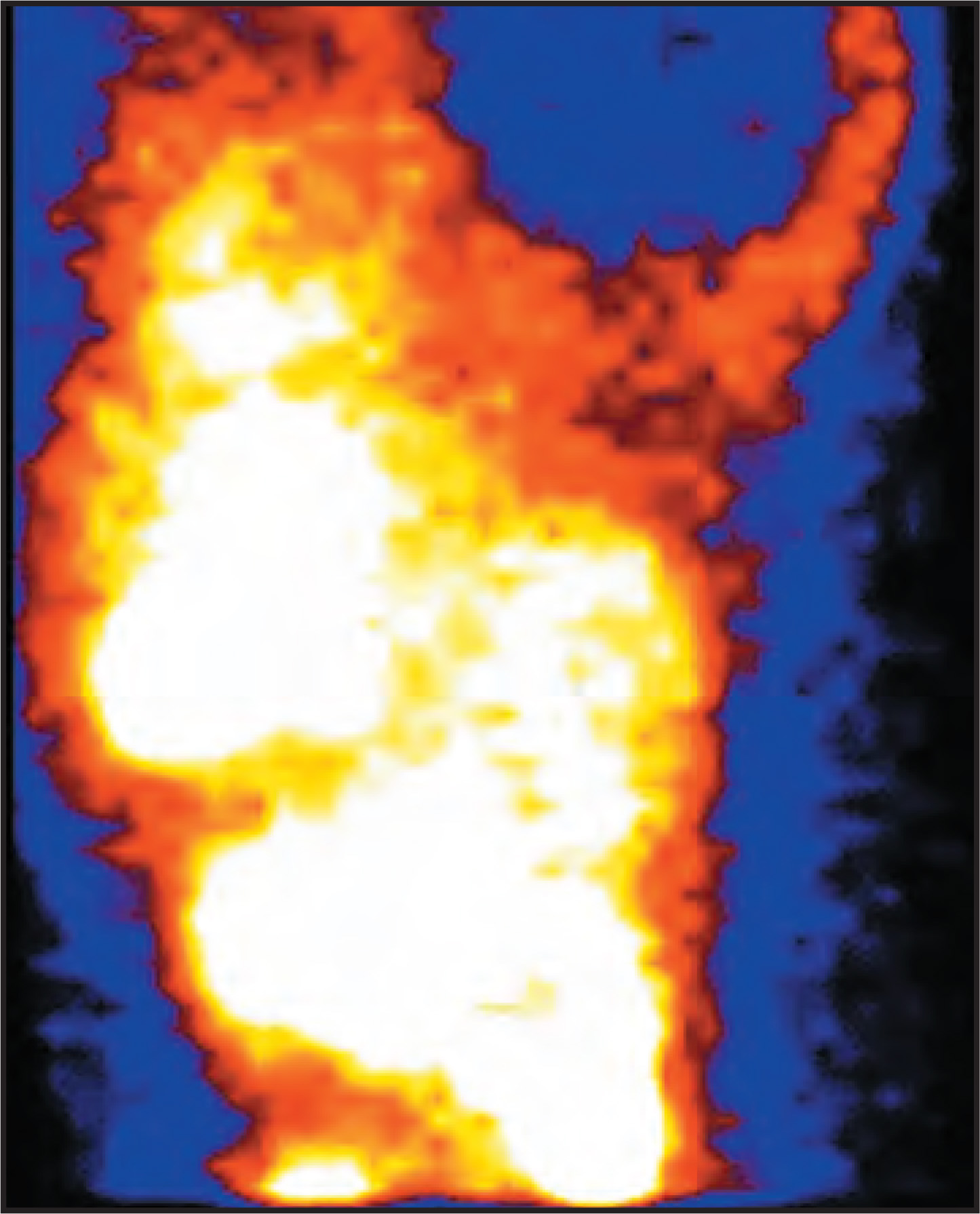
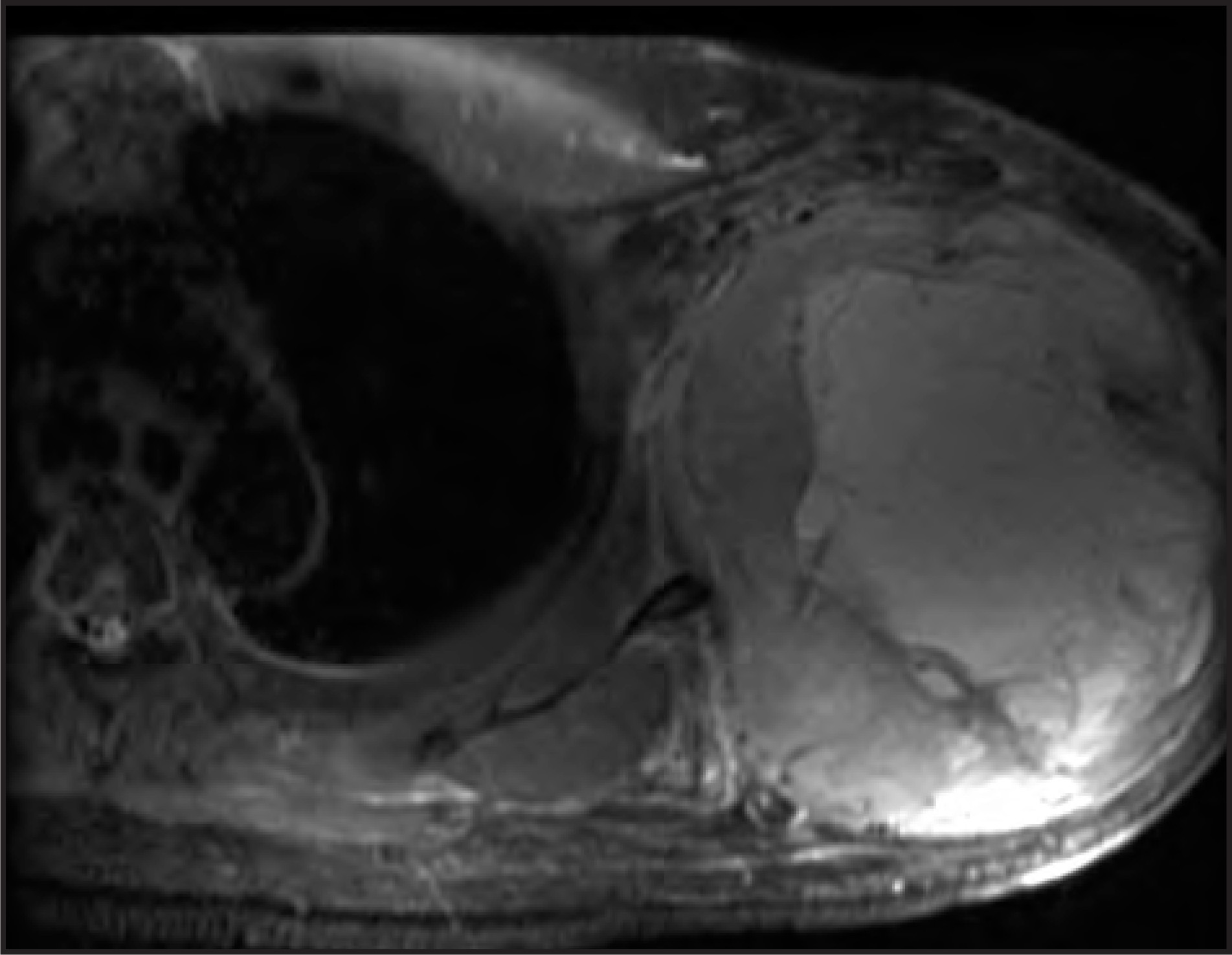
A 57-year-old man with a history of smoking, hypertension, and diabetes presented with chest pain and an enlarging left axillary mass (LAM). Physical examination was significant for a large LAM. Myocardial perfusion imaging (MPI) (Figure 1) and magnetic resonance imaging (MRI) of the left axilla and chest (Figure 2) were performed because of his cardiac risk factors and to evaluate the LAM.
IMAGING FINDINGS
DIAGNOSIS
Malignant fibrous histiocytoma (MFH)
PATHOLOGIC FINDINGS
Fibrous histiocytoma was suspected based on the results of a core needle biopsy. The patient underwent amputation of the left upper extremity and dissection of a LAM.
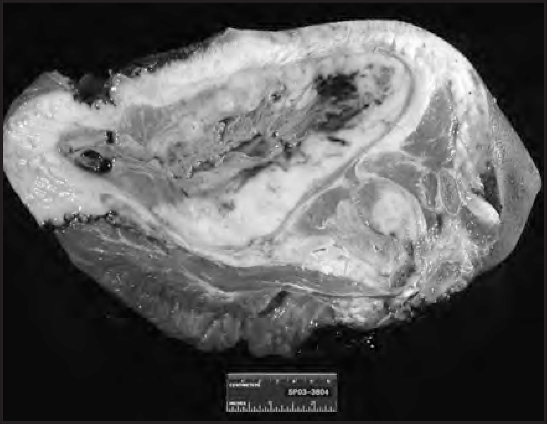
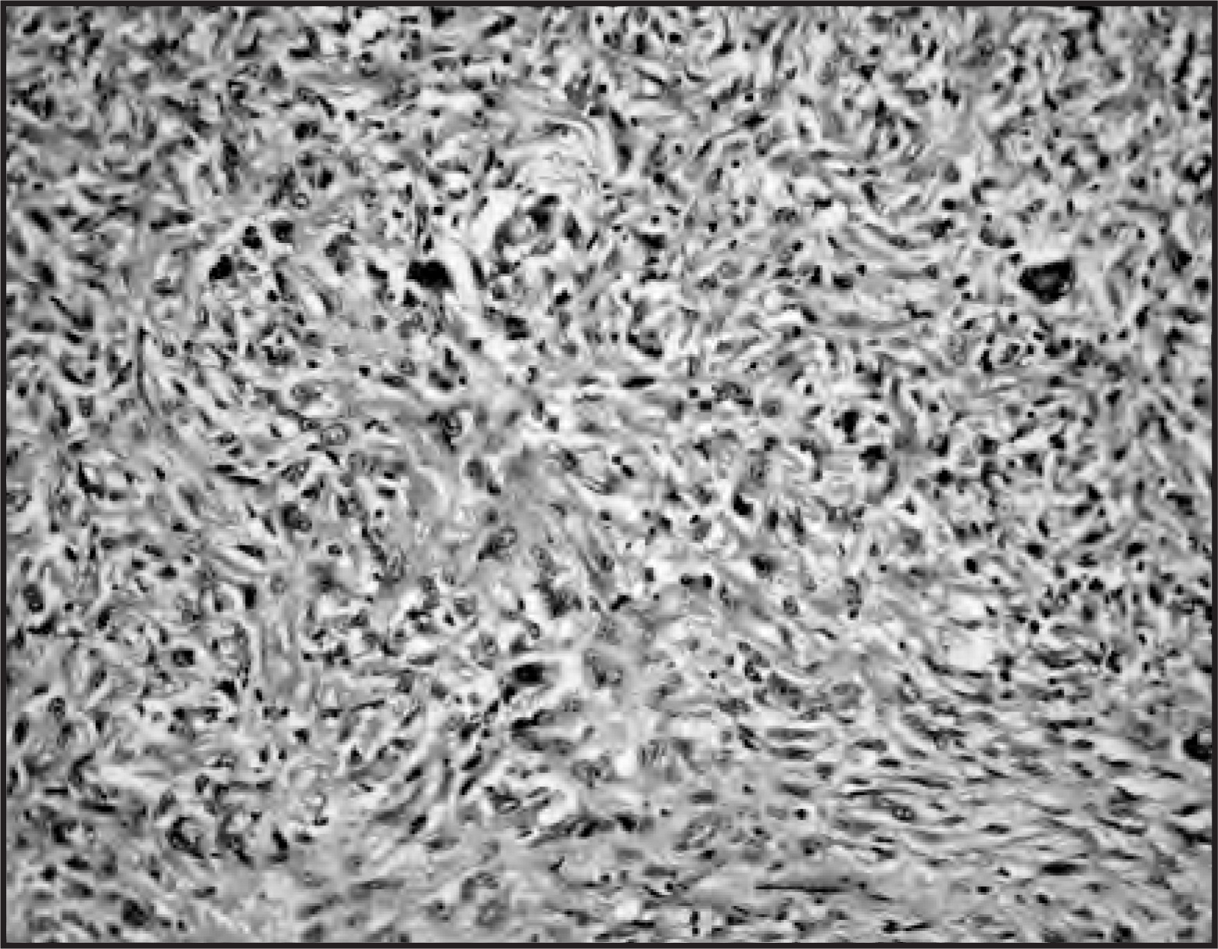
The gross specimen and histopathology of the LAM confirmed the diagnosis of malignant fibrous histiocytoma. A gross photomacrograph section of the tumor, which involved the left shoulder, shows the grayish white mass with areas of hemorrhage and necrosis, which are typical of MFH (Figure 3). A photomicrograph of the tumor shows storiform-pleomorphic pattern with anaplastic cells arranged haphazardly (Figure 4).
The patient received external radiation with subsequent disease progression with the development of lung metastases.
DISCUSSION
Malignant fibrous histiocytoma is a tumor of late adulthood that occurs in men more commonly than women.1,2 Computed tomography (CT) and MRI have been widely used in the diagnosis and staging of MFH.3-5 Several radiopharmaceuticals have been shown to have avid uptake by MFH, including Tc-99m methylene diphosphonate (MDP), gallium-67 (Ga-67) citrate, Tc-99m sestamibi, Tc-99m hexamethylpropyleneamine oxime (HMPAO), indium-111(In-111)–labeled leukocytes, Tc-99m sulfur colloid, and Tc-99m–labeled antifibrin monoclonal antibody.6-12 Imaging with Ga-67 is useful for the evaluation of primary and metastatic MFH. A Tc-99m MDP bone scan is helpful only for the evaluation of bone involvement,6 although Tc-99m MDP has been shown to accumulate in soft tissue MFH because of the calcification or the iron content of the tumor itself.7,8 Phagocytosis by functional histiocytes is believed to be responsible for the accumulation of In-111–labeled leukocytes and Tc-99m sulfur colloid in MFH.9 Cross-linked fibrin deposits by Tc-99m–labeled Fab′ antifibrin monoclonal antibody accounted for MFH localization.10 Bleeding and inflammatory reaction with necrosis was noted in the accumulation of Tc-99m HMPAO in malignant abdominal tumors.11 However, imaging findings in MFH using Tc-99m tetrosfosmin have not been reported before. The unique feature of the Tc-99m tetrofosmin images reported here (Figure 1) is the rim of intense activity in the wall with a large pho-ton-deficient central area. This phenomenon is best explained by the necrosis of the core of the lesion and the presence of hemorrhagic fluid in the cystic mass. These findings are in contrast to those reported in MFH images with Tc-99m sestamibi in which an intense increased uptake had been noted throughout the tumor.12
CONCLUSION
Uptake of Tc-99m tetrofosmin in various tumors is well known. In this case, we report the appearance of MFH with a circular rim of increased radiotracer uptake and a central necrotic photopenic area.
References
- Enzinger F, Weiss S, Enzinger F, Weiss S. Soft Tissue Tumors. 1995:351-380.
- O’Brien J, Stout A. Malignant fibrous xanthomas. Cancer. 1964;17:1445-1455.
- Tateishi U, Kusumoto M, Hasegawa T. Primary malignant fibrous histiocytoma of the chest wall: CT and MR appearance. J Comp Assist Tomogr. 2002;26:558-563.
- Miller T, Hermann G, Abdelwahab I. MRI of malignant fibrous histiocytoma of soft tissue: Analysis of 13 cases with pathologic correlation. Skelet Radiol. 1994;23:271-275.
- Munk P, Sallomi D, Janzen D. Malignant fibrous histiocytoma of soft tissue imaging with emphasis on MRI. J Comput Assist Tomogr. 1998;22:819-826.
- Lin W, Kao C, Hsu C. The role of Tc-99m MDP and Ga-67 imaging in the clinical evaluation of malignant fibrous histiocytoma. Clin Nucl Med. 1994;19:996-1000.
- Spencer R, Evans D, Forouher F. Tc-99m diphosphonate uptake in malignant fibrous histiocytoma: A possible iron-related effect. Clin Nucl Med. 1988;13:734-735.
- Rosenthall L. 99MTc-methylene diphosphonate concentration in soft tissue malignant fibrous histiocytoma. Clin Nucl Med. 1978;3:58-61.
- Palestro C, Klein M, Kim C. Indium-111-labeled leukocyte and technetium-99m-sulfur colloid uptake by a malignant fibrous histiocytoma: Phagocytosis by tumor cells?. J Nuc Med. 1990;31:1548-1551.
- Angelides S, Lee F, Basten A. Detection of malignant tumors using Tc-99m labeled Fab' fragments from a monoclonal antibody with specificity for D-dimer of cross-linked fibrin. Clin Nuc Med. 1996;21:242-244.
- Lantto E, Jarvi K, Lantto T. Accumulation of leucocytes labelled with technetium-99m hexamethylpropylene amine oxime in malignant abdominal tumours. Eur J Nuc Med. 1991;18:824-828.
- Aktolun C, Berk F, Demir H, Ercin C. Tc-99m MIBI imaging in malignant fibrous histiocytoma. Ann Nuc Med. 2001;15:451-453.
Citation
. RCOM RADIOLOGICAL CASE OF THE MONTH. Applied Radiology. 2006;35(3):40-43. doi:10.37549/AR1412.