RCOM RADIOLOGICAL CASE OF THE MONTH
Applied Radiology — Vol. 35 , Issue 4 , pp. 46 -47
DOI: 10.37549/AR1413
Published: April 1, 2006
Categories
CASE SUMMARY
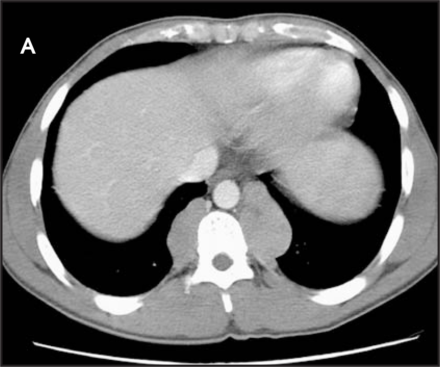
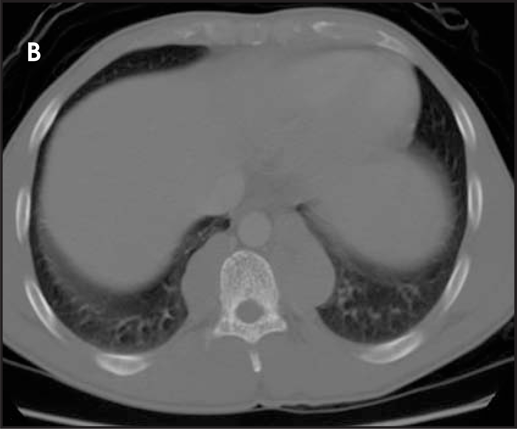
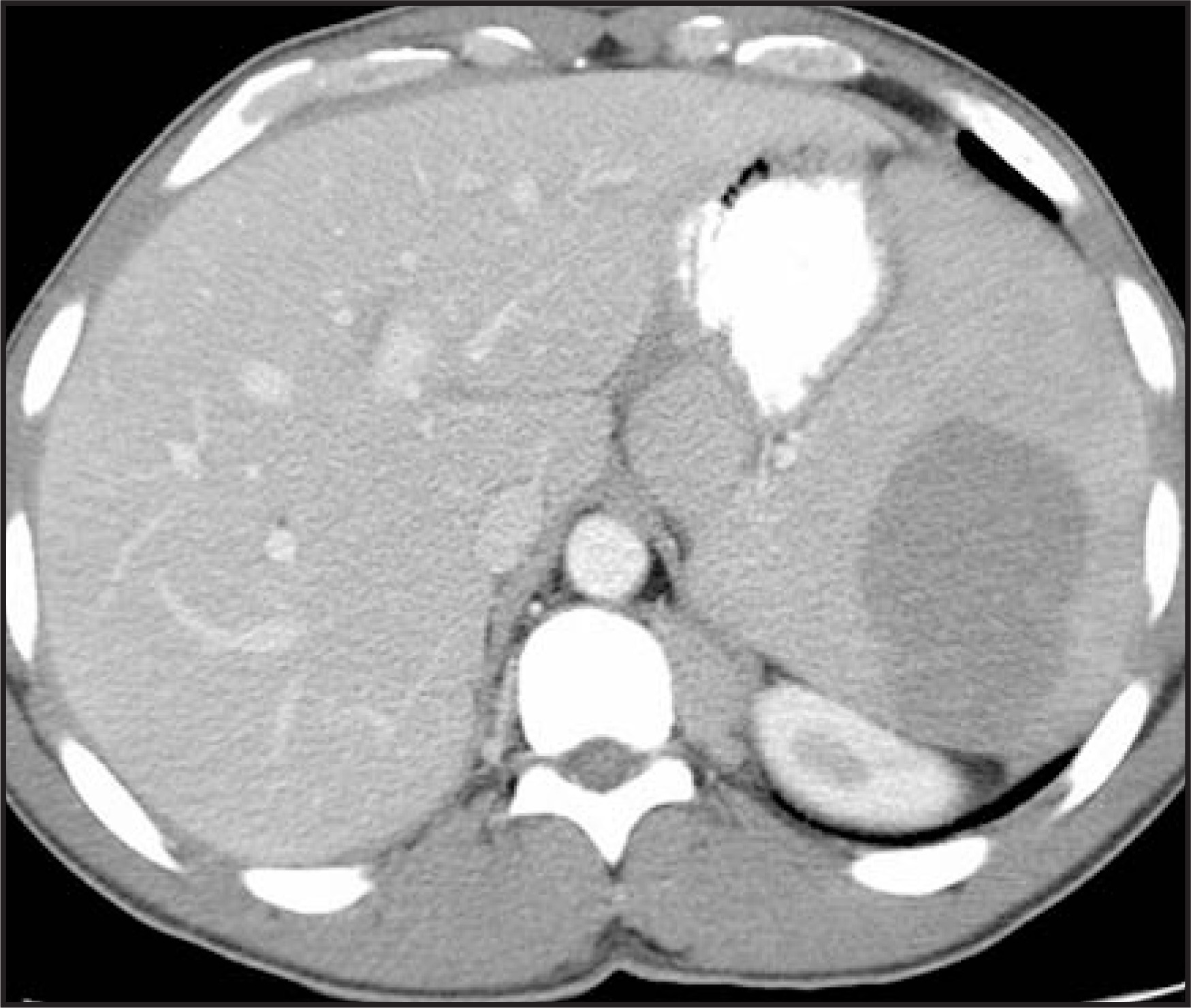
A 42-year-old man presented to the emergency department with back pain. He denied any trauma to the back. He had a history of thalassemia intermedia. Physical examination revealed an enlarged liver and spleen. Computed tomographic (CT) scans of the chest (Figures 1 and 2) and abdomen (Figure 3) were obtained.
DIAGNOSIS
Extramedullary hematopoiesis (EMH)
IMAGING FINDINGS
The CT scan of the chest revealed well-defined bilateral paraspinal masses in the lower thoracic regions with no calcification (Figure 1). Focal areas of low density were also seen in the masses. There were no erosive or extrinsic pressure changes on the adjacent vertebrae or ribs. The vertebrae, however, showed increased trabeculation (Figure 2). There was hepatosplenomegaly. A large hypodense lesion was seen in the spleen (Figure 3). The diagnosis of EMH was confirmed by fine-needle biopsy of the paraspinal masses.
DISCUSSION
Extramedullary hematopoiesis is the production of blood elements outside the bone marrow cavity. Until 20 weeks of prenatal life, blood-cell production occurs in the yolk sac and in the reticuloendothelial (RE) system. Thereafter, the bone marrow is the primary site of hematopoiesis. Normal blood production is maintained by the pluripotent stem cells.1 Extramedullary hematopoiesis occurs in severe chronic anemia, which can arise from deficiency disorders such as vitamin B12 or folate deficiency2 or from disorders affecting pluripotent stem cells. The latter are classified as: a) stem cell dysplasia, as in thalassemia, sickle cell hemoglobinopathy, and hereditary spherocytosis; b) stem cell failure resulting in aplastic anemia; c) uncontrolled stem cell proliferation, as in polycythemia vera; and d) malignant transformation and replacement, as in leukemia, lymphoma, multiple myeloma, and metastasis.1 Extramedullary hematopoiesis mainly involves the RE system (liver, spleen, and lymph nodes) but is also known to occur in every organ of the body, including the thyroid, prostate, pericardium, kidney, and lungs.3 Most often, it is microscopic and asymptomatic, but it can sometimes manifest as organomegaly and tumor-like masses.2 Rarely, it can cause cord compression, pleural effusion, massive hemothorax, and respiratory failure.4 Intrathoracic EMH is a rare cause of paraspinal mass5 and should be differentiated from other common causes, such as neurogenic tumors, lymphoma, metastasis, paravertebral abscess, and lateral meningocele.
Bone changes are due to marrow expansion and reconversion. The latter is best seen by magnetic resonance imaging examination. There is thinning of the cortices, underconstriction of long bones, and widening of ribs. The trabeculae are resorbed, giving the bones a coarsened appearance (Figure 2). The tumor-like masses are usually slow-growing and do not cause bone erosion or pressure changes. Older, inactive masses may reveal iron deposits and/or fatty replacement (Figure 1)5 and do not calcify. Radionucleide imaging with technetium-99m sulfur colloid may reveal extrahepatosplenic uptake of colloid. Fine-needle biopsy can confirm the diagnosis.
CONCLUSION
Extramedullary hematopoiesis occurs as a compensatory mechanism for abnormal hematopoiesis when normal red marrow is unable to function because of deficiency disorders or because of various pluripotent stem cell disorders. Most often, it is microscopic and asymptomatic but can sometimes produce organomegaly, tumor-like masses and even life-threatening symptoms. Extramedullary hematopoiesis should be considered in the differential diagnosis in a patient with chronic severe anemia and a paraspinal mass.
References
- Steiner R, Mitchell D, Rao V. Magnetic resonance imaging of bone marrow: Diagnostic value in diffuse hematologic disorders. Magn Reson Q. 1990;6:17-34.
- Bolaman Z, Polatli M, Cildag O. Intrathoracic extramedullary hematopoiesis resembling posterior mediastinal tumor. Am J Med. 2002;112:739-741.
- Yusen R, Kollef M. Acute respiratory failure due to extra-medullary hematopoiesis. Chest. 1995;108:1170-1172.
- Turgut B, Pamuk G, Vural O. An interesting presentation of intrathoracic extramedullary hematopoiesis In a patient with thalassemia intermedia. Clin Lab Haematol. 2003;25:409-412.
- Lall C, Payne D. A patient with anemia and a paraspinal chest mass. Chest. 2003;124:732-734.
Citation
. RCOM RADIOLOGICAL CASE OF THE MONTH. Applied Radiology. 2006;35(4):46-47. doi:10.37549/AR1413.