RCOM RADIOLOGICAL CASE OF THE MONTH
Applied Radiology — Vol. 35 , Issue 4 , pp. 53 -55
DOI: 10.37549/AR1418
Published: April 1, 2006
Categories
CASE SUMMARY
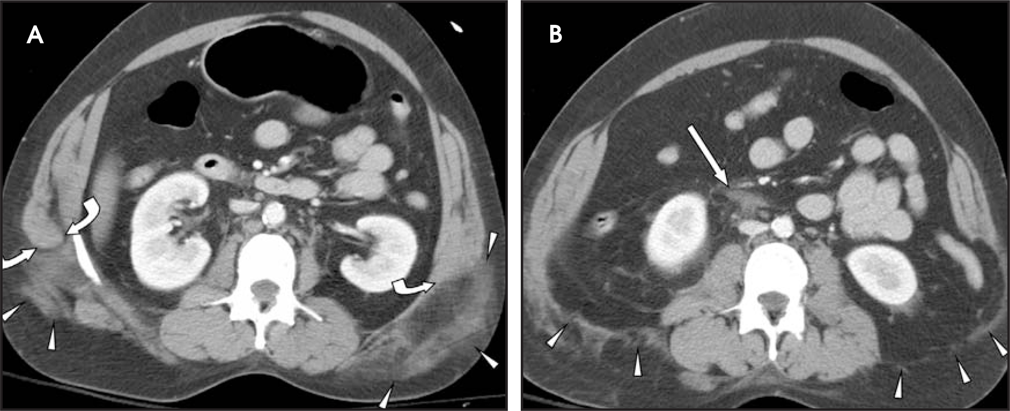
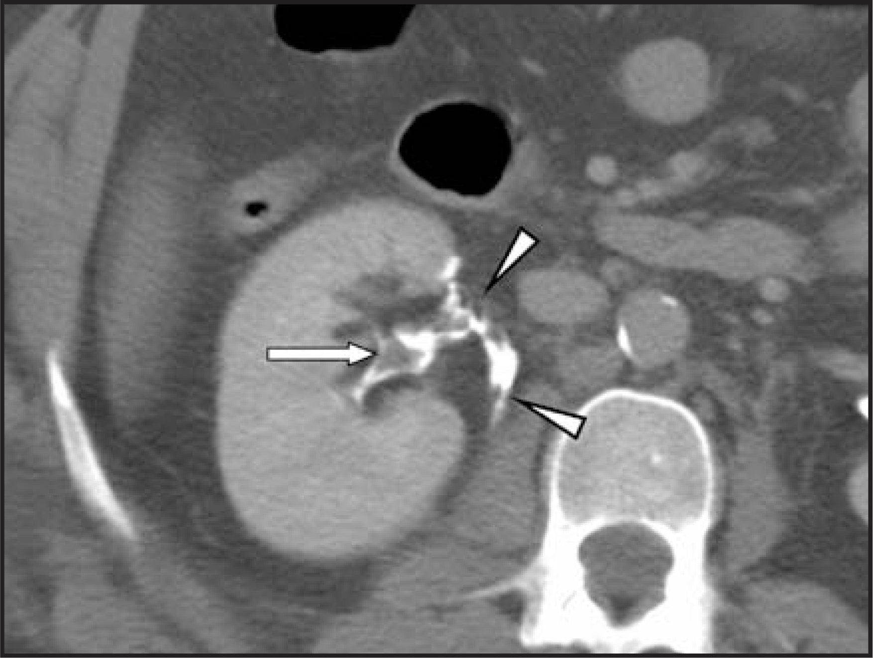
A 39-year-old restrained driver who had been involved in a motor vehicle collision presented to the University of Maryland R. Adams Cowley Shock Trauma Center, Baltimore, MD, with an abdominal “seatbelt contusion,” and a deep right lower quadrant soft tissue laceration. The patient also complained of low back pain. Computed tomography (CT) was performed on admission (Figures 1 and 2).
DIAGNOSIS
Bilateral traumatic lumbar hernias with injuries of the right renal pelvis and ureteropelvic junction (UPJ)
IMAGING FINDINGS
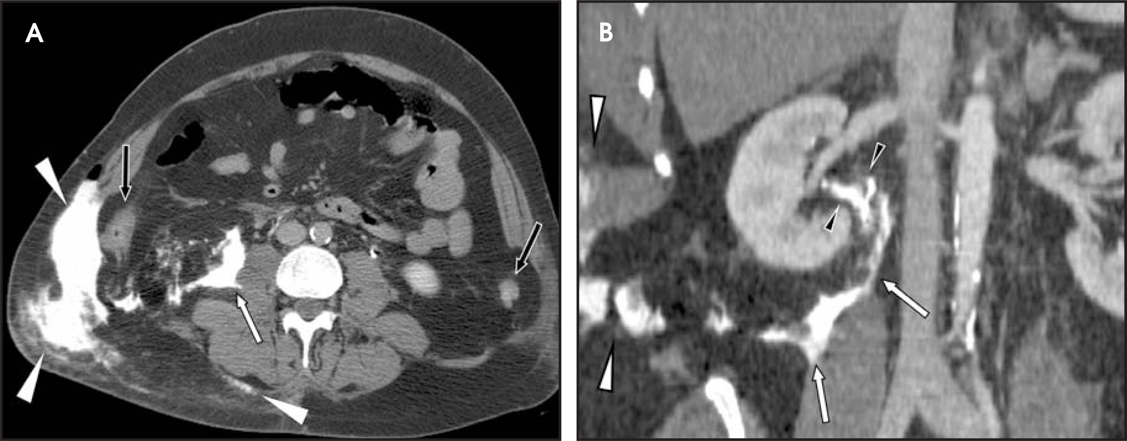
Multidetector (16-slice) CT images of the abdomen were obtained with both renal corticomedullary- and excretory-phase acquisitions. Early corticomedullary-phase images revealed bilateral traumatic lumbar hernias and minimal right-sided retroperitoneal fluid or hemorrhage (Figure 1), while the excretory-phase images revealed extravasation of contrast material near the right renal hilum (Figure 2). Scanning performed 7 hours later showed progressive urinary contrast extravasation into the retroperitoneum and the right lumbar hernia (Figure 3).
CLINICAL COURSE
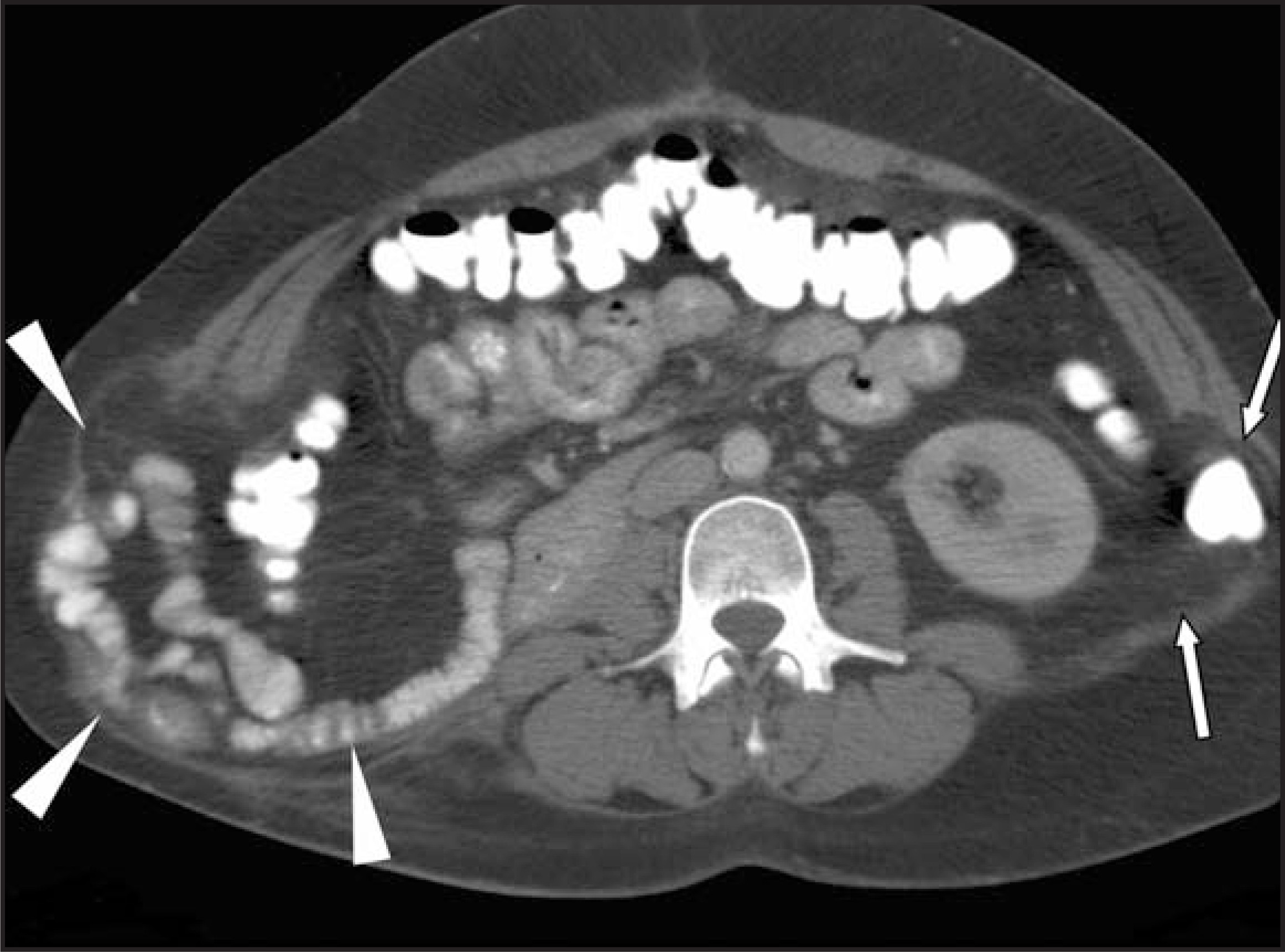
Emergent surgical exploration of the abdomen was performed to address the abdominal wall and UPJ injuries, in addition to other injuries. The attempted UPJ repair was unsuccessful, and, ultimately, a right nephrectomy was necessary. Herniated bowel and peritoneal fat were reduced, and the retroperitoneum was repaired. The abdominal wall musculature was macerated at the injured sites, and acute repair of their attachments was impossible. Following initial recovery and discharge, the patient returned 4 months later with a bulging right flank mass. Repeat CT scans of the abdomen revealed recurrent bilateral lumbar hernias containing bowel and intraperitoneal fat (Figure 4) that necessitated definitive repair of the abdominal wall defects.
DISCUSSION
Traumatic lumbar hernias are uncommon injuries that typically result from high-speed motor vehicular trauma. The proposed mechanism of injury involves transmission of force from a seatbelt to the abdomen during sudden deceleration. The resulting increase in intrabdominal pressure exerts sufficient force to rupture the abdominal wall musculature at a point of weakness.1,2 Injury typically occurs at either the superior lumbar (Grynfeltt-Lesshaft) triangle, which is bordered by the 12th rib superiorly, erector spinae muscle posteriorly, and internal oblique muscle anteriorly; or the inferior lumbar (Petit’s) triangle, which is bordered by the iliac crest inferiorly, external oblique muscle anteriorly, and latissimus dorsi muscle posteriorly.3 However, the injury may occur at other locations. Early recognition is important, since without surgical repair, the hernia can enlarge, thereby increasing long-term morbidity. An index of suspicion is necessary, since the high incidence of other intra-abdominal injuries may distract the radiologist from the correct diagnosis.4
Renal collecting system injuries are often unsuspected or initially overlooked. This can sometimes be the result of inadequate renal imaging, as major renal pelvic injuries may not be evident on routine medullary- or corticomedullary-phase imaging.6 As in this case, evidence of excreted urinary contrast extra-vasation on delayed (renal-excretory) phase imaging is often necessary for the diagnosis. While urine extravasation is not necessarily an indication for surgery, it is nonetheless important to recognize, since a delay in diagnosis can lead to increased morbidity due to urinoma formation, increased rate of nephrectomy, and longer hospital stays.7 The importance of delayed-phase imaging is not limited to evaluation of the urinary tract, since other injuries, such as slow active hemorrhage, may manifest only on delayed-phase images. For this reason, renal excretory-phase images are routinely acquired during initial CT evaluation of all high-impact, blunt abdominal trauma patients at the Shock Trauma Center.
CONCLUSION
Traumatic lumbar hernias are uncommon injuries that are typically associated with other intra-abdominal injuries that may garner greater immediate attention from the radiologist and trauma surgeon. Similarly, UPJ injuries may be easily overlooked, with delayed contrast-enhanced CT imaging improving sensitivity for this and other injuries. Since early repair reduces the morbidity associated with both injuries, a high index of suspicion should be maintained when assessing patients with signs of high-energy blunt trauma to the abdomen.
References
- Balkan M, Kozak O, Gulec B. Traumatic lumbar hernia due to seat belt injury: Case report. J Trauma. 1999;47:154-155.
- McCarthy M, Lemmon G. Traumatic lumbar hernia: A seat belt injury. J Trauma. 1996;40:121-122.
- Shanmuganathan K, Killeen K, Mirvis S, Shanmuganathan K. Imaging in Trauma and Critical Care. 2003:369-481.
- Killeen K, Girard S, DeMeo J. Using CT to diagnose traumatic lumbar hernia. AJR Am J Roentgenol. 2000;174:1413-1415.
- Barden B, Maull K. Traumatic lumbar hernia. South Med J. 2000;93:1067-1069.
- Mulligan J, Cagiannos I, Collins J, Millward S. Ureteropelvic junction disruption secondary to blunt trauma: Excretory phase imaging (delayed films) should help prevent a missed diagnosis. J Urol. 1998;159:67-70.
- Harris A, Zwirewich C, Lyburn I. CT findings in blunt renal trauma. RadioGraphics. 2001;21(Spec No):S201-S214.
Citation
. RCOM RADIOLOGICAL CASE OF THE MONTH. Applied Radiology. 2006;35(4):53-55. doi:10.37549/AR1418.