RCOM RADIOLOGICAL CASE OF THE MONTH
Applied Radiology — Vol. 35 , Issue 7 , pp. 53 -55
DOI: 10.37549/AR1441
Published: July 1, 2006
Categories
CASE SUMMARY
A 38-year-old woman with Child-Pugh class C alcoholic liver disease presented with recurrent bright red blood from her colostomy site, which had been placed as a result of long-standing Crohn’s disease. This 2-L bleed could not be controlled with manual compression. She had a transjugular intrahepatic portosystemic shunt (TIPS) implanted 2 years previously for refractory ascites. Her repeated episodes of encephalopathy had been best managed with diet and lactulose. Endoscopic evaluation identified enlarged varices proximal to a colonic stricture. These did not show recent bleeding. Despite active bleeding at the stoma site, endoscopy failed to identify a bleeding source. Thus, sclerotherapy and ligation were not attempted. Although surgical revision of the colostomy had been performed 3 years earlier, current depletion of clotting factors and the extent of varices precluded safe surgical revision at this time. A duplex ultrasound evaluation revealed a patent TIPS with a 47 cm/sec velocity across the TIPS. Interventional radiology was contacted for TIPS evaluation and potential variceal embolization.
DIAGNOSIS
Stomal bleed in a TIPS patient
IMAGING FINDINGS
The right internal jugular vein was accessed using ultrasound guidance with a micropuncture needle, and a 5F sheath was placed.
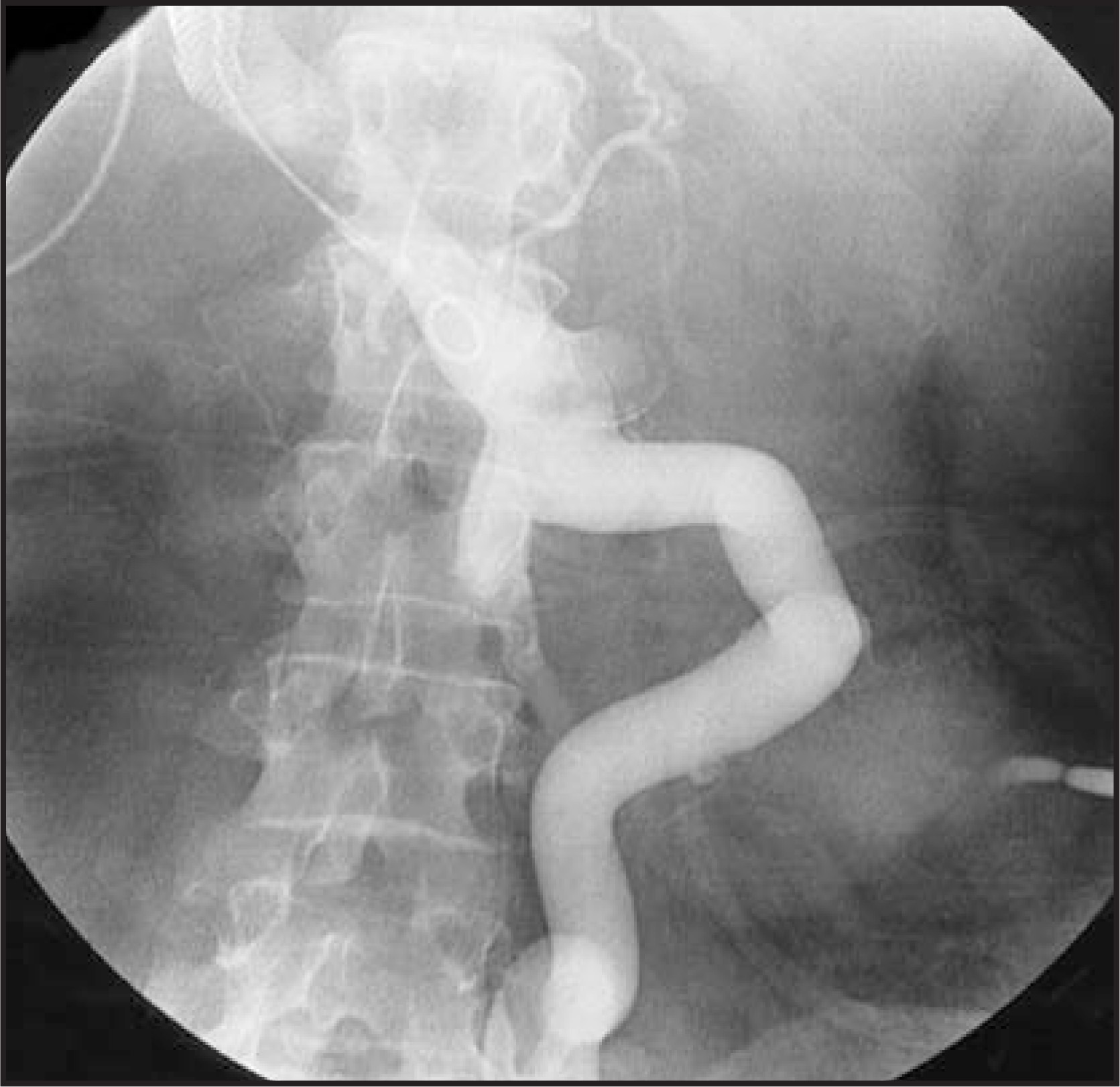
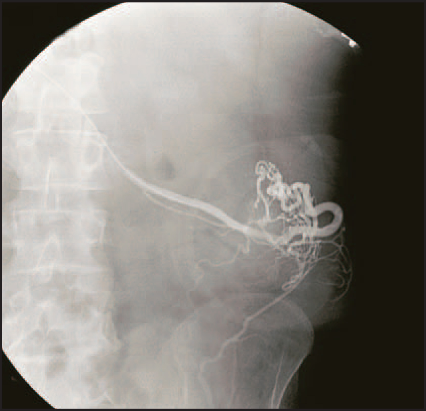
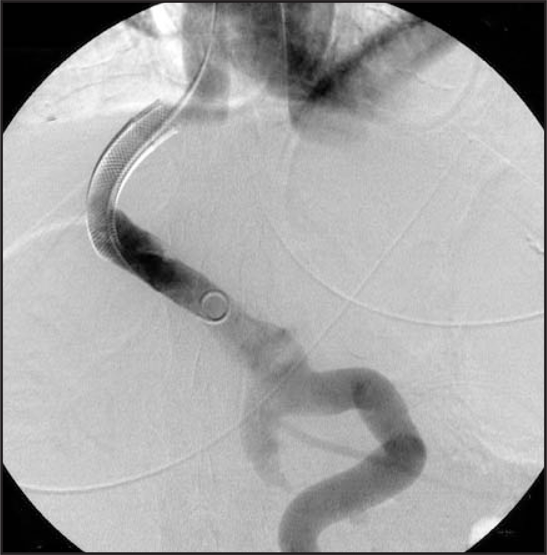
Over a 0.035-inch angled glide wire, a 5F Cobra 2 catheter (AngioDynamics Inc., Queensbury, NY) was passed into the right atrium, inferior vena cava (IVC), and portal vein, and venous pressures were recorded. The right hepatic vein to right portal vein TIPS stent was patent. A digital portogram was obtained (Figure 1). The pressures measured were as follows: portal vein 14 mm Hg, proximal shunt 9 mm Hg, mid shunt 6 mm Hg, and IVC 5 mm Hg. The portosystemic gradient was 9 mm Hg. Hepatofugal flow into a large inferior mesenteric vein and stomal varices was identified. The main stomal varix was selected using a 5F H1 catheter (Cook Inc., Bloomington, IN) (Figure 2). The H1 was exchanged for a 65-cm, 5F angled glide catheter, and embolization of the stomal varices was performed. A total of 4 mL of absolute ethanol was infused until hemostasis was achieved in the stomal varices (Figure 3). The catheter was removed, and the right internal jugular venotomy site was closed with 2-0 Ethilon suture (Ethicon, a Johnson & Johnson Co., Somerville, NJ). Immediate hemostasis was achieved, and the patient tolerated the procedure well.
The patient remained hemodynamically stable, and no further variceal hemorrhage was identified. The patient was discharged from the hospital and is currently living in a nursing facility. At 8-week follow-up after the variceal embolization, the patient had not had recurrent bleeding.
DISCUSSION
Patients with portal hypertension and surgical ostomies may occasionally develop stomal varices. The risk of death from bleeding in these patients is 3% to 4%.1 The varices form because of the anastomosis between the high-pressure portal system and the relatively low-pressure venous system of the abdominal wall.2 In 1968, Resnick3 first described stomal hemorrhage from ostomy varices in 3 patients who had undergone colostomies.
Stomal variceal bleeding is difficult to definitively treat. Even with endoscopy, the bleeding site may be difficult to locate and treat, and mesenteric angiography is often needed to pinpoint the bleeding site. Treatment options include suture ligation, colostomy revision, sclerotherapy, TIPS, surgical shunt creation, and percutaneous embolization.4 Stomal surgery may yield temporary success in the control of bleeding. However, multiple transfusions may be required. In addition, local surgical corrective measures can lead to scarring or stomal dysfunction.5 Surgical decompression of the portal venous system with a surgical shunt or TIPS placement is usually effective in preventing and controlling variceal hemorrhage. However, this treatment option may exacerbate liver failure or hepatic encephalopathy.6
Our patient had undergone prior colostomy revision and sclerotherapy treatments but was rejected for a liver transplant because of alcohol abuse. To our knowledge, this is the only patient who has a patent TIPS who failed all other treatment options with continued hemorrhage from the ostomy site. As a result, transjugular variceal embolization was performed with infusion of absolute ethanol until thrombosis of colostomy varices was achieved. Embolization with absolute ethanol was performed––rather than coils or gelfoam––because of its relative toxicity to the endothelium and greater prevention of angiogenesis, thus preventing recanalization and subsequent bleeding.
The transjugular route was used since a patent TIPS was in place. This route is safer than the transhepatic route used in patients without TIPS because of the risks of infection and liver and biliary trauma associated with transhepatic access. Usually, patients with portal hypertension and variceal bleeding have significant reduction in bleeding with the TIPS procedure as a result of venous decompression. In this patient, there was no decompression of the stomal veins despite normal portal pressures. We hypothesize that given the moderate portal vein pressures, the patient’s colonic stricture proximal to the bleeding varices may have created an increased pressure gradient within the stoma, contributing to varices development. Embolization with absolute ethanol resulted in direct thrombosis of the stomal varices, with no evidence of bleeding.
CONCLUSION
In a patient with a refractory stomal bleed and an existing TIPS, transjugular embolization of stomal varices is an effective treatment. Prior to treatment, a transjugular portovenogram with venous pressure and TIPS assessment should be performed. This approach to diagnosis and treatment of a stomal bleed proved to be successful in this patient.
References
- Ackerman N, Graeber G, Fey J. Enterostomal varices secondary to portal hypertension: Progression of disease in conservatively managed cases. Arch Surg. 1980;115:1454-1455.
- Samaraweera R, Feldman L, Widrich W. Stomal varices: Percutaneous transhepatic embolization. Radiology. 1989;170:779-782.
- Resnick R, Ishihara A, Chalmers T, Schimmel E. A controlled trial of colon bypass in chronic hepatic encephalopathy. Gastroenterology. 1968;54:1057-1069.
- Ahari H, Feldman L, Kaufman J, Gianturco L. Vascular and interventional case of the day. Peristomal varices. AJR Am J Roentgenol. 1999;173.
- Samaraweera R, Feldman L, Widrich W. Stomal varices: Percutaneous transhepatic embolization. Radiology. 1989;170(3 Pt 1):779-782.
- Kishimoto K, Hara A, Arita T. Stomal varices: Treatment by percutaneous transhepatic coil embolization. Cardiovasc Intervent Radiol. 1999;22:523-525.
Citation
. RCOM RADIOLOGICAL CASE OF THE MONTH. Applied Radiology. 2006;35(7):53-55. doi:10.37549/AR1441.