RCOM RADIOLOGICAL CASE OF THE MONTH
Applied Radiology — Vol. 35 , Issue 8 , pp. 44 -45
DOI: 10.37549/AR1445
Published: August 1, 2006
Categories
CASE SUMMARY
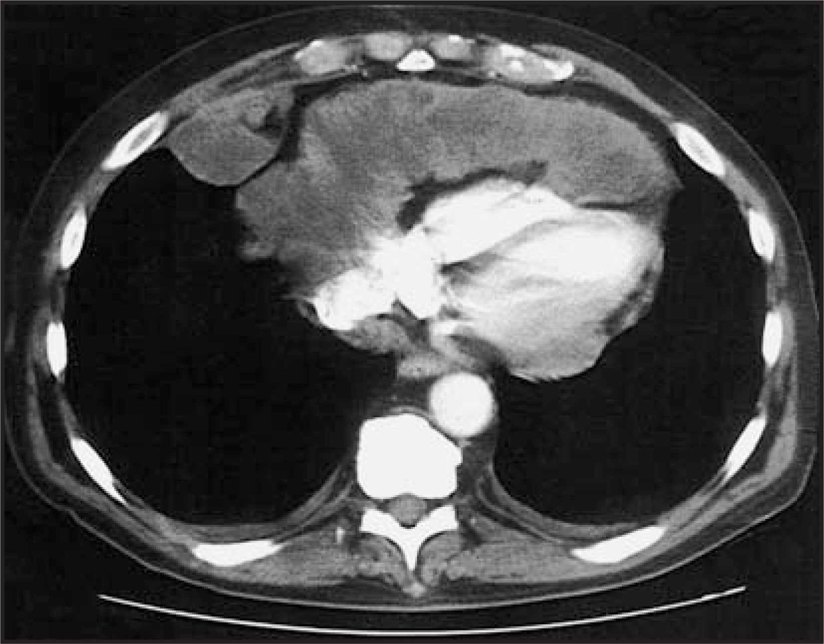
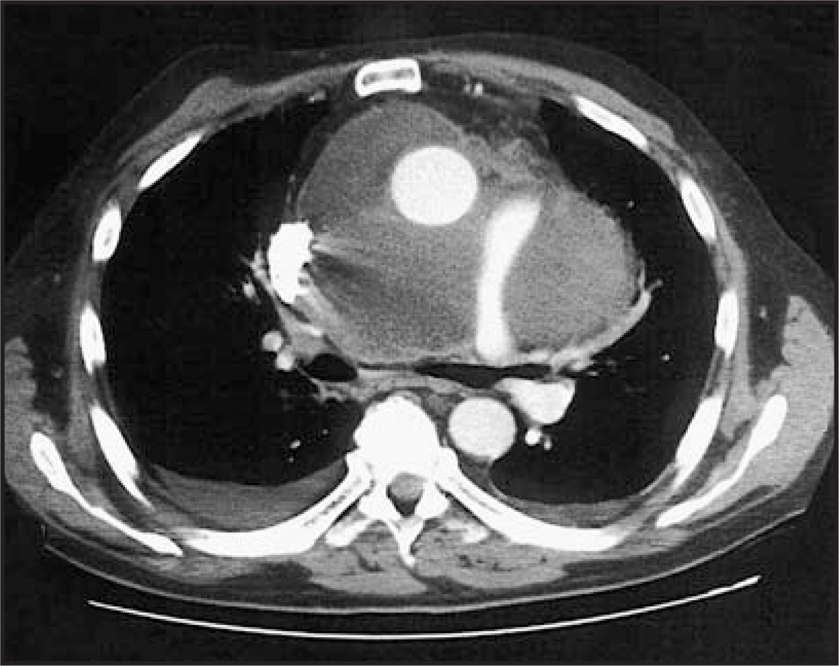
A 61-year-old white man presented with shortness of breath, nonproductive cough, fatigue, dysphagia, weakness, and chest tightness. A diagnostic workup revealed a pericardial effusion and massive mediastinal adenopathy encasing the aorta and pulmonary arteries. The patient underwent a short course of radiation treatment. The patient presented 3 weeks later with increasing shortness of breath. A chest X-ray revealed further widening of the mediastinum (not shown). Subsequent computed tomography (CT) revealed massive mediastinal and pericardiophrenic adenopathy, persistent pericardial effusion, compression of the main and left pulmonary arteries, and bilateral pleural effusions (Figures 1 and 2). Two-dimensional (2D) echocardiography revealed cardiac tamponade. The patient underwent palliative surgical debulking secondary to rapid tumor enlargement.
IMAGING FINDINGS
Multidetector (16-slice) CT axial images of the chest were obtained after the administration of intravenous contrast (GE Medical Systems, Waukesha, WI). Imaging revealed a rind of partially necrotic soft tissue completely replacing the normal pericardium (Figure 1). Massive contiguous pathologic adenopathy was also noted in the right cardiophrenic region. Tumor extended superiorly to surround the great vessels leading to compression of the left pulmonary artery (Figure 2). The left mainstem bronchus was also narrowed by significant tumor mass effect. A large volume of fluid was noted within the right lateral portion of the superior aortic recess. The patient progressed rapidly to the point of cardiac tamponade, presumably as a result of these combined factors.
DIAGNOSIS
Pleomorphic rhabdomyosarcoma involving the pericardium and lung pleura
DISCUSSION
Rhabdomyosarcoma (RMS) is a malignant skeletal muscle neoplasm that is histologically subclassified into the embryonal, alveolar, and pleomorphic variants. Typically a childhood malignancy, RMS is exceedingly rare in adults. Soft tissue sarcomas comprise <1% of all adult malignancies, and RMS accounts for 3% of all soft tissue sarcomas.1 Pleomorphic RMS is characterized by bizarre polygonal, round, and spindle cells displaying evidence of skeletal muscle differentiation without identifiable alveolar or embryonal components. Pleomorphic RMS is the most common RMS subtype reported in adults, typically occurring in men in their sixth decade of life and usually found within the deep skeletal muscles of the extremities and trunk.
Currently, the diagnosis of pleomorphic RMS is best suggested by clinical presentation, radiographic findings, and light microscopy, but this diagnosis requires either immunohistochemical or ultrastructural confirmation. Pleomorphic RMS is variably immunoreactive for desmin and always reactive for vimentin and actin isoforms.2 On ultrastructural exam, diagnosis is confirmed with identification of the spectrum of rhabdomyoblastic differentiation and rudimentary sarcomere formation consisting of Z bands or Z band material.3 Radiographic analysis usually shows large masses of variable attenuation due to necrosis and cystic components.4
The histologic variant and location of adult RMS influences prognosis. Treatment must be aggressive because of the nature of this neoplasm, generally combining surgery and chemotherapy, with or without radiation. As shown in previous studies, the prognosis for adults with RMS is quite poor, with a mortality rate >70%, in comparison with the prognosis for children with RMS, with a mortality rate of <45%.5-7 This patient died 3 months after the initial diagnosis.
The mediastinal tumor samples taken from our patient stained positively for vimentin, muscle-specific actin, and desmin and were negative for cytokeratin AE 1/3, cytokeratin 7, cytokeratin 20, epithelial membrane antigen (EMA), calretinin, S100 protein, melanoma antibody (HMB45), and leukocyte common antigen. Tumor samples revealed high-grade large-cell malignancy with extensive necrosis. Sections of tumor showed areas of large “giant” multinucleated anaplastic rhabdomyoblasts, thus making the diagnosis of pleomorphic RMS involving the pericardium and lung pleura.
In adults, pleomorphic RMS rarely arises in the mediastinum.4 To date, few instances of mediastinal pleomorphic RMS have been reported, thus it is imperative that new cases are documented as treatment options are further defined. Therapeutic success in treating adults with pleomorphic RMS has been limited, and long-term survival rates remain poor, in the range of 35% to 45%.4 Clinical experience is limited to case reports, small series, and nonrandomized therapeutic approaches.3,7 The purpose of this report is to highlight the aggressive nature of adult RMS with the intention of stimulating further research into the appropriate management of these patients.
References
- Ferrari A, Dileo P, Casanova M. Rhabdomyosarcoma in adults: A retrospective analysis of 171 patients treated at a single institution. Cancer. 2003;98:571-580.
- Gaffney E, Dervan P, Fletcher C. Pleomorphic rhabdomyosarcoma in adulthood. Analysis of 11 cases with definition of diagnostic criteria. Am J Surg Pathol. 1993;17:601-609.
- Montgomery E, Barr F, Fletcher C, Unni K, Mertens F. World Health Organization Classification of Tumours.Pathology & Genetics, Tumours of Soft Tissue and Bone. 2002:153.
- Gladish G, Sabloff B, Munden R. Primary thoracic sarcomas. RadioGraphics. 2002;22:621-637.
- Furlong M, Mentzel T, Fanburg-Smith J. Pleomorphic rhabdomyosarcoma in adults: A clinicopathologic study of 38 cases with emphasis on morphologic variants and recent skeletal muscle-specific markers. Mod Pathol. 2001;14:595-603.
- Prestidge B, Donaldson S. Treatment results among adults with childhood tumors: A 20-year experience. Int J Radiation Oncology Biol Phys. 1989;17:507-514.
- Little D, Ballo M, Zagars G. Adult rhabdomyosarcoma: Outcome following multimodality treatment. Cancer. 2002;95:377-388.
Citation
. RCOM RADIOLOGICAL CASE OF THE MONTH. Applied Radiology. 2006;35(8):44-45. doi:10.37549/AR1445.