RCOM RADIOLOGICAL CASE OF THE MONTH
Applied Radiology — Vol. 35 , Issue 8 , pp. 40 -43
DOI: 10.37549/AR1446
Published: August 1, 2006
Categories
CASE SUMMARY
A 54-year-old man presented with acute worsening of joint pain involving the hands and wrists. A careful history revealed an approximately 20-year period of mild, yet slowly progressive, multiple joint arthralgias, most notably in the fingers of both hands and in the wrists, knees, and shoulders. The patient reported that he had previously received intermittent treatment with over-the-counter analgesics for what he was told was “arthritis of old age.”
IMAGING FINDINGS
Radiographic imaging of both hands and wrists revealed joint-space narrowing and sclerosis, involving the metacarpal (MCP) joints, 1st carpometacarpal (CMC) joints, and multiple interphalangeal (IP) joints (Figure 1). Subchondral cysts were noted to involve the bilateral capitates, the left hamate, and the right scaphoid. Furthermore, prominent beak-like osteophytes were observed on the radial aspect of the metacarpals, bilaterally. Plain-film imaging of the knee (Figure 2) and shoulders (Figure 3) revealed degenerative changes in the bilateral knees and the left glenohumeral joint.
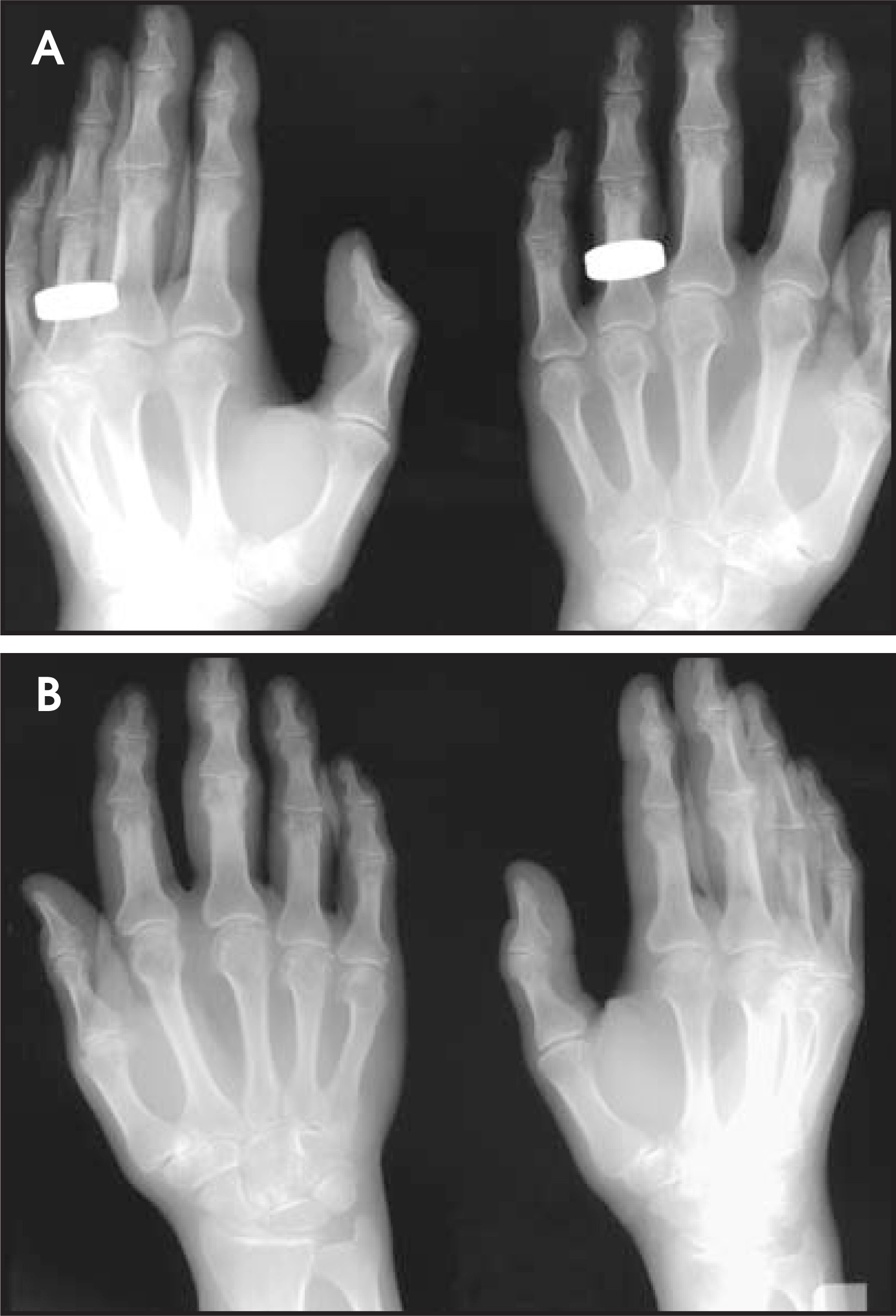
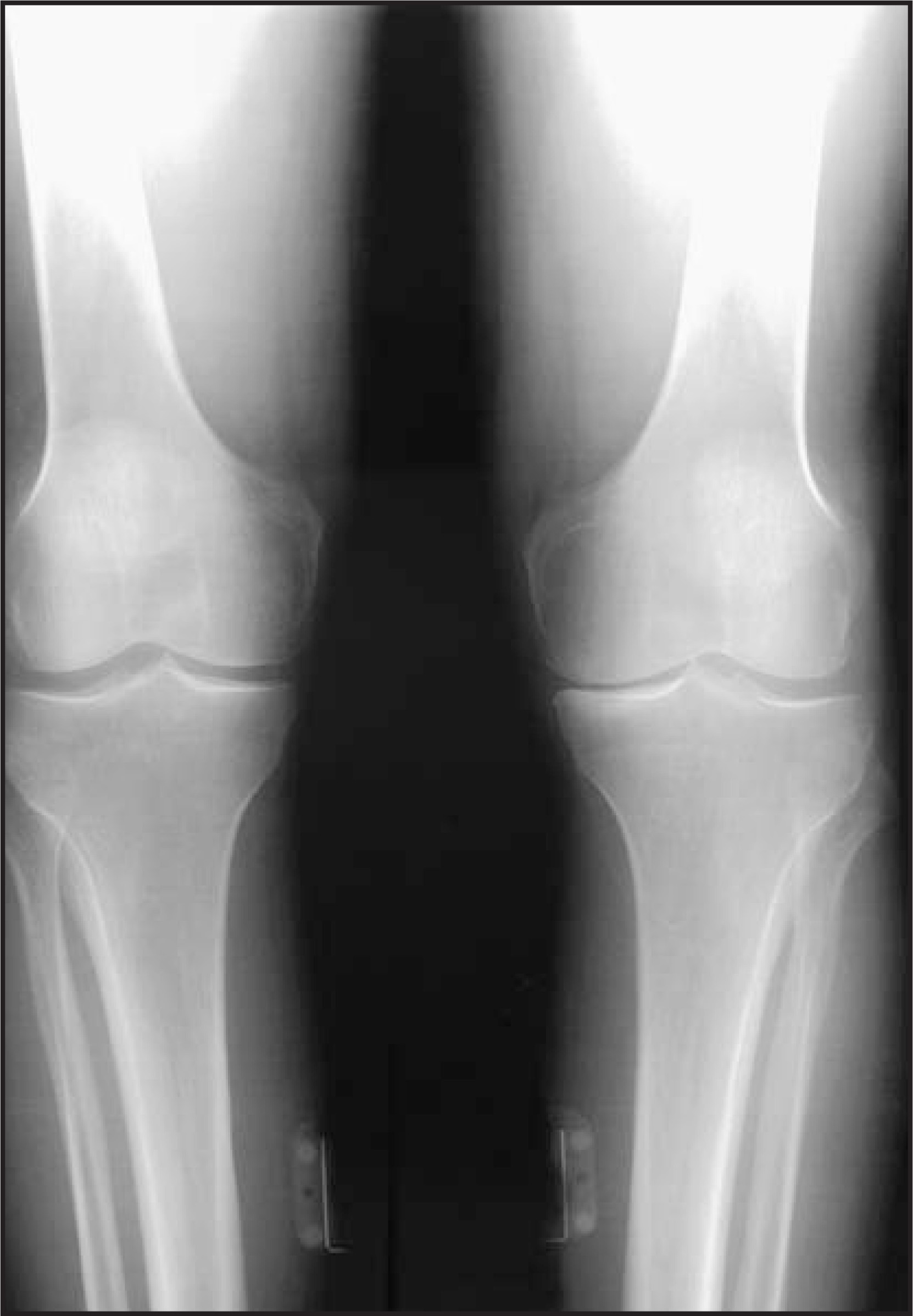
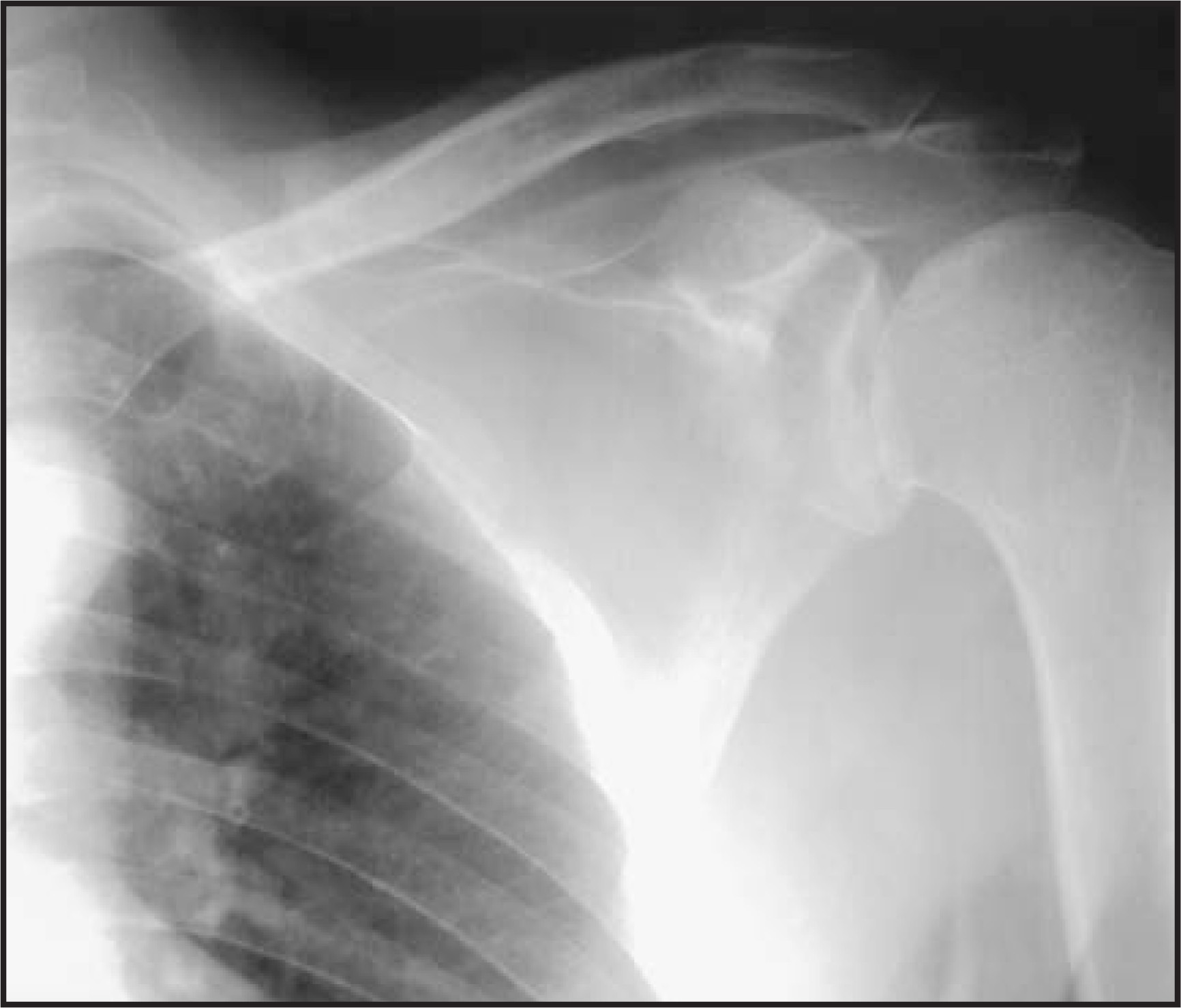
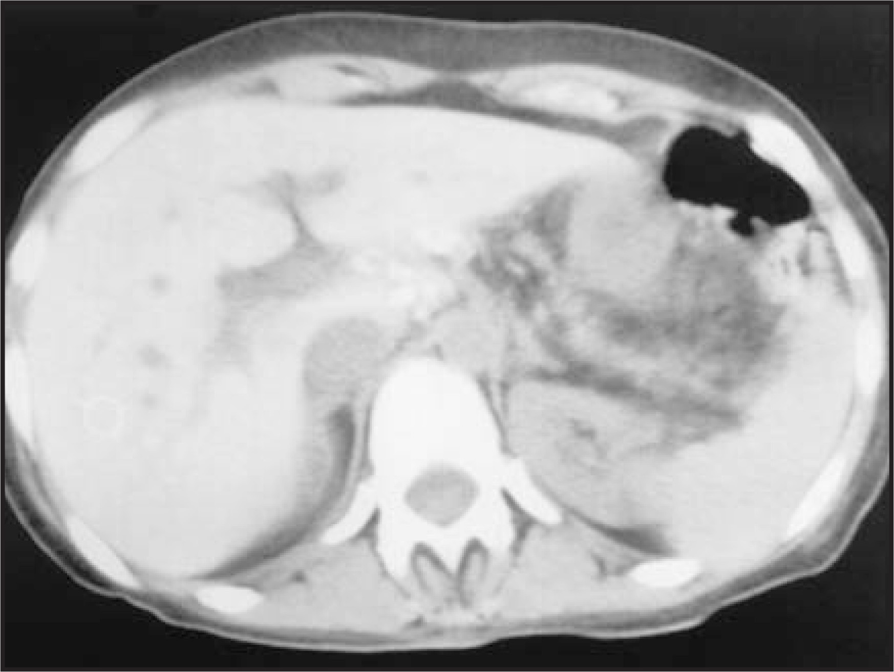
Laboratory work-up revealed elevations of both serum ferritin and transferring saturation (63%). Subsequently, a noncontrast CT of the abdomen was obtained (Figure 4), which showed marked increase in the attenuation of the liver and spleen (110 to 130 HU) as well as a 2-cm low-density solitary mass in the caudate lobe (which was found to represent a regenerative nodule). The patient underwent open liver biopsy, and the findings were consistent with hemochromatosis.
DIAGNOSIS
Hemochromatosis arthropathy
DISCUSSION
Hemochromatosis is a rare disorder characterized pathologically by tissue damage produced by iron deposition. Specific clinical manifestations relate to the site of abnormal iron accumulation: Iron within the parenchymal cells of the liver is associated with hypertrophy and cirrhosis; iron deposits in the pancreas result in diabetes; iron and melanin accumulations in the skin produce abnormal pigmentation; and cardiac deposition of iron results in heart failure.1 The disease can be classified as either primary (endogenous or idiopathic) or secondary hemochromatosis. Primary hemochromatosis is believed to be a consequence of a genetically determined error of metabolism in which an unexplained increased absorption of iron occurs from the gastrointestinal tract. It is widely accepted that the disease occurs secondary to a dominant gene that shows poor penetrance and is intimately linked to histocompatibility antigens HLA-A3, HLA-B7, and HLA-B14.2,3 Secondary hemochromatosis is associated with increased intake and accumulation of iron of known cause, such as alcoholic cirrhosis, multiple blood transfusions, refractory anemia, and chronic excess oral iron ingestion.1 Significant iron overload occurs only after many years, so the onset of disease is usually between 40 and 60 years of age. Men are affected 10 to 20 times more often than women, presumably because of menstrual blood loss.4 Serologic markers for the disease include increased ferritin and transferrin saturation.5 Definitive diagnosis is made by liver biopsy, which reveals histologic evidence of iron overload.
The arthritis of hemochromatosis was first described by Schumacher in 1964.1 It is insidious in onset and may occur at any stage during the course of the disease; in rare cases, it precedes other features.6 The arthropathy associated with hemochromatosis is manifested as a noninflammatory condition that initially involves the small joints of the hands, particularly the second and third metacarpophalangeal joints and eventually the large articulations, such as the knees, hips, and shoulders. Symptoms and signs include mild joint pain, swelling, and stiffness, without evidence of increased warmth, erythema, or deformity. Involvement usually is symmetric and progressive.2 Attacks of acute arthritis may be related to the presence of calcium pyrophosphate dihydrate (CPPD) crystals, which may be superimposed on the chronic progressive arthritic changes (that resemble primary degenerative joint disease) in as many as 30% of patients with hemochromatosis.1
The development of arthropathy appears to be intimately related to the deposition of small amounts of iron or hemosiderin within affected joints. Schumacher1 has proposed that iron alters cartilage matrix either directly or indirectly by impairing chondrocyte function and that synovial iron deposition indirectly impairs cartilage nutrition. Furthermore, ferric salts promote the formation and deposition of intra-articular CPPD crystals by inhibiting the activity of synovial pyrophosphatase (manifested as chondrocalcinosis on radiographs) and by decreasing the clearance of intra-articular immune complexes by inhibiting the activity of synovial reticuloendothelial cells.7
The arthropathy superficially resembles degenerative joint disease, with joint-space narrowing, sclerosis, and osteophytosis; but, in hemochromatosis, it reveals definite characteristics in its distribution and appearance that enable it to be recognized on radiographic examination.1 Arthropathy may produce abnormalities at joints that are not commonly involved in degenerative joint disease, such as the metacarpophalangeal joints, wrists, elbows, and glenohumeral articulations. The metacarpophalangeal joints are the most characteristic sites of involvement. Abnormalities are particularly frequent in the second and third metacarpophalangeal joints and peculiar hook-like osteophytes on the radial aspect of the metacarpal heads are characteristic.8 Widespread abnormalities of the wrist can be seen with involvement of the carpal bones in 30% to 50% of patients. Occasionally, the radiocarpal compartment may be unaffected. Arthropathy may be associated with multiple cysts in the subchondral bone, which can occasionally reach a large size. Furthermore, hemochromatosis arthropathy may be characterized by symmetric loss of articular space, an unusual finding in a degenerative joint disease. The joint-space loss is associated with subchondral bony eburnation and cyst formation. Although changes may be more rapid, in general, the arthropathy of hemochromatosis usually progresses slowly.
If untreated, death may occur by congestive heart failure (30%), liver failure or portal hypertension (25%), or hepatoma (30%). The best current treatment is vigorous phlebotomy, with the goal of removing 500 mL of whole blood 1 to 3 times a week until the hematocrit level drops to 32 to 25. Chelating agents (desferoxamine) are not as effective, but can serve as alternative therapy for patients with anemia or hypoproteinemia severe enough to contraindicate phlebotomy. With therapy, the 5-year survival rate increases from 33% to 89%.8
CONCLUSION
Hemochromatosis is an uncommon disease of iron metabolism that often manifests in middle age as a slowly progressive and symmetric arthropathy. This case illustrates the typical degenerative features and distribution. Knowledge of the plain-film findings and distribution of the disease can result in detection early enough to modify the course of the arthropathy.
References
- Schumacher H. Hemochromatosis and arthritis. Arthritis Rheum. 1964;7:41-50.
- Resnick D. Diagnosis of Bone and Joint Disorders. 1995:1649-1661.
- Hirsch J, Killien F, Troupin R. The arthropathy of hemochromatosis. Radiology. 1976;118:591-596.
- Powell L, Bassett M, Halliday J. 1980 update. Gastroenterology. 1980;78:374-381.
- Eustace S, Baker N, Lan H. Hemochromatosis arthropathy. Radiol Clin North Am. 1996;34:441-445.
- Kempis J. Arthropathy in hereditary hemochromatosis. Curr Opin Rheumatol. 2001;13:80-83.
- Dabbagh A, Trenam C, Morris C. Iron in joint inflammation. Ann Rheum Dis. 1993;51:67-73.
- Sartoris D. Rheumatologic disorders. Musculoskel Imaging. 1996;3:129-141.
Citation
. RCOM RADIOLOGICAL CASE OF THE MONTH. Applied Radiology. 2006;35(8):40-43. doi:10.37549/AR1446.