RCOM RADIOLOGICAL CASE OF THE MONTH
Applied Radiology — Vol. 35 , Issue 10 , pp. 55 -55
DOI: 10.37549/AR1456
Published: October 1, 2006
Categories
CASE SUMMARY
A young, sexually active woman presented for a pelvic magnetic resonance imaging (MRI) study as a component of an infertility workup following an abnormal physical examination and nondiagnostic ultrasound study (images not available). At the time of the examination, the patient reported no further medical conditions, and a review of systems was negative. No constitutional symptoms were present.
IMAGING FINDINGS
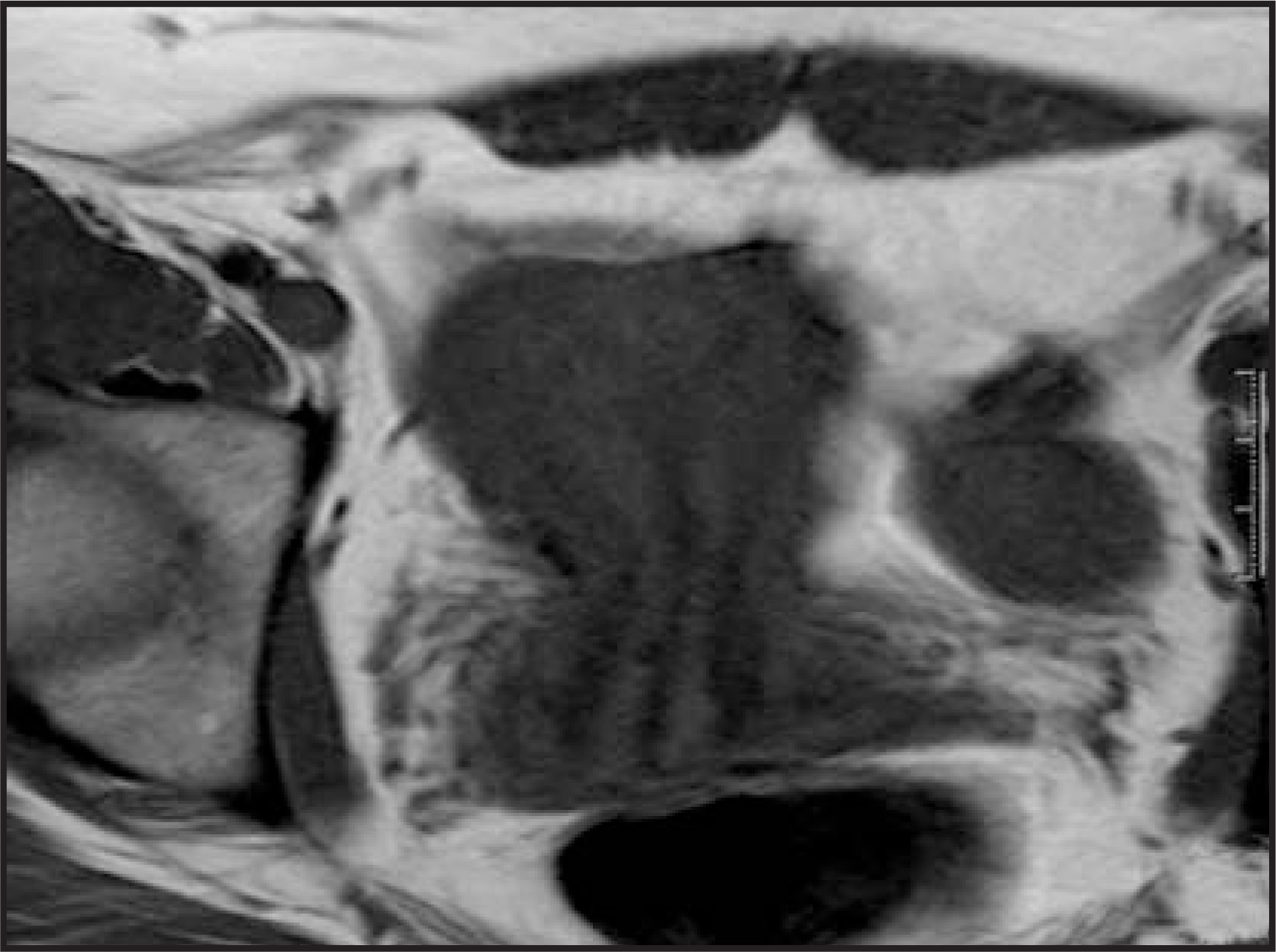
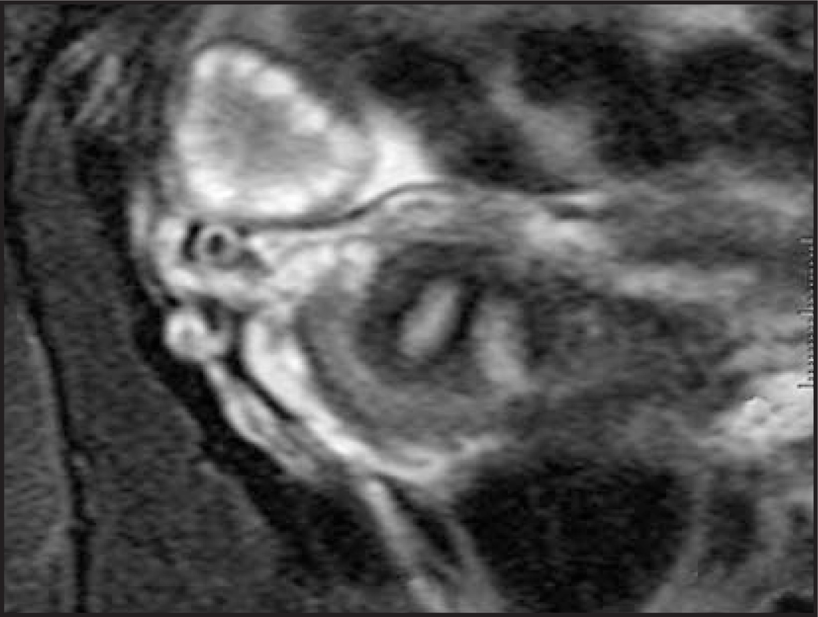
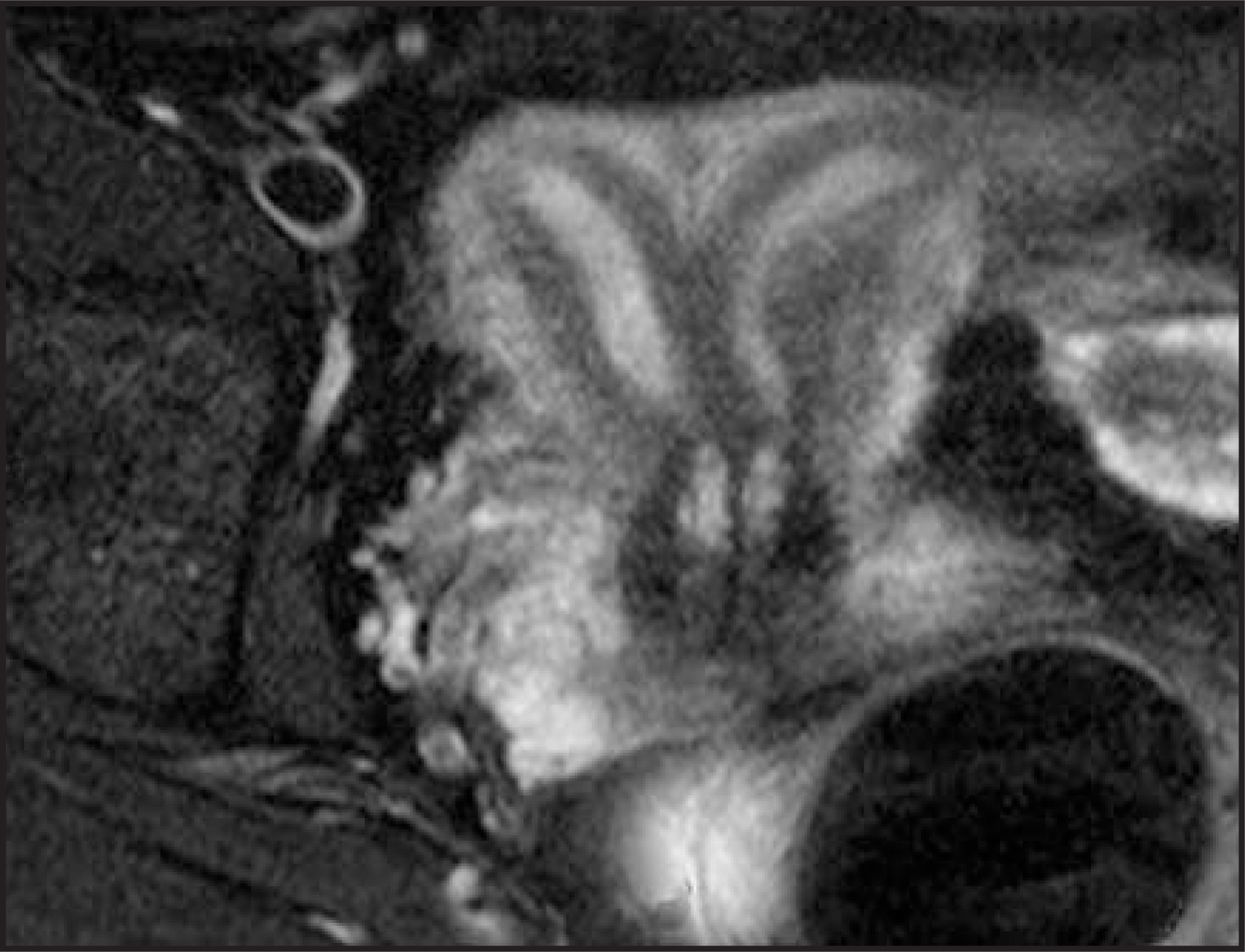
MRI shows a normal external contour of the uterine fundus (Figures 1 and 3) and 2 endometrial canals (Figures 2 and 3). The uterine division extends to the level of the cervix (Figure 3).
DIAGNOSIS
Septate uterus
DISCUSSION
Müllerian duct anomalies in the female patient include a lack of development (hypoplasia, aplasia, or the unicornuate uterus), lack of fusion at the midline (didelphys or bicornuate uterus), or lack of resorption of midline tissue after fusion (septate and arcuate uteri). Those anomalies that result in the appearance of 2 symmetric endometrial cavities (septate, bicornuate, and didelphys uteri) may be difficult to distinguish. This distinction is important if infertility warrants surgical repair. No surgery is undertaken in didelphys uteri, whereas the approach in bicornuate uteri is laparoscopic and hysteroscopic in septate uteri.1, 2
Uterine didelphys is complete nonfusion of the müllerian ducts with 2 uteri, 2 cervices, and duplication of the upper third of the vagina. The bicornuate uterus results from partial nonfusion with 1 cervix and vagina. In a septate uterus, there is complete fusion with a single uterus, but lack of resorption of the midline septum. Septal resorption occurs from caudal to cranial and may be arrested at any point, resulting in variable length of the septum. In the most severe case, the septum extends to the level of the cervix, or even perhaps the upper vagina, resulting in a similar appearance to a didelphys uterus.2
Characterization of the intervening uterine tissue and presence or absence of cervical duplication are not reliable distinguishing features. The key to diagnosis is the contour of the fundus. The fundal contour is normal in the septate uterus, with a deep concavity (>1 cm) in didelphys and bicornuate uteri.3,4,5
CONCLUSION
MRI is the most accurate modality for characterizing this type of abnormality and is used when other imaging studies are inconclusive. The most important sequence is a T2-weighted acquisition in the coronal oblique plane, parallel to the long axis of the uterus. This case of a septate uterus shows 2 endometrial canals and cervices and a normal external uterine contour.
References
- Zagoria R. Genitourinary Radiology:Radiology Requisites Series. 2004.
- Troiano R, McCarthy S. Mullerian duct anomalies: Imaging and clinical issues. Radiology. 2004;233:19-34.
- Imaoka I, Wada A, Matsuo M. MR imaging of disorders associated with female infertility: Use in diagnosis, treatment, and management. RadioGraphics. 2003;23:1401-1421.
- Saleem S. MR imaging diagnosis of uterovaginal anomalies: Current state of the art. RadioGraphics. 2003;23.
- Salim R, Regan L, Woelfer B. A comparative study of the morphology of congenital uterine anomalies in women with and without a history of recurrent first trimester miscarriage. Hum Reprod. 2003;18:162-166.
Citation
. RCOM RADIOLOGICAL CASE OF THE MONTH. Applied Radiology. 2006;35(10):55-55. doi:10.37549/AR1456.