RCOM RADIOLOGICAL CASE OF THE MONTH
Applied Radiology — Vol. 35 , Issue 10 , pp. 50 -52
DOI: 10.37549/AR1457
Published: October 1, 2006
Categories
CASE SUMMARY
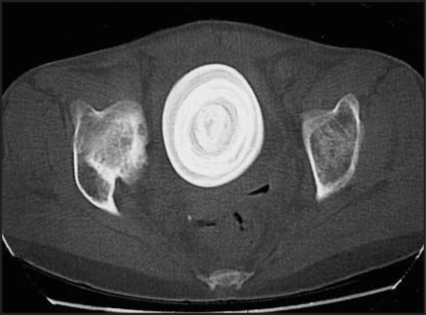
A 37-year-old woman with a history of illicit polysubstance abuse was transferred to our institution after a large calcified pelvic mass was found on computed tomography (CT) (Figure 1). Gross painless hematuria of several months’ duration, prompted her emergency department visit. The patient denied any previous abdominal or pelvic surgery, previous urologic instrumentation, urinary difficulty, or incontinence. Clinically, a rock-hard suprapubic mass was appreciated.
IMAGING FINDINGS
Pelvic CT showed “concentric ring” vesical calculus and normal upper urinary tracts (Figure 1).
DIAGNOSIS
Giant bladder calculus
CASE FOLLOW-UP
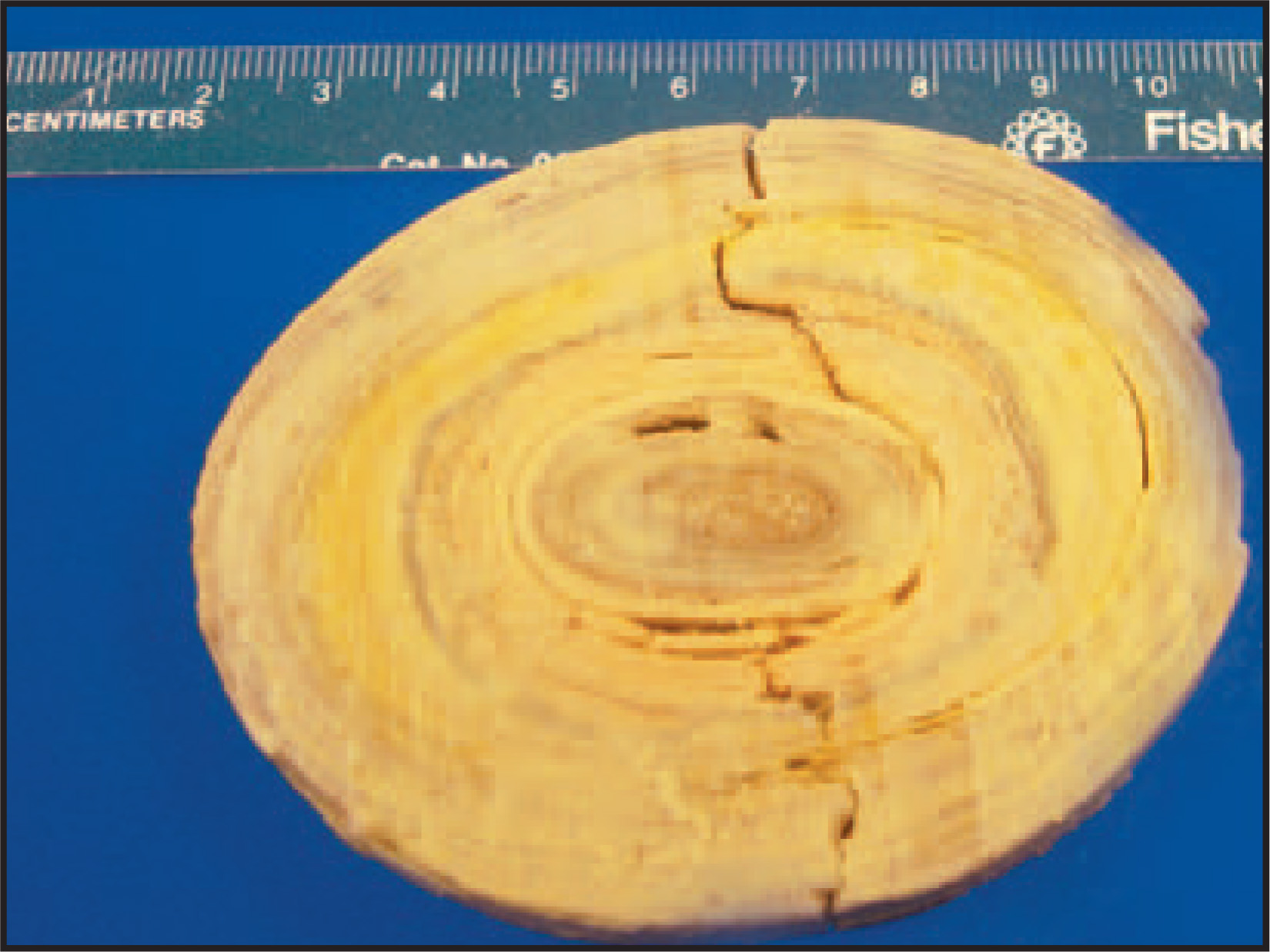
In view of the size of the calculus, an open cystolithotomy with suprapubic catheter placement was performed. No foreign objects, diverticula, sutures, or any other nidus for infection were seen within the bladder. The stone was composed mainly of magnesium ammonium phosphate (struvite) with calcium oxalate monohydrate and calcium phosphate components (Figure 2). The stone had a dry weight of 400 g and measured 9 × 8 × 5 cm.
Based on calculus composition, we presume the patient had a chronic urinary tract infection by a urease-splitting organism, possibly in the context of a neurogenic bladder. The patient failed to return for further follow-up and evaluation.
DISCUSSION
Bladder calculi have beleaguered man for thousands of years, as documented by the ancient Greeks and discovered by an archeologist in a 7000-year-old Egyptian skeleton. The incidence of vesical calculi has decreased in the Western world over the past few decades and females are generally less commonly affected than males. Bladder calculi are most often seen in the setting of obstruction, foreign bodies, or an infection1; furthermore, these factors are by no means mutually exclusive and may each contribute to stone formation.2 Among females, those at higher risk include women who have undergone anti-incontinence surgeries and those with genital prolapse. Several procedures that can predispose a patient to stone formation include vesical neck suspension procedures or bladder augmentations. The risk lies in the procedural type and technique and may often be due to exposure to suspension sutures, polypropylene pledgets, or mesh––all of which may serve as a nidus for crystallization.1 Prolonged Foley catheterization or another foreign body may also serve as a source of infection or nidus of crystallization.1 Additionally, there are many documented reports of foreign body migration into the bladder, including intrauterine devices and intravaginal gynecologic accessories, such as diaphragms, pessaries, and cerclages, as well as items used in autoerotic behavior.1 Anatomic anomalies that may aid in stone formation include bladder diverticuli3 and genital prolapse. In cases of genital prolapse, patients suffer from bladder obstruction as a consequence of urethral kinking,1 which may prompt urinary retention or chronic urinary tract infection and may lead to stone formation.
Bladder calculi may often present as they did in this patient, with gross hematuria. Patients may or may not complain of discomfort, pain, or lower urinary tract symptoms – depending on the underlying cause as well as the severity of disease. Obstruction caused by a vesical calculus can lead to further infection, urosepsis, and, although rare, bladder perforation,4 hydronephrosis, and acute renal failure.1
Bladder stones may often be multilayered, as in the case of this patient. Studies have shown that the stone nucleus often does not contain struvite or calcium phosphate, whereas subsequent concentric layers contain large amounts of these substances. This indicates that infection may not be the inciting factor in stone formation, but may play a major role in further stone crystallization.1
Most bladder calculi, by nature of their composition, are radiopaque. Further, as in this case, the concentric nature is evidenced in radiologic studies, such as the CT scan. This may correlate pathologically with the mixed composition of the stone, and with the mechanism of stone formation. A host of radiologic studies can show bladder calculi, including CT, magnetic resonance imaging, ultrasound, and intravenous urogram, but contrast-enhanced CT is the test of choice because of its remarkable sensitivity in detecting urinary tract stones, including those composed of uric acid.
References
- Schwartz B, Stoller M. The vesical calculus. Urol Clin North Am. 2000;27:333-346.
- Leach G, Fitzpatrick T. Giant vesical calculi in the female. Urology. 1981;17:274-275.
- Di Tonno F, Forte M, Guidoni E. A giant bladder stone. Br J Urol. 1988;62:90-91.
- Basu A, Mojahid I, Williamson E. Spontaneous bladder rupture resulting from giant vesical calculus. Br J Urol. 1994;74:385-386.
Citation
. RCOM RADIOLOGICAL CASE OF THE MONTH. Applied Radiology. 2006;35(10):50-52. doi:10.37549/AR1457.